 Movie
Movie Controller

[English] 日本語
 Yorodumi
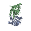
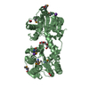
- PDB-1amh: UNCOMPLEXED RAT TRYPSIN MUTANT WITH ASP 189 REPLACED WITH SER (D189S) -
+ Open data

- Basic information
| Entry | Database: PDB / ID: 1amh | ||||||
|---|---|---|---|---|---|---|---|
| Title | UNCOMPLEXED RAT TRYPSIN MUTANT WITH ASP 189 REPLACED WITH SER (D189S) | ||||||
 Components Components | ANIONIC TRYPSIN | ||||||
 Keywords Keywords |  HYDROLASE / SERINE PROTEASE / ACTIVATION DOMAIN / SUBSTRATE SPECIFICITY HYDROLASE HYDROLASE / SERINE PROTEASE / ACTIVATION DOMAIN / SUBSTRATE SPECIFICITY HYDROLASE | ||||||
| Function / homology |  Function and homology informationAntimicrobial peptides / Alpha-defensins / Activation of Matrix Metalloproteinases / Collagen degradation / Neutrophil degranulation / collagen catabolic process / trypsin / digestion / response to nutrient / serine-type endopeptidase activity ...Antimicrobial peptides / Alpha-defensins / Activation of Matrix Metalloproteinases / Collagen degradation / Neutrophil degranulation / collagen catabolic process / trypsin / digestion / response to nutrient / serine-type endopeptidase activity / calcium ion binding / proteolysis / extracellular space / extracellular region Function and homology informationAntimicrobial peptides / Alpha-defensins / Activation of Matrix Metalloproteinases / Collagen degradation / Neutrophil degranulation / collagen catabolic process / trypsin / digestion / response to nutrient / serine-type endopeptidase activity ...Antimicrobial peptides / Alpha-defensins / Activation of Matrix Metalloproteinases / Collagen degradation / Neutrophil degranulation / collagen catabolic process / trypsin / digestion / response to nutrient / serine-type endopeptidase activity / calcium ion binding / proteolysis / extracellular space / extracellular regionSimilarity search - Function | ||||||
| Biological species |  Rattus rattus (black rat) Rattus rattus (black rat) | ||||||
| Method | X-RAY DIFFRACTION / MOLECULAR REPLACEMENT / Resolution: 2.5 Å | ||||||
 Authors Authors | Szabo, E. / Bocskei, Z.S. / Naray-Szabo, G. / Graf, L. | ||||||
 Citation Citation | Journal: Eur.J.Biochem. / Year: 1999 Title: The three-dimensional structure of Asp189Ser trypsin provides evidence for an inherent structural plasticity of the protease. Authors: Szabo, E. / Bocskei, Z. / Naray-Szabo, G. / Graf, L. #1: Journal: Biochemistry / Year: 1994Title: Exogenous Acetate Reconstitutes the Enzymatic Activity of Trypsin Asp189Ser Authors: Perona, J.J. / Hedstrom, L. / Wagner, R.L. / Rutter, W.J. / Craik, C.S. / Fletterick, R.J. #2: Journal: Proc.Natl.Acad.Sci.USA / Year: 1988Title: Electrostatic Complementarity within the Substrate-Binding Pocket of Trypsin Authors: Graf, L. / Jancso, A. / Szilagyi, L. / Hegyi, G. / Pinter, K. / Naray-Szabo, G. / Hepp, J. / Medzihradszky, K. / Rutter, W.J. #3: Journal: Acc.Chem.Res. / Year: 1978Title: Structural Basis of the Activation and Action of Trypsin Authors: Huber, R. / Bode, W. | ||||||
| History |
|
- Structure visualization
| Structure viewer | Molecule: MolmilJmol/JSmol |
|---|
- Downloads & links
-Download
| PDBx/mmCIF format | 1amh.cif.gz | 93.8 KB | Display | PDBx/mmCIF format |
|---|---|---|---|---|
| PDB format | pdb1amh.ent.gz | 74.1 KB | Display | PDB format |
| PDBx/mmJSON format | 1amh.json.gz | Tree view | PDBx/mmJSON format | |
| Others |  Other downloads Other downloads |
-Validation report
| Arichive directory | https://data.pdbj.org/pub/pdb/validation_reports/am/1amhftp://data.pdbj.org/pub/pdb/validation_reports/am/1amh | HTTPS FTP |
|---|
-Related structure data
| Related structure data |  1brbS S: Starting model for refinement |
|---|---|
| Similar structure data |


-Links
 PDBj
PDBj


- Assembly
| Deposited unit | 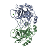
| |||||||||
|---|---|---|---|---|---|---|---|---|---|---|
| 1 |
| |||||||||
| Unit cell |
| |||||||||
| Noncrystallographic symmetry (NCS) | NCS domain:
NCS oper: (Code: given Matrix: (0.739109, 0.012922, -0.673462), Vector : |
-Components
| #1: Protein | Mass: 23786.828 Da / Num. of mol.: 2 / Mutation: D189S Source method: isolated from a genetically manipulated source Source: (gene. exp.) Rattus rattus (black rat) / Organ: PANCREAS / Plasmid: PYES2 / Cellular location (production host): SECRETION / Production host:  Saccharomyces cerevisiae (brewer's yeast) / Strain (production host): INVSC1 / References: UniProt: P00763, trypsin Saccharomyces cerevisiae (brewer's yeast) / Strain (production host): INVSC1 / References: UniProt: P00763, trypsin#2: Chemical |   Mass: 40.078 Da / Num. of mol.: 2 / Source method: obtained synthetically / Formula: Ca Mass: 40.078 Da / Num. of mol.: 2 / Source method: obtained synthetically / Formula: Ca#3: Water | ChemComp-HOH / | Water Mass: 18.015 Da / Num. of mol.: 128 / Source method: isolated from a natural source / Formula: H2O Mass: 18.015 Da / Num. of mol.: 128 / Source method: isolated from a natural source / Formula: H2O |
|---|
-Experimental details
-Experiment
| Experiment | Method: X-RAY DIFFRACTION / Number of used crystals: 1 |
|---|
- Sample preparation
| Crystal | Density Matthews: 2.07 Å3/Da / Density % sol: 61 % | ||||||||||||||||||||||||||||||||||||
|---|---|---|---|---|---|---|---|---|---|---|---|---|---|---|---|---|---|---|---|---|---|---|---|---|---|---|---|---|---|---|---|---|---|---|---|---|---|
| Crystal grow | Temperature: 277 K / pH: 5.6 Details: CRYSTALLIZED AT 277 K FROM 10 MG/ML PROTEIN SOLUTION USING 22% PEG 3350, 10 MM CACL2 AND 0.15M NH4CL AS PRECIPITANT AT PH=5.6 (0.1 M SODIUM CITRATE). | ||||||||||||||||||||||||||||||||||||
| Crystal grow | *PLUS Temperature: 4 ℃ / Method: vapor diffusion, hanging drop | ||||||||||||||||||||||||||||||||||||
| Components of the solutions | *PLUS
|
-Data collection
| Diffraction | Mean temperature: 293 K |
|---|---|
| Diffraction source | Source: ROTATING ANODE / Type: RIGAKU RUH2R / Wavelength: 1.5418 |
| Detector | Type: RIGAKU / Detector: IMAGE PLATE / Date: Dec 1, 1995 |
| Radiation | Monochromator: GRAPHITE(002) / Monochromatic (M) / Laue (L): M / Scattering type: x-ray |
| Radiation wavelength | Wavelength: 1.5418 Å / Relative weight: 1 |
| Reflection | Resolution: 2.52→100 Å / Num. obs: 13312 / % possible obs: 94.5 % / Observed criterion σ(I): 0 / Redundancy: 3.1 % / Biso Wilson estimate: 36.25 Å2 / Rmerge(I) obs: 0.12 / Rsym value: 0.131 / Net I/σ(I): 7.7 |
| Reflection shell | Resolution: 2.5→2.6 Å / Redundancy: 3 % / Rmerge(I) obs: 0.296 / Mean I/σ(I) obs: 2.63 / Rsym value: 0.312 / % possible all: 91.9 |
| Reflection shell | *PLUS Highest resolution: 2.54 Å / Lowest resolution: 2.59 Å / % possible obs: 91.9 % / Num. unique obs: 470 / Mean I/σ(I) obs: 2.6 |
- Processing
| Software |
| ||||||||||||||||||||||||||||||||||||||||||||||||||||||||||||||||||||||||||||||||
|---|---|---|---|---|---|---|---|---|---|---|---|---|---|---|---|---|---|---|---|---|---|---|---|---|---|---|---|---|---|---|---|---|---|---|---|---|---|---|---|---|---|---|---|---|---|---|---|---|---|---|---|---|---|---|---|---|---|---|---|---|---|---|---|---|---|---|---|---|---|---|---|---|---|---|---|---|---|---|---|---|---|
| Refinement | Method to determine structure: MOLECULAR REPLACEMENT Starting model: PDB ENTRY 1BRB Resolution: 2.5→100 Å / Rfactor Rfree error: 0.007 / Data cutoff high absF: 100000 / Data cutoff low absF: 0.1 / Isotropic thermal model: RESTRAINED / Cross valid method: THROUGHOUT / σ(F): 2
| ||||||||||||||||||||||||||||||||||||||||||||||||||||||||||||||||||||||||||||||||
| Displacement parameters | Biso mean: 18.64 Å2 | ||||||||||||||||||||||||||||||||||||||||||||||||||||||||||||||||||||||||||||||||
| Refine analyze |
| ||||||||||||||||||||||||||||||||||||||||||||||||||||||||||||||||||||||||||||||||
| Refinement step | Cycle: LAST / Resolution: 2.5→100 Å
| ||||||||||||||||||||||||||||||||||||||||||||||||||||||||||||||||||||||||||||||||
| Refine LS restraints |
| ||||||||||||||||||||||||||||||||||||||||||||||||||||||||||||||||||||||||||||||||
| Refine LS restraints NCS | Refine-ID: X-RAY DIFFRACTION / Weight Biso : 3
| ||||||||||||||||||||||||||||||||||||||||||||||||||||||||||||||||||||||||||||||||
| LS refinement shell | Resolution: 2.54→2.59 Å / Rfactor Rfree error: 0.05 / Total num. of bins used: 20
| ||||||||||||||||||||||||||||||||||||||||||||||||||||||||||||||||||||||||||||||||
| Xplor file |
| ||||||||||||||||||||||||||||||||||||||||||||||||||||||||||||||||||||||||||||||||
| Software | *PLUS Name: X-PLOR / Version: 3.851 / Classification: refinement | ||||||||||||||||||||||||||||||||||||||||||||||||||||||||||||||||||||||||||||||||
| Refinement | *PLUS Num. reflection obs: 12103 | ||||||||||||||||||||||||||||||||||||||||||||||||||||||||||||||||||||||||||||||||
| Solvent computation | *PLUS | ||||||||||||||||||||||||||||||||||||||||||||||||||||||||||||||||||||||||||||||||
| Displacement parameters | *PLUS | ||||||||||||||||||||||||||||||||||||||||||||||||||||||||||||||||||||||||||||||||
| Refine LS restraints | *PLUS
| ||||||||||||||||||||||||||||||||||||||||||||||||||||||||||||||||||||||||||||||||
| LS refinement shell | *PLUS Rfactor obs: 0.259 |
