 Movie
Movie Controller

[English] 日本語
 Yorodumi
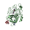
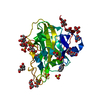
- PDB-1a5h: CATALYTIC DOMAIN OF HUMAN TWO-CHAIN TISSUE PLASMINOGEN ACTIVATOR ... -
+ Open data

- Basic information
| Entry | Database: PDB / ID: 1a5h | ||||||
|---|---|---|---|---|---|---|---|
| Title | CATALYTIC DOMAIN OF HUMAN TWO-CHAIN TISSUE PLASMINOGEN ACTIVATOR COMPLEX OF A BIS-BENZAMIDINE | ||||||
 Components Components | (TISSUE PLASMINOGEN ACTIVATOR ) x 2 ) x 2 | ||||||
 Keywords Keywords | HYDROLASE / TRYPSIN LIKE SERINE PROTEASE / FIBRINOLYTIC ENZYME | ||||||
| Function / homology |  Function and homology informationt-plasminogen activator / prevention of polyspermy / trans-synaptic signaling by BDNF, modulating synaptic transmission / Signaling by PDGF / negative regulation of plasminogen activation / Dissolution of Fibrin Clot / smooth muscle cell migration / plasminogen activation / platelet-derived growth factor receptor signaling pathway / negative regulation of fibrinolysis ...t-plasminogen activator / prevention of polyspermy / trans-synaptic signaling by BDNF, modulating synaptic transmission / Signaling by PDGF / negative regulation of plasminogen activation / Dissolution of Fibrin Clot / smooth muscle cell migration / plasminogen activation / platelet-derived growth factor receptor signaling pathway / negative regulation of fibrinolysis / serine protease inhibitor complex / fibrinolysis / secretory granule / negative regulation of proteolysis / phosphoprotein binding / Schaffer collateral - CA1 synapse / protein modification process / blood coagulation / apical part of cell / response to hypoxia / signaling receptor binding / serine-type endopeptidase activity / glutamatergic synapse / cell surface / proteolysis / extracellular space / extracellular exosome / extracellular region / cytoplasm Function and homology informationt-plasminogen activator / prevention of polyspermy / trans-synaptic signaling by BDNF, modulating synaptic transmission / Signaling by PDGF / negative regulation of plasminogen activation / Dissolution of Fibrin Clot / smooth muscle cell migration / plasminogen activation / platelet-derived growth factor receptor signaling pathway / negative regulation of fibrinolysis ...t-plasminogen activator / prevention of polyspermy / trans-synaptic signaling by BDNF, modulating synaptic transmission / Signaling by PDGF / negative regulation of plasminogen activation / Dissolution of Fibrin Clot / smooth muscle cell migration / plasminogen activation / platelet-derived growth factor receptor signaling pathway / negative regulation of fibrinolysis / serine protease inhibitor complex / fibrinolysis / secretory granule / negative regulation of proteolysis / phosphoprotein binding / Schaffer collateral - CA1 synapse / protein modification process / blood coagulation / apical part of cell / response to hypoxia / signaling receptor binding / serine-type endopeptidase activity / glutamatergic synapse / cell surface / proteolysis / extracellular space / extracellular exosome / extracellular region / cytoplasmSimilarity search - Function | ||||||
| Biological species |  Homo sapiens (human) Homo sapiens (human) | ||||||
| Method | X-RAY DIFFRACTION / MOLECULAR REPLACEMENT / Resolution: 2.9 Å | ||||||
 Authors Authors | Renatus, M. / Bode, W. / Stubbs, M.T. | ||||||
 Citation Citation | Journal: J.Biol.Chem. / Year: 1997 Title: Structural mapping of the active site specificity determinants of human tissue-type plasminogen activator. Implications for the design of low molecular weight substrates and inhibitors. Authors: Renatus, M. / Bode, W. / Huber, R. / Sturzebecher, J. / Prasa, D. / Fischer, S. / Kohnert, U. / Stubbs, M.T. #1: Journal: Curr.Opin.Struct.Biol. / Year: 1997Title: Tissue-Type Plasminogen Activator: Variants and Crystal/Solution Structures Demarcate Structural Determinants of Function Authors: Bode, W. / Renatus, M. #2: Journal: J.Mol.Biol. / Year: 1996Title: The 2.3 A Crystal Structure of the Catalytic Domain of Recombinant Two-Chain Human Tissue-Type Plasminogen Activator Authors: Lamba, D. / Bauer, M. / Huber, R. / Fischer, S. / Rudolph, R. / Kohnert, U. / Bode, W. | ||||||
| History |
|
- Structure visualization
| Structure viewer | Molecule: MolmilJmol/JSmol |
|---|
- Downloads & links
-Download
| PDBx/mmCIF format | 1a5h.cif.gz | 133.5 KB | Display | PDBx/mmCIF format |
|---|---|---|---|---|
| PDB format | pdb1a5h.ent.gz | 108.4 KB | Display | PDB format |
| PDBx/mmJSON format | 1a5h.json.gz | Tree view | PDBx/mmJSON format | |
| Others |  Other downloads Other downloads |
-Validation report
| Arichive directory | https://data.pdbj.org/pub/pdb/validation_reports/a5/1a5hftp://data.pdbj.org/pub/pdb/validation_reports/a5/1a5h | HTTPS FTP |
|---|
-Related structure data
| Related structure data |  1rtfS S: Starting model for refinement |
|---|---|
| Similar structure data |


-Links
 PDBj
PDBj


- Assembly
| Deposited unit | 
| ||||||||||||
|---|---|---|---|---|---|---|---|---|---|---|---|---|---|
| 1 | 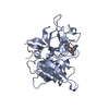
| ||||||||||||
| 2 | 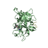
| ||||||||||||
| Unit cell |
| ||||||||||||
| Noncrystallographic symmetry (NCS) | NCS oper:
|
-Components
| #1: Protein/peptide | Mass: 840.968 Da / Num. of mol.: 2 / Fragment: HEAVY CHAIN FRAGMENT, CATALYTIC DOMAIN Source method: isolated from a genetically manipulated source Source: (gene. exp.) Homo sapiens (human) / Production host:  Escherichia coli (E. coli) / References: UniProt: P00750, t-plasminogen activator Escherichia coli (E. coli) / References: UniProt: P00750, t-plasminogen activator#2: Protein | / TC-TPA(BISB)Mass: 28157.883 Da / Num. of mol.: 2 / Fragment: LIGHT CHAIN, CATALYTIC DOMAIN Source method: isolated from a genetically manipulated source Source: (gene. exp.) Homo sapiens (human) / Production host: Escherichia coli (E. coli) / References: UniProt: P00750, t-plasminogen activator#3: Chemical | 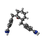  Mass: 376.495 Da / Num. of mol.: 2 / Source method: obtained synthetically / Formula: C23H28N4O Mass: 376.495 Da / Num. of mol.: 2 / Source method: obtained synthetically / Formula: C23H28N4O#4: Water | ChemComp-HOH / | Water Mass: 18.015 Da / Num. of mol.: 91 / Source method: isolated from a natural source / Formula: H2O Mass: 18.015 Da / Num. of mol.: 91 / Source method: isolated from a natural source / Formula: H2O |
|---|
-Experimental details
-Experiment
| Experiment | Method: X-RAY DIFFRACTION / Number of used crystals: 1 |
|---|
- Sample preparation
| Crystal | Density Matthews: 2.24 Å3/Da / Density % sol: 45 % | |||||||||||||||||||||||||||||||||||
|---|---|---|---|---|---|---|---|---|---|---|---|---|---|---|---|---|---|---|---|---|---|---|---|---|---|---|---|---|---|---|---|---|---|---|---|---|
| Crystal grow | pH: 4.5 / Details: pH 4.5 | |||||||||||||||||||||||||||||||||||
| Crystal grow | *PLUS Temperature: 23 ℃ / pH: 5 / Method: vapor diffusion, sitting drop | |||||||||||||||||||||||||||||||||||
| Components of the solutions | *PLUS
|
-Data collection
| Diffraction | Mean temperature: 280 K |
|---|---|
| Diffraction source | Source: ROTATING ANODE / Type: RIGAKU RUH2R / Wavelength: 1.5418 |
| Detector | Type: SIEMENS / Detector: AREA DETECTOR / Date: Sep 28, 1995 |
| Radiation | Monochromator: GRAPHITE(002) / Monochromatic (M) / Laue (L): M / Scattering type: x-ray |
| Radiation wavelength | Wavelength: 1.5418 Å / Relative weight: 1 |
| Reflection | Highest resolution: 2.9 Å / Num. obs: 12586 / % possible obs: 94.4 % / Observed criterion σ(I): 0 / Redundancy: 2 % / Rmerge(I) obs: 0.088 |
| Reflection shell | Resolution: 2.8→3.1 Å / Redundancy: 1.5 % / Rmerge(I) obs: 0.25 / % possible all: 81.2 |
| Reflection | *PLUS Lowest resolution: 9999 Å / Num. measured all: 25355 |
| Reflection shell | *PLUS % possible obs: 86.2 % |
- Processing
| Software |
| ||||||||||||||||||||||||||||||||||||||||||||||||||||||||||||
|---|---|---|---|---|---|---|---|---|---|---|---|---|---|---|---|---|---|---|---|---|---|---|---|---|---|---|---|---|---|---|---|---|---|---|---|---|---|---|---|---|---|---|---|---|---|---|---|---|---|---|---|---|---|---|---|---|---|---|---|---|---|
| Refinement | Method to determine structure: MOLECULAR REPLACEMENT Starting model: 1RTF Resolution: 2.9→7 Å / Data cutoff high absF: 100000 / Data cutoff low absF: 0.25 / σ(F): 2
| ||||||||||||||||||||||||||||||||||||||||||||||||||||||||||||
| Displacement parameters | Biso mean: 14.2 Å2 | ||||||||||||||||||||||||||||||||||||||||||||||||||||||||||||
| Refinement step | Cycle: LAST / Resolution: 2.9→7 Å
| ||||||||||||||||||||||||||||||||||||||||||||||||||||||||||||
| Refine LS restraints |
| ||||||||||||||||||||||||||||||||||||||||||||||||||||||||||||
| LS refinement shell | Resolution: 2.9→3 Å / Total num. of bins used: 10
| ||||||||||||||||||||||||||||||||||||||||||||||||||||||||||||
| Xplor file |
| ||||||||||||||||||||||||||||||||||||||||||||||||||||||||||||
| Software | *PLUS Name: X-PLOR / Version: 3.1 / Classification: refinement | ||||||||||||||||||||||||||||||||||||||||||||||||||||||||||||
| Refine LS restraints | *PLUS
| ||||||||||||||||||||||||||||||||||||||||||||||||||||||||||||
| LS refinement shell | *PLUS Lowest resolution: 3 Å |
