 Movie
Movie Controller

[English] 日本語
 Yorodumi
- EMDB-6517: Structure and function of outer dynein intermediate and light cha... -
+ Open data

- Basic information
| Entry | Database: EMDB / ID: EMD-6517 | |||||||||
|---|---|---|---|---|---|---|---|---|---|---|
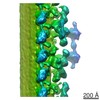
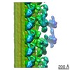
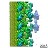
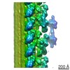
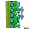
| Title | Structure and function of outer dynein intermediate and light chain complex | |||||||||
 Map data Map data | Streptavidin-labeled IC1248BCCP axoneme | |||||||||
 Sample Sample |
| |||||||||
 Keywords Keywords | cilia and flagella /  axoneme / outer dynein arm / intermediate chain / light chain axoneme / outer dynein arm / intermediate chain / light chain | |||||||||
| Biological species |   Chlamydomonas reinhardtii (plant) Chlamydomonas reinhardtii (plant) | |||||||||
| Method | subtomogram averaging / cryo EM / Resolution: 48.0 Å | |||||||||
 Authors Authors | Oda T / Abe T / Yanagisawa H / Kikkawa M | |||||||||
 Citation Citation | Journal: Mol Biol Cell / Year: 2016 Title: Structure and function of outer dynein arm intermediate and light chain complex. Authors: Toshiyuki Oda / Tatsuki Abe / Haruaki Yanagisawa / Masahide Kikkawa /  Abstract: The outer dynein arm (ODA) is a molecular complex that drives the beating motion of cilia/flagella. Chlamydomonas ODA is composed of three heavy chains (HCs), two ICs, and 11 light chains (LCs). ...The outer dynein arm (ODA) is a molecular complex that drives the beating motion of cilia/flagella. Chlamydomonas ODA is composed of three heavy chains (HCs), two ICs, and 11 light chains (LCs). Although the three-dimensional (3D) structure of the whole ODA complex has been investigated, the 3D configurations of the ICs and LCs are largely unknown. Here we identified the 3D positions of the two ICs and three LCs using cryo-electron tomography and structural labeling. We found that these ICs and LCs were all localized at the root of the outer-inner dynein (OID) linker, designated the ODA-Beak complex. Of interest, the coiled-coil domain of IC2 extended from the ODA-Beak to the outer surface of ODA. Furthermore, we investigated the molecular mechanisms of how the OID linker transmits signals to the ODA-Beak, by manipulating the interaction within the OID linker using a chemically induced dimerization system. We showed that the cross-linking of the OID linker strongly suppresses flagellar motility in vivo. These results suggest that the ICs and LCs of the ODA form the ODA-Beak, which may be involved in mechanosignaling from the OID linker to the HCs. | |||||||||
| History |
|
- Structure visualization
| Movie |
 Movie viewer Movie viewer |
|---|---|
| Structure viewer | EM map: SurfViewMolmilJmol/JSmol |
| Supplemental images |
- Downloads & links
-EMDB archive
| Map data | emd_6517.map.gz | 16.5 MB | EMDB map data format | |
|---|---|---|---|---|
| Header (meta data) | emd-6517-v30.xmlemd-6517.xml | 8.5 KB 8.5 KB | Display Display | EMDB header |
| Images |  400_6517.gif 400_6517.gif 80_6517.gif 80_6517.gif | 54 KB 4.1 KB | ||
| Archive directory |  http://ftp.pdbj.org/pub/emdb/structures/EMD-6517ftp://ftp.pdbj.org/pub/emdb/structures/EMD-6517 http://ftp.pdbj.org/pub/emdb/structures/EMD-6517ftp://ftp.pdbj.org/pub/emdb/structures/EMD-6517 | HTTPS FTP |
-Related structure data
| Related structure data | 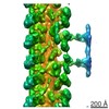 6515C 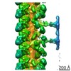 6516C 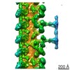 6518C 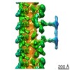 6519C 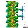 6520C 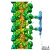 6521C 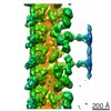 6522C 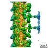 6523C 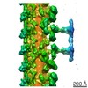 6524C C: citing same article ( |
|---|---|
| Similar structure data |

-Links
| EMDB pages | EMDB (EBI/PDBe) / EMDataResource |
|---|
-Map
| File | Download / File: emd_6517.map.gz / Format: CCP4 / Size: 19 MB / Type: IMAGE STORED AS FLOATING POINT NUMBER (4 BYTES) | ||||||||||||||||||||||||||||||||||||||||||||||||||||||||||||
|---|---|---|---|---|---|---|---|---|---|---|---|---|---|---|---|---|---|---|---|---|---|---|---|---|---|---|---|---|---|---|---|---|---|---|---|---|---|---|---|---|---|---|---|---|---|---|---|---|---|---|---|---|---|---|---|---|---|---|---|---|---|
| Annotation | Streptavidin-labeled IC1248BCCP axoneme | ||||||||||||||||||||||||||||||||||||||||||||||||||||||||||||
| Voxel size | X=Y=Z: 6.07 Å | ||||||||||||||||||||||||||||||||||||||||||||||||||||||||||||
| Density |
| ||||||||||||||||||||||||||||||||||||||||||||||||||||||||||||
| Symmetry | Space group: 1 | ||||||||||||||||||||||||||||||||||||||||||||||||||||||||||||
| Details | EMDB XML:
CCP4 map header:
| ||||||||||||||||||||||||||||||||||||||||||||||||||||||||||||
-Supplemental data
- Sample components
-Entire : Streptavidin-labeled IC1M248BCCP axoneme
| Entire | Name: Streptavidin-labeled IC1M248BCCP axoneme |
|---|---|
| Components |
|
-Supramolecule #1000: Streptavidin-labeled IC1M248BCCP axoneme
| Supramolecule | Name: Streptavidin-labeled IC1M248BCCP axoneme / type: sample / ID: 1000 / Number unique components: 1 |
|---|
-Supramolecule #1: axoneme
| Supramolecule | Name: axoneme / type: organelle_or_cellular_component / ID: 1 / Recombinant expression: No / Database: NCBI |
|---|---|
| Source (natural) | Organism: Chlamydomonas reinhardtii (plant) / Organelle: flagella |
-Experimental details
-Structure determination
| Method | cryo EM |
|---|---|
 Processing Processing | subtomogram averaging |
| Aggregation state | cell |
-Sample preparation
| Concentration | 0.01 mg/mL |
|---|---|
| Buffer | pH: 7.2 Details: 30 mM Hepes-NaOH pH 7.2, 5 mM MgCl2, 1 mM dithiothreitol, 1 mM EGTA, 50 mM K-acetate |
| Grid | Details: 300 mesh copper grid, holey carbon |
| Vitrification | Cryogen name: ETHANE / Chamber humidity: 90 % / Chamber temperature: 93 K / Instrument: LEICA EM GP / Method: Blot for 5 seconds before plunging |
- Electron microscopy
| Microscope | OTHER |
|---|---|
| Electron beam | Acceleration voltage: 300 kV / Electron source: FIELD EMISSION GUN |
| Electron optics | Illumination mode: FLOOD BEAM / Imaging mode: BRIGHT FIELDBright-field microscopy / Cs: 2.2 mm / Nominal defocus max: 9.0 µm / Nominal defocus min: 6.0 µm / Nominal magnification: 25700 |
| Specialist optics | Energy filter - Name: Omega / Energy filter - Lower energy threshold: 0.0 eV / Energy filter - Upper energy threshold: 20.0 eV |
| Sample stage | Specimen holder model: GATAN LIQUID NITROGEN / Tilt series - Axis1 - Min angle: -60 ° / Tilt series - Axis1 - Max angle: 60 ° |
| Date | May 14, 2015 |
| Image recording | Category: CCD / Film or detector model: TVIPS TEMCAM-F416 (4k x 4k) / Average electron dose: 100 e/Å2 |
-Image processing
| Final reconstruction | Resolution.type: BY AUTHOR / Resolution: 48.0 Å / Resolution method: OTHER / Software - Name: IMOD, PEET / Number subtomograms used: 984 |
|---|---|
| Details | Number of tilts (projections) used in 3D reconstruction: 60 Tomographic tilt angle increment: 2 |
