 Movie
Movie Controller

[English] 日本語
 Yorodumi
- EMDB-5493: Reconstruction of the Microtubule Alone on CCD for Difference Map... -
+ Open data

- Basic information
| Entry | Database: EMDB / ID: EMD-5493 | |||||||||
|---|---|---|---|---|---|---|---|---|---|---|
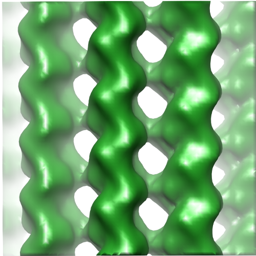
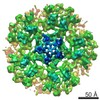
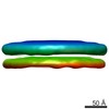
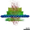
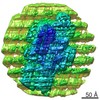
| Title | Reconstruction of the Microtubule Alone on CCD for Difference Map Calculation | |||||||||
 Map data Map data | Reconstruction of undecorated 14 protofilament microtubule for difference map calculation | |||||||||
 Sample Sample |
| |||||||||
 Keywords Keywords |  microtubule / Ndc80 / Hec1 / kinetochore / mitosis microtubule / Ndc80 / Hec1 / kinetochore / mitosis | |||||||||
| Biological species |  Bos taurus (cattle) Bos taurus (cattle) | |||||||||
| Method | helical reconstruction / cryo EM / Resolution: 10.8 Å | |||||||||
 Authors Authors | Alushin GM / Musinipally V / Matson D / Tooley J / Stukenberg PT / Nogales E | |||||||||
 Citation Citation | Journal: Nat Struct Mol Biol / Year: 2012 Title: Multimodal microtubule binding by the Ndc80 kinetochore complex. Authors: Gregory M Alushin / Vivek Musinipally / Daniel Matson / John Tooley / P Todd Stukenberg / Eva Nogales /  Abstract: The Ndc80 complex is a key site of kinetochore-microtubule attachment during cell division. The human complex engages microtubules with a globular 'head' formed by tandem calponin-homology domains ...The Ndc80 complex is a key site of kinetochore-microtubule attachment during cell division. The human complex engages microtubules with a globular 'head' formed by tandem calponin-homology domains and an 80-amino-acid unstructured 'tail' that contains sites of phosphoregulation by the Aurora B kinase. Using biochemical, cell biological and electron microscopy analyses, we dissected the roles of the tail in binding of microtubules and mediation of cooperative interactions between Ndc80 complexes. Two segments of the tail that contain Aurora B phosphorylation sites become ordered at interfaces; one with tubulin and the second with an adjacent Ndc80 head on the microtubule surface, forming interactions that are disrupted by phosphorylation. We propose a model in which Ndc80's interaction with either growing or shrinking microtubule ends can be tuned by the phosphorylation state of its tail. | |||||||||
| History |
|
- Structure visualization
| Movie |
 Movie viewer Movie viewer |
|---|---|
| Structure viewer | EM map: SurfViewMolmilJmol/JSmol |
| Supplemental images |
- Downloads & links
-EMDB archive
| Map data | emd_5493.map.gz | 1.2 MB | EMDB map data format | |
|---|---|---|---|---|
| Header (meta data) | emd-5493-v30.xmlemd-5493.xml | 10.2 KB 10.2 KB | Display Display | EMDB header |
| Images |  emd_5493.png emd_5493.png | 225.1 KB | ||
| Archive directory |  http://ftp.pdbj.org/pub/emdb/structures/EMD-5493ftp://ftp.pdbj.org/pub/emdb/structures/EMD-5493 http://ftp.pdbj.org/pub/emdb/structures/EMD-5493ftp://ftp.pdbj.org/pub/emdb/structures/EMD-5493 | HTTPS FTP |
-Related structure data


-Links
| EMDB pages | EMDB (EBI/PDBe) / EMDataResource |
|---|---|
| Related items in Molecule of the Month |

-Map
| File | Download / File: emd_5493.map.gz / Format: CCP4 / Size: 1.3 MB / Type: IMAGE STORED AS FLOATING POINT NUMBER (4 BYTES) | ||||||||||||||||||||||||||||||||||||||||||||||||||||||||||||||||||||
|---|---|---|---|---|---|---|---|---|---|---|---|---|---|---|---|---|---|---|---|---|---|---|---|---|---|---|---|---|---|---|---|---|---|---|---|---|---|---|---|---|---|---|---|---|---|---|---|---|---|---|---|---|---|---|---|---|---|---|---|---|---|---|---|---|---|---|---|---|---|
| Annotation | Reconstruction of undecorated 14 protofilament microtubule for difference map calculation | ||||||||||||||||||||||||||||||||||||||||||||||||||||||||||||||||||||
| Voxel size | X=Y=Z: 2.74 Å | ||||||||||||||||||||||||||||||||||||||||||||||||||||||||||||||||||||
| Density |
| ||||||||||||||||||||||||||||||||||||||||||||||||||||||||||||||||||||
| Symmetry | Space group: 1 | ||||||||||||||||||||||||||||||||||||||||||||||||||||||||||||||||||||
| Details | EMDB XML:
CCP4 map header:
| ||||||||||||||||||||||||||||||||||||||||||||||||||||||||||||||||||||
-Supplemental data
- Sample components
-Entire : microtubule
| Entire | Name: microtubule |
|---|---|
| Components |
|
-Supramolecule #1000: microtubule
| Supramolecule | Name: microtubule / type: sample / ID: 1000 Oligomeric state: tubulin is a heterodimer of alpha- and beta- tubulin Number unique components: 1 |
|---|---|
| Molecular weight | Theoretical: 110 KDa |
-Macromolecule #1: tubulin
| Macromolecule | Name: tubulin / type: protein_or_peptide / ID: 1 / Name.synonym: tubulin / Number of copies: 1 / Oligomeric state: heterodimer / Recombinant expression: No / Database: NCBI |
|---|---|
| Source (natural) | Organism: Bos taurus (cattle) / synonym: bovine / Tissue: brain / Location in cell: cytoskeleton |
| Molecular weight | Theoretical: 110 KDa |
-Experimental details
-Structure determination
| Method | cryo EM |
|---|---|
 Processing Processing | helical reconstruction |
| Aggregation state | filament |
-Sample preparation
| Concentration | 0.25 mg/mL |
|---|---|
| Buffer | pH: 6.8 Details: 80mM PIPES, 1mM MgCl2, 1mM EGTA, 1mM DTT, 0.05% Nonidet P-40, 20uM taxol |
| Grid | Details: C-flat 1.2/1.3 |
| Vitrification | Cryogen name: ETHANE / Chamber humidity: 100 % / Instrument: FEI VITROBOT MARK II Method: 2 uL of 0.25 mg/mL MTs applied to grid for 1 minute, 4 uL of 0.7 mg/mL Ndc80 bonsai added, manually blot 1 minute, another 4 uL of Ndc80 applied for 1 minute, 2 uL removed with pipettor, blot ...Method: 2 uL of 0.25 mg/mL MTs applied to grid for 1 minute, 4 uL of 0.7 mg/mL Ndc80 bonsai added, manually blot 1 minute, another 4 uL of Ndc80 applied for 1 minute, 2 uL removed with pipettor, blot for 2 seconds before plunging, 0 mm offset |
- Electron microscopy
| Microscope | FEI TECNAI F20 |
|---|---|
| Electron beam | Acceleration voltage: 120 kV / Electron source: FIELD EMISSION GUN |
| Electron optics | Illumination mode: FLOOD BEAM / Imaging mode: BRIGHT FIELDBright-field microscopy / Cs: 2.2 mm / Nominal defocus max: 2.8 µm / Nominal defocus min: 0.8 µm / Nominal magnification: 80000 |
| Sample stage | Specimen holder: side-entry / Specimen holder model: GATAN LIQUID NITROGEN |
| Alignment procedure | Legacy - Astigmatism: objective lens astigmatism corrected at 100Kx mag |
| Date | Aug 17, 2011 |
| Image recording | Category: CCD / Film or detector model: GATAN ULTRASCAN 4000 (4k x 4k) / Digitization - Sampling interval: 15 µm / Number real images: 146 / Average electron dose: 20 e/Å2 |
| Experimental equipment |  Model: Tecnai F20 / Image courtesy: FEI Company |
-Image processing
| CTF correction | Details: Phase-flipping each image |
|---|---|
| Final reconstruction | Applied symmetry - Helical parameters - Δz: 8.90136 Å Applied symmetry - Helical parameters - Δ&Phi: 25.75775 ° Algorithm: OTHER / Resolution.type: BY AUTHOR / Resolution: 10.8 Å / Resolution method: FSC 0.5 CUT-OFF / Software - Name: EMAN2/SPARX Details: Particles were aligned using multi-model IHRSR protocol in EMAN2/SPARX with naked 13 and 14 protofilament microtubules as references. The deposited map is a segmented region for difference ...Details: Particles were aligned using multi-model IHRSR protocol in EMAN2/SPARX with naked 13 and 14 protofilament microtubules as references. The deposited map is a segmented region for difference map calculation. No amplitude scaling was applied. |
| Details | The phase-flipped particles were aligned using IHRSR in EMAN2/SPARX. |
