 ムービー
ムービー コントローラー


+ データを開く

- 基本情報
基本情報
| 登録情報 | データベース: EMDB / ID: EMD-5493 | |||||||||
|---|---|---|---|---|---|---|---|---|---|---|
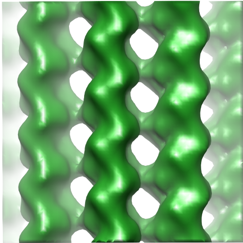
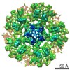
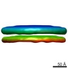
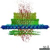
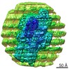
| タイトル | Reconstruction of the Microtubule Alone on CCD for Difference Map Calculation | |||||||||
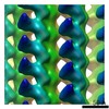
 マップデータ マップデータ | Reconstruction of undecorated 14 protofilament microtubule for difference map calculation | |||||||||
 試料 試料 |
| |||||||||
 キーワード キーワード |  microtubule (微小管) / Ndc80 / Hec1 / kinetochore (動原体) / mitosis (有糸分裂) microtubule (微小管) / Ndc80 / Hec1 / kinetochore (動原体) / mitosis (有糸分裂) | |||||||||
| 生物種 |  Bos taurus (ウシ) Bos taurus (ウシ) | |||||||||
| 手法 | らせん対称体再構成法 / クライオ電子顕微鏡法 / 解像度: 10.8 Å | |||||||||
 データ登録者 データ登録者 | Alushin GM / Musinipally V / Matson D / Tooley J / Stukenberg PT / Nogales E | |||||||||
 引用 引用 | ジャーナル: Nat Struct Mol Biol / 年: 2012 タイトル: Multimodal microtubule binding by the Ndc80 kinetochore complex. 著者: Gregory M Alushin / Vivek Musinipally / Daniel Matson / John Tooley / P Todd Stukenberg / Eva Nogales /  要旨: The Ndc80 complex is a key site of kinetochore-microtubule attachment during cell division. The human complex engages microtubules with a globular 'head' formed by tandem calponin-homology domains ...The Ndc80 complex is a key site of kinetochore-microtubule attachment during cell division. The human complex engages microtubules with a globular 'head' formed by tandem calponin-homology domains and an 80-amino-acid unstructured 'tail' that contains sites of phosphoregulation by the Aurora B kinase. Using biochemical, cell biological and electron microscopy analyses, we dissected the roles of the tail in binding of microtubules and mediation of cooperative interactions between Ndc80 complexes. Two segments of the tail that contain Aurora B phosphorylation sites become ordered at interfaces; one with tubulin and the second with an adjacent Ndc80 head on the microtubule surface, forming interactions that are disrupted by phosphorylation. We propose a model in which Ndc80's interaction with either growing or shrinking microtubule ends can be tuned by the phosphorylation state of its tail. | |||||||||
| 履歴 |
|
- 構造の表示
構造の表示
| ムービー |
 ムービービューア ムービービューア |
|---|---|
| 構造ビューア | EMマップ: SurfViewMolmilJmol/JSmol |
| 添付画像 |
- ダウンロードとリンク
-EMDBアーカイブ
| マップデータ | emd_5493.map.gz | 1.2 MB | EMDBマップデータ形式 | |
|---|---|---|---|---|
| ヘッダ (付随情報) | emd-5493-v30.xmlemd-5493.xml | 10.2 KB 10.2 KB | 表示 表示 | EMDBヘッダ |
| 画像 |  emd_5493.png emd_5493.png | 225.1 KB | ||
| アーカイブディレクトリ |  http://ftp.pdbj.org/pub/emdb/structures/EMD-5493ftp://ftp.pdbj.org/pub/emdb/structures/EMD-5493 http://ftp.pdbj.org/pub/emdb/structures/EMD-5493ftp://ftp.pdbj.org/pub/emdb/structures/EMD-5493 | HTTPS FTP |
-関連構造データ

-リンク
| EMDBのページ | EMDB (EBI/PDBe) / EMDataResource |
|---|---|
| 「今月の分子」の関連する項目 |

-マップ
| ファイル | ダウンロード / ファイル: emd_5493.map.gz / 形式: CCP4 / 大きさ: 1.3 MB / タイプ: IMAGE STORED AS FLOATING POINT NUMBER (4 BYTES) | ||||||||||||||||||||||||||||||||||||||||||||||||||||||||||||||||||||
|---|---|---|---|---|---|---|---|---|---|---|---|---|---|---|---|---|---|---|---|---|---|---|---|---|---|---|---|---|---|---|---|---|---|---|---|---|---|---|---|---|---|---|---|---|---|---|---|---|---|---|---|---|---|---|---|---|---|---|---|---|---|---|---|---|---|---|---|---|---|
| 注釈 | Reconstruction of undecorated 14 protofilament microtubule for difference map calculation | ||||||||||||||||||||||||||||||||||||||||||||||||||||||||||||||||||||
| ボクセルのサイズ | X=Y=Z: 2.74 Å | ||||||||||||||||||||||||||||||||||||||||||||||||||||||||||||||||||||
| 密度 |
| ||||||||||||||||||||||||||||||||||||||||||||||||||||||||||||||||||||
| 対称性 | 空間群: 1 | ||||||||||||||||||||||||||||||||||||||||||||||||||||||||||||||||||||
| 詳細 | EMDB XML:
CCP4マップ ヘッダ情報:
| ||||||||||||||||||||||||||||||||||||||||||||||||||||||||||||||||||||
-添付データ
- 試料の構成要素
-全体 : microtubule
| 全体 | 名称: microtubule微小管 |
|---|---|
| 要素 |
|
-超分子 #1000: microtubule
| 超分子 | 名称: microtubule / タイプ: sample / ID: 1000 集合状態: tubulin is a heterodimer of alpha- and beta- tubulin Number unique components: 1 |
|---|---|
| 分子量 | 理論値: 110 KDa |
-分子 #1: tubulin
| 分子 | 名称: tubulin / タイプ: protein_or_peptide / ID: 1 / Name.synonym: tubulin / コピー数: 1 / 集合状態: heterodimer / 組換発現: No / データベース: NCBI |
|---|---|
| 由来(天然) | 生物種: Bos taurus (ウシ) / 別称: bovine / 組織: brain / 細胞中の位置: cytoskeleton |
| 分子量 | 理論値: 110 KDa |
-実験情報
-構造解析
| 手法 | クライオ電子顕微鏡法 |
|---|---|
 解析 解析 | らせん対称体再構成法 |
| 試料の集合状態 | filament |
-試料調製
| 濃度 | 0.25 mg/mL |
|---|---|
| 緩衝液 | pH: 6.8 詳細: 80mM PIPES, 1mM MgCl2, 1mM EGTA, 1mM DTT, 0.05% Nonidet P-40, 20uM taxol |
| グリッド | 詳細: C-flat 1.2/1.3 |
| 凍結 | 凍結剤: ETHANE / チャンバー内湿度: 100 % / 装置: FEI VITROBOT MARK II 手法: 2 uL of 0.25 mg/mL MTs applied to grid for 1 minute, 4 uL of 0.7 mg/mL Ndc80 bonsai added, manually blot 1 minute, another 4 uL of Ndc80 applied for 1 minute, 2 uL removed with pipettor, blot ...手法: 2 uL of 0.25 mg/mL MTs applied to grid for 1 minute, 4 uL of 0.7 mg/mL Ndc80 bonsai added, manually blot 1 minute, another 4 uL of Ndc80 applied for 1 minute, 2 uL removed with pipettor, blot for 2 seconds before plunging, 0 mm offset |
- 電子顕微鏡法
| 顕微鏡 | FEI TECNAI F20 |
|---|---|
| 電子線 | 加速電圧: 120 kV / 電子線源: FIELD EMISSION GUN |
| 電子光学系 | 照射モード: FLOOD BEAM / 撮影モード: BRIGHT FIELDBright-field microscopy / Cs: 2.2 mm / 最大 デフォーカス(公称値): 2.8 µm / 最小 デフォーカス(公称値): 0.8 µm / 倍率(公称値): 80000 |
| 試料ステージ | 試料ホルダー: side-entry / 試料ホルダーモデル: GATAN LIQUID NITROGEN |
| アライメント法 | Legacy - 非点収差: objective lens astigmatism corrected at 100Kx mag |
| 日付 | 2011年8月17日 |
| 撮影 | カテゴリ: CCD フィルム・検出器のモデル: GATAN ULTRASCAN 4000 (4k x 4k) デジタル化 - サンプリング間隔: 15 µm / 実像数: 146 / 平均電子線量: 20 e/Å2 |
| 実験機器 |  モデル: Tecnai F20 / 画像提供: FEI Company |
-画像解析
| CTF補正 | 詳細: Phase-flipping each image |
|---|---|
| 最終 再構成 | 想定した対称性 - らせんパラメータ - Δz: 8.90136 Å 想定した対称性 - らせんパラメータ - ΔΦ: 25.75775 ° アルゴリズム: OTHER / 解像度のタイプ: BY AUTHOR / 解像度: 10.8 Å / 解像度の算出法: FSC 0.5 CUT-OFF / ソフトウェア - 名称: EMAN2/SPARX 詳細: Particles were aligned using multi-model IHRSR protocol in EMAN2/SPARX with naked 13 and 14 protofilament microtubules as references. The deposited map is a segmented region for difference ...詳細: Particles were aligned using multi-model IHRSR protocol in EMAN2/SPARX with naked 13 and 14 protofilament microtubules as references. The deposited map is a segmented region for difference map calculation. No amplitude scaling was applied. |
| 詳細 | The phase-flipped particles were aligned using IHRSR in EMAN2/SPARX. |
