 Movie
Movie Controller

[English] 日本語
 Yorodumi
- EMDB-5048: CLIC2-RyR1 interaction and structural characterization by cryo-el... -
+ Open data

- Basic information
| Entry | Database: EMDB / ID: EMD-5048 | |||||||||
|---|---|---|---|---|---|---|---|---|---|---|
| Title | CLIC2-RyR1 interaction and structural characterization by cryo-electron microscopy | |||||||||
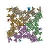
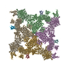
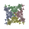
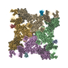
 Map data Map data | This is an image of a surface rendered side view of RyR1-CLIC2 complex | |||||||||
 Sample Sample |
| |||||||||
 Keywords Keywords | Ca2+-release channel / Ca2+ signaling /  Chloride intracellular channel 2 / cryo-electron microscopy / ryanodine receptor Chloride intracellular channel 2 / cryo-electron microscopy / ryanodine receptor | |||||||||
| Biological species | unidentified (others) | |||||||||
| Method | single particle reconstruction / cryo EM / Resolution: 25.0 Å | |||||||||
 Authors Authors | Meng X / Wang G / Viero C / Wang Q / Mi W / Su X / Wagenknecht T / Williams A / Liu Z / Yin C-C | |||||||||
 Citation Citation | Journal: J Mol Biol / Year: 2009 Title: CLIC2-RyR1 interaction and structural characterization by cryo-electron microscopy. Authors: Xing Meng / Guoliang Wang / Cedric Viero / Qiongling Wang / Wei Mi / Xiao-Dong Su / Terence Wagenknecht / Alan J Williams / Zheng Liu / Chang-Cheng Yin /  Abstract: Chloride intracellular channel 2 (CLIC2), a newly discovered small protein distantly related to the glutathione transferase (GST) structural family, is highly expressed in cardiac and skeletal ...Chloride intracellular channel 2 (CLIC2), a newly discovered small protein distantly related to the glutathione transferase (GST) structural family, is highly expressed in cardiac and skeletal muscle, although its physiological function in these tissues has not been established. In the present study, [3H]ryanodine binding, Ca2+ efflux from skeletal sarcoplasmic reticulum (SR) vesicles, single channel recording, and cryo-electron microscopy were employed to investigate whether CLIC2 can interact with skeletal ryanodine receptor (RyR1) and modulate its channel activity. We found that: (1) CLIC2 facilitated [3H]ryanodine binding to skeletal SR and purified RyR1, by increasing the binding affinity of ryanodine for its receptor without significantly changing the apparent maximal binding capacity; (2) CLIC2 reduced the maximal Ca2+ efflux rate from skeletal SR vesicles; (3) CLIC2 decreased the open probability of RyR1 channel, through increasing the mean closed time of the channel; (4) CLIC2 bound to a region between domains 5 and 6 in the clamp-shaped region of RyR1; (5) and in the same clamp region, domains 9 and 10 became separated after CLIC2 binding, indicating CLIC2 induced a conformational change of RyR1. These data suggest that CLIC2 can interact with RyR1 and modulate its channel activity. We propose that CLIC2 functions as an intrinsic stabilizer of the closed state of RyR channels. | |||||||||
| History |
|
- Structure visualization
| Movie |
 Movie viewer Movie viewer |
|---|---|
| Structure viewer | EM map: SurfViewMolmilJmol/JSmol |
| Supplemental images |

- Downloads & links
-EMDB archive
| Map data | emd_5048.map.gz | 4.2 MB | EMDB map data format | |
|---|---|---|---|---|
| Header (meta data) | emd-5048-v30.xmlemd-5048.xml | 10.6 KB 10.6 KB | Display Display | EMDB header |
| Images | 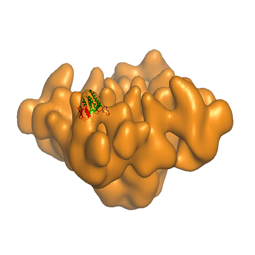 emd_5048_1.jpg emd_5048_1.jpg | 103.9 KB | ||
| Archive directory |  http://ftp.pdbj.org/pub/emdb/structures/EMD-5048ftp://ftp.pdbj.org/pub/emdb/structures/EMD-5048 http://ftp.pdbj.org/pub/emdb/structures/EMD-5048ftp://ftp.pdbj.org/pub/emdb/structures/EMD-5048 | HTTPS FTP |
-Related structure data
| Similar structure data |
|---|

-Links
| EMDB pages | EMDB (EBI/PDBe) / EMDataResource |
|---|
-Map
| File | Download / File: emd_5048.map.gz / Format: CCP4 / Size: 4.3 MB / Type: IMAGE STORED AS FLOATING POINT NUMBER (4 BYTES) | ||||||||||||||||||||||||||||||||||||||||||||||||||||||||||||||||||||
|---|---|---|---|---|---|---|---|---|---|---|---|---|---|---|---|---|---|---|---|---|---|---|---|---|---|---|---|---|---|---|---|---|---|---|---|---|---|---|---|---|---|---|---|---|---|---|---|---|---|---|---|---|---|---|---|---|---|---|---|---|---|---|---|---|---|---|---|---|---|
| Annotation | This is an image of a surface rendered side view of RyR1-CLIC2 complex | ||||||||||||||||||||||||||||||||||||||||||||||||||||||||||||||||||||
| Voxel size | X=Y=Z: 5.52 Å | ||||||||||||||||||||||||||||||||||||||||||||||||||||||||||||||||||||
| Density |
| ||||||||||||||||||||||||||||||||||||||||||||||||||||||||||||||||||||
| Symmetry | Space group: 1 | ||||||||||||||||||||||||||||||||||||||||||||||||||||||||||||||||||||
| Details | EMDB XML:
CCP4 map header:
| ||||||||||||||||||||||||||||||||||||||||||||||||||||||||||||||||||||
-Supplemental data
- Sample components
-Entire : Skeletal muscle ryanodine receptor and chloride intracellular cha...
| Entire | Name: Skeletal muscle ryanodine receptor and chloride intracellular channel 2 complex |
|---|---|
| Components |
|
-Supramolecule #1000: Skeletal muscle ryanodine receptor and chloride intracellular cha...
| Supramolecule | Name: Skeletal muscle ryanodine receptor and chloride intracellular channel 2 complex type: sample / ID: 1000 Oligomeric state: Four CLIC2 molecules binds to one homotetramer of RyR1 Number unique components: 2 |
|---|---|
| Molecular weight | Experimental: 565 KDa / Theoretical: 565 KDa / Method: Sequencing |
-Supramolecule #1: sarcoplasmic reticulum
| Supramolecule | Name: sarcoplasmic reticulum / type: organelle_or_cellular_component / ID: 1 / Name.synonym: SR / Number of copies: 1 / Oligomeric state: Tetramer / Recombinant expression: No / Database: NCBI |
|---|---|
| Source (natural) | Organism: unidentified (others) / synonym: rabbit / Tissue: skeletal muscle / Organelle: sarcoplasmic reticulum / Location in cell: sarcoplasmic reticulum membrane |
| Molecular weight | Experimental: 2.3 MDa / Theoretical: 2.3 MDa |
-Experimental details
-Structure determination
| Method | cryo EM |
|---|---|
 Processing Processing | single particle reconstruction |
| Aggregation state | particle |
-Sample preparation
| Concentration | 0.05 mg/mL |
|---|---|
| Buffer | pH: 7.4 Details: 20 mM Na-MOPS, pH7.4, 200 mM NaCl, 2.0 mM EGTA, 0.5% CHAPS, 2 mM DTT, and 2.0 ug/ml leupeptin |
| Grid | Details: 300 mesh copper grid |
| Vitrification | Cryogen name: ETHANE / Chamber humidity: 85 % / Instrument: OTHER / Details: Vitrification instrument: FEI Vitrobot / Method: Blot for 2 seconds before plunging |
- Electron microscopy
| Microscope | FEI TECNAI F20 |
|---|---|
| Electron beam | Acceleration voltage: 200 kV / Electron source: FIELD EMISSION GUN |
| Electron optics | Calibrated magnification: 50760 / Illumination mode: FLOOD BEAM / Imaging mode: BRIGHT FIELDBright-field microscopy / Cs: 2.0 mm / Nominal defocus max: 4.5 µm / Nominal defocus min: 1.5 µm / Nominal magnification: 50000 |
| Sample stage | Specimen holder: Oxford CT3500 / Specimen holder model: OTHER / Tilt angle max: 50 |
| Temperature | Average: 90 K |
| Alignment procedure | Legacy - Astigmatism: objective lens astigmatism was corrected at 100,000 times magnification |
| Image recording | Category: FILM / Film or detector model: KODAK SO-163 FILM / Digitization - Scanner: ZEISS SCAI / Digitization - Sampling interval: 7.0 µm / Number real images: 258 / Average electron dose: 10 e/Å2 |
| Tilt angle min | 0 |
| Experimental equipment |  Model: Tecnai F20 / Image courtesy: FEI Company |
-Image processing
| CTF correction | Details: Each particle |
|---|---|
| Final two d classification | Number classes: 1 |
| Final reconstruction | Algorithm: OTHER / Resolution.type: BY AUTHOR / Resolution: 25.0 Å / Resolution method: FSC 0.5 CUT-OFF / Software - Name: Spider / Number images used: 15889 |
