 Movie
Movie Controller

[English] 日本語
 Yorodumi
- EMDB-42987: Structure of the Human Respirovirus 3 Fusion Protein Bound to Cam... -
+ Open data

- Basic information
| Entry |  | |||||||||
|---|---|---|---|---|---|---|---|---|---|---|
| Title | Structure of the Human Respirovirus 3 Fusion Protein Bound to Camelid Nanobodies 1D10 and 4C06 | |||||||||
 Map data Map data | Final refined volume. DeepEMhancer sharpened. Used to build coordinates. | |||||||||
 Sample Sample |
| |||||||||
 Keywords Keywords | viral fusion protein / camelid nanobodies / viral glycoprotein /  membrane fusion / VIRAL PROTEIN / VIRAL PROTEIN-IMMUNE SYSTEM complex membrane fusion / VIRAL PROTEIN / VIRAL PROTEIN-IMMUNE SYSTEM complex | |||||||||
| Function / homology | Precursor fusion glycoprotein F0, Paramyxoviridae / Fusion glycoprotein F0 / fusion of virus membrane with host plasma membrane / viral envelope / host cell plasma membrane / virion membrane / plasma membrane / Fusion glycoprotein F0 Function and homology information Function and homology information | |||||||||
| Biological species |  Human respirovirus 3 / Human respirovirus 3 /  Lama glama (llama) Lama glama (llama) | |||||||||
| Method | single particle reconstruction / cryo EM / Resolution: 3.4 Å | |||||||||
 Authors Authors | Johnson NJ / Ramamohan AR / McLellan JS | |||||||||
| Funding support | 1 items
| |||||||||
 Citation Citation | Journal: To Be Published Title: Structural basis for potent neutralization of human respirovirus type 3 by single-domain camelid antibodies Authors: Johnson NV / van Scherpenzeel RC / Bakkers MJG / Ramamohan AR / van Overveld D / Le L / Langedijk JPM / Kolkman JA / McLellan JS | |||||||||
| History |
|
- Structure visualization
| Supplemental images |
|---|

- Downloads & links
-EMDB archive
| Map data | emd_42987.map.gz | 108.2 MB | EMDB map data format | |
|---|---|---|---|---|
| Header (meta data) | emd-42987-v30.xmlemd-42987.xml | 18 KB 18 KB | Display Display | EMDB header |
| FSC (resolution estimation) | emd_42987_fsc.xml | 10.6 KB | Display | FSC data file |
| Images | 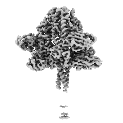 emd_42987.png emd_42987.png | 65.8 KB | ||
| Filedesc metadata | emd-42987.cif.gz | 5.9 KB | ||
| Others | emd_42987_additional_1.map.gzemd_42987_half_map_1.map.gzemd_42987_half_map_2.map.gz | 118 MB 116.1 MB 116.1 MB | ||
| Archive directory |  http://ftp.pdbj.org/pub/emdb/structures/EMD-42987ftp://ftp.pdbj.org/pub/emdb/structures/EMD-42987 http://ftp.pdbj.org/pub/emdb/structures/EMD-42987ftp://ftp.pdbj.org/pub/emdb/structures/EMD-42987 | HTTPS FTP |
-Related structure data
| Related structure data | 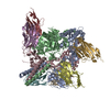 8v62MC 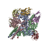 8v5kC C: citing same article ( M: atomic model generated by this map |
|---|---|
| Similar structure data |

-Links
| EMDB pages | EMDB (EBI/PDBe) / EMDataResource |
|---|---|
| Related items in Molecule of the Month |


-Map
| File | Download / File: emd_42987.map.gz / Format: CCP4 / Size: 125 MB / Type: IMAGE STORED AS FLOATING POINT NUMBER (4 BYTES) | ||||||||||||||||||||
|---|---|---|---|---|---|---|---|---|---|---|---|---|---|---|---|---|---|---|---|---|---|
| Annotation | Final refined volume. DeepEMhancer sharpened. Used to build coordinates. | ||||||||||||||||||||
| Voxel size | X=Y=Z: 0.94 Å | ||||||||||||||||||||
| Density |
| ||||||||||||||||||||
| Symmetry | Space group: 1 | ||||||||||||||||||||
| Details | EMDB XML:
|
-Supplemental data
-Additional map: Final refined volume. Sharpened map, not DeepEMhancer sharpened.
| File | emd_42987_additional_1.map | ||||||||||||
|---|---|---|---|---|---|---|---|---|---|---|---|---|---|
| Annotation | Final refined volume. Sharpened map, not DeepEMhancer sharpened. | ||||||||||||
| Projections & Slices |
| ||||||||||||
| Density Histograms |
 Z
Z Y
Y X
X


-Half map: half map A
| File | emd_42987_half_map_1.map | ||||||||||||
|---|---|---|---|---|---|---|---|---|---|---|---|---|---|
| Annotation | half map A | ||||||||||||
| Projections & Slices |
| ||||||||||||
| Density Histograms |


-Half map: half map B
| File | emd_42987_half_map_2.map | ||||||||||||
|---|---|---|---|---|---|---|---|---|---|---|---|---|---|
| Annotation | half map B | ||||||||||||
| Projections & Slices |
| ||||||||||||
| Density Histograms |


- Sample components
-Entire : Prefusion Human Respirovirus 3 Fusion Protein Bound to Camelid Na...
| Entire | Name: Prefusion Human Respirovirus 3 Fusion Protein Bound to Camelid Nanobodies 1D10 and 4C06 |
|---|---|
| Components |
|
-Supramolecule #1: Prefusion Human Respirovirus 3 Fusion Protein Bound to Camelid Na...
| Supramolecule | Name: Prefusion Human Respirovirus 3 Fusion Protein Bound to Camelid Nanobodies 1D10 and 4C06 type: complex / ID: 1 / Parent: 0 / Macromolecule list: all |
|---|---|
| Source (natural) | Organism: Human respirovirus 3 |
| Molecular weight | Theoretical: 277.5 KDa |
-Macromolecule #1: Fusion glycoprotein F0
| Macromolecule | Name: Fusion glycoprotein F0 / type: protein_or_peptide / ID: 1 / Number of copies: 3 / Enantiomer: LEVO |
|---|---|
| Source (natural) | Organism: Human respirovirus 3 |
| Molecular weight | Theoretical: 57.336508 KDa |
| Recombinant expression | Organism:  Homo sapiens (human) Homo sapiens (human) |
| Sequence | String: MPISILLIIT TMIMASHCQI DITKLQHVGV LVNSPKGMKI SQNFETRYLI LSLIPKIEDS NSCGDQQIKQ YKRLLDRLII PLYDGLRLQ KDVIVTNQES NENTDPRTER FFGGVIGTIA LGVATSAQIT AAVALVEAKQ ARSDIEKLKE AIRDTNKAVQ S VQSSPGNL ...String: MPISILLIIT TMIMASHCQI DITKLQHVGV LVNSPKGMKI SQNFETRYLI LSLIPKIEDS NSCGDQQIKQ YKRLLDRLII PLYDGLRLQ KDVIVTNQES NENTDPRTER FFGGVIGTIA LGVATSAQIT AAVALVEAKQ ARSDIEKLKE AIRDTNKAVQ S VQSSPGNL IVAIKSVQDY VNKEIVPCIA RLGCEACGLL LGLALDQHYS ELTNIFGDNI GSLQEKGIKL QGIASLYRTN IT EIFTTST VDKYDIYDLL FTESIKVRVI DVDLNDYSIT LQVRLPLLTR LLNTQIYKVD SISYNIQNRE WYIPLPSHIM TKG AFLGGA DVKECIEAFS SYICPSDPGF VLNHEMESCL SGNISQCPRT TVTSDIVPRY AFVNGGVVAN CITTTCTCNG IGNR INQPP DQGVKIITHK ECNTIGINGM LFNTNKEGTL AFYTPDDITL NNSVALNPID ISIELNKAKS DLEESKEWIR RSNQK LDSI EDKIEEILSK IYHIENEIAR IKKLIGEAEP EA UniProtKB: Fusion glycoprotein F0 |
-Macromolecule #2: Camelid nanobody 1D10
| Macromolecule | Name: Camelid nanobody 1D10 / type: protein_or_peptide / ID: 2 / Number of copies: 3 / Enantiomer: LEVO |
|---|---|
| Source (natural) | Organism: Lama glama (llama) |
| Molecular weight | Theoretical: 12.436922 KDa |
| Recombinant expression | Organism:  Escherichia coli (E. coli) Escherichia coli (E. coli) |
| Sequence | String: EVQLVESGGG LVQTGDSLRL SCAASGSIFG ENAMAWFRQA PGKQRELVAR VSTGGTLFYA DFAKVRFTIS RDTAKQTVYL QMSSLRPED TAVYYCAVAV GTRNYWGQGT QVTVSS |
-Macromolecule #3: Camelid nanobody 4C06
| Macromolecule | Name: Camelid nanobody 4C06 / type: protein_or_peptide / ID: 3 / Number of copies: 3 / Enantiomer: LEVO |
|---|---|
| Source (natural) | Organism: Lama glama (llama) |
| Molecular weight | Theoretical: 12.487816 KDa |
| Recombinant expression | Organism: Escherichia coli (E. coli) |
| Sequence | String: EVQLVESGGG LVQPGGSLRL SCSASGSLST IKALGWYRRA PGRERELVAS ITSAGETNYA DSAKGRFTVS TDNAKNTVDL RMNSLKPED TAVYYCYAES FVLNIYWGQG TQVTVSSG |
-Experimental details
-Structure determination
| Method | cryo EM |
|---|---|
 Processing Processing | single particle reconstruction |
| Aggregation state | particle |
-Sample preparation
| Buffer | pH: 8 |
|---|---|
| Vitrification | Cryogen name: ETHANE |
- Electron microscopy
| Microscope | TFS GLACIOS |
|---|---|
| Electron beam | Acceleration voltage: 200 kV / Electron source: FIELD EMISSION GUN |
| Electron optics | Illumination mode: FLOOD BEAM / Imaging mode: BRIGHT FIELDBright-field microscopy / Nominal defocus max: 3.0 µm / Nominal defocus min: 1.0 µm |
| Image recording | Film or detector model: FEI FALCON IV (4k x 4k) / Average electron dose: 50.0 e/Å2 |
-Image processing
| Startup model | Type of model: NONE |
|---|---|
| Initial angle assignment | Type: MAXIMUM LIKELIHOOD |
| Final angle assignment | Type: MAXIMUM LIKELIHOOD |
| Final reconstruction | Resolution.type: BY AUTHOR / Resolution: 3.4 Å / Resolution method: FSC 0.143 CUT-OFF / Number images used: 56650 |
| FSC plot (resolution estimation) |  |
