Movie
Movie Controller

[English] 日本語
 Yorodumi
- EMDB-41299: Structural basis of peptidoglycan synthesis by E. coli RodA-PBP2 ... -
+ Open data

- Basic information
| Entry | 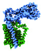 | |||||||||
|---|---|---|---|---|---|---|---|---|---|---|
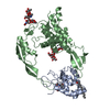
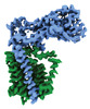
| Title | Structural basis of peptidoglycan synthesis by E. coli RodA-PBP2 complex | |||||||||
 Map data Map data | ||||||||||
 Sample Sample |
| |||||||||
 Keywords Keywords |  Peptidoglycan / glycosyltransferase / enzyme / MEMBRANE PROTEIN Peptidoglycan / glycosyltransferase / enzyme / MEMBRANE PROTEIN | |||||||||
| Function / homology |  Function and homology informationpeptidoglycan glycosyltransferase / peptidoglycan glycosyltransferase activity / serine-type D-Ala-D-Ala carboxypeptidase / serine-type D-Ala-D-Ala carboxypeptidase activity / acyltransferase activity / glycosyltransferase activity / penicillin binding / peptidoglycan biosynthetic process / cell wall organization / regulation of cell shape ...peptidoglycan glycosyltransferase / peptidoglycan glycosyltransferase activity / serine-type D-Ala-D-Ala carboxypeptidase / serine-type D-Ala-D-Ala carboxypeptidase activity / acyltransferase activity / glycosyltransferase activity / penicillin binding / peptidoglycan biosynthetic process / cell wall organization / regulation of cell shape / cell division / proteolysis / plasma membrane Function and homology informationpeptidoglycan glycosyltransferase / peptidoglycan glycosyltransferase activity / serine-type D-Ala-D-Ala carboxypeptidase / serine-type D-Ala-D-Ala carboxypeptidase activity / acyltransferase activity / glycosyltransferase activity / penicillin binding / peptidoglycan biosynthetic process / cell wall organization / regulation of cell shape ...peptidoglycan glycosyltransferase / peptidoglycan glycosyltransferase activity / serine-type D-Ala-D-Ala carboxypeptidase / serine-type D-Ala-D-Ala carboxypeptidase activity / acyltransferase activity / glycosyltransferase activity / penicillin binding / peptidoglycan biosynthetic process / cell wall organization / regulation of cell shape / cell division / proteolysis / plasma membraneSimilarity search - Function | |||||||||
| Biological species |  Escherichia coli (E. coli) Escherichia coli (E. coli) | |||||||||
| Method | single particle reconstruction / cryo EM / Resolution: 3.2 Å | |||||||||
 Authors Authors | Nygaard R / Mancia F | |||||||||
| Funding support |  United States, 1 items United States, 1 items
| |||||||||
 Citation Citation | Journal: Nat Commun / Year: 2023 Title: Structural basis of peptidoglycan synthesis by E. coli RodA-PBP2 complex Authors: Nygaard R / Graham CLB / Belcher Dufrisne M / Colburn JD / Pepe J / Hydorn MA / Corradi S / Brown CM / Ashraf KU / Vickery ON / Briggs NS / Deering JJ / Kloss B / Botta B / Clarke OB / ...Authors: Nygaard R / Graham CLB / Belcher Dufrisne M / Colburn JD / Pepe J / Hydorn MA / Corradi S / Brown CM / Ashraf KU / Vickery ON / Briggs NS / Deering JJ / Kloss B / Botta B / Clarke OB / Columbus L / Dworkin J / Stansfeld PJ / Roper DI / Mancia F | |||||||||
| History |
|
- Structure visualization
| Supplemental images |
|---|

- Downloads & links
-EMDB archive
| Map data | emd_41299.map.gz | 4.2 MB | EMDB map data format | |
|---|---|---|---|---|
| Header (meta data) | emd-41299-v30.xmlemd-41299.xml | 14 KB 14 KB | Display Display | EMDB header |
| Images | 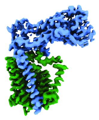 emd_41299.png emd_41299.png | 494.6 KB | ||
| Archive directory |  http://ftp.pdbj.org/pub/emdb/structures/EMD-41299ftp://ftp.pdbj.org/pub/emdb/structures/EMD-41299 http://ftp.pdbj.org/pub/emdb/structures/EMD-41299ftp://ftp.pdbj.org/pub/emdb/structures/EMD-41299 | HTTPS FTP |
-Related structure data
| Related structure data | 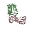 8tj3MC C: citing same article ( M: atomic model generated by this map |
|---|---|
| Similar structure data |

-Links
| EMDB pages | EMDB (EBI/PDBe) / EMDataResource |
|---|---|
| Related items in Molecule of the Month |

-Map
| File | Download / File: emd_41299.map.gz / Format: CCP4 / Size: 244.1 MB / Type: IMAGE STORED AS FLOATING POINT NUMBER (4 BYTES) | ||||||||||||||||||||
|---|---|---|---|---|---|---|---|---|---|---|---|---|---|---|---|---|---|---|---|---|---|
| Voxel size | X=Y=Z: 0.83 Å | ||||||||||||||||||||
| Density |
| ||||||||||||||||||||
| Symmetry | Space group: 1 | ||||||||||||||||||||
| Details | EMDB XML:
|
-Supplemental data
- Sample components
-Entire : RodA-PBP2
| Entire | Name: RodA-PBP2 |
|---|---|
| Components |
|
-Supramolecule #1: RodA-PBP2
| Supramolecule | Name: RodA-PBP2 / type: complex / ID: 1 / Parent: 0 / Macromolecule list: all / Details: Nanodisc were formed using MSP1E3D1 and POPG lipid |
|---|---|
| Source (natural) | Organism: Escherichia coli (E. coli) |
| Molecular weight | Theoretical: 111.803 KDa |
-Macromolecule #1: Peptidoglycan glycosyltransferase MrdB
| Macromolecule | Name: Peptidoglycan glycosyltransferase MrdB / type: protein_or_peptide / ID: 1 / Number of copies: 1 / Enantiomer: LEVO |
|---|---|
| Source (natural) | Organism: Escherichia coli (E. coli) |
| Molecular weight | Theoretical: 40.508766 KDa |
| Recombinant expression | Organism: Escherichia coli (E. coli) |
| Sequence | String: MTDNPNKKTF WDKVHLDPTM LLILLALLVY SALVIWSASG QDIGMMERKI GQIAMGLVIM VVMAQIPPRV YEGWAPYLYI ICIILLVAV DAFGAISKGA QRWLDLGIVR FQPSEIAKIA VPLMVARFIN RDVCPPSLKN TGIALVLIFM PTLLVAAQPD L GTSILVAL ...String: MTDNPNKKTF WDKVHLDPTM LLILLALLVY SALVIWSASG QDIGMMERKI GQIAMGLVIM VVMAQIPPRV YEGWAPYLYI ICIILLVAV DAFGAISKGA QRWLDLGIVR FQPSEIAKIA VPLMVARFIN RDVCPPSLKN TGIALVLIFM PTLLVAAQPD L GTSILVAL SGLFVLFLSG LSWRLIGVAV VLVAAFIPIL WFFLMHDYQR QRVMMLLDPE SDPLGAGYHI IQSKIAIGSG GL RGKGWLH GTQSQLEFLP ERHTDFIFAV LAEELGLVGI LILLALYILL IMRGLWIAAR AQTTFGRVMA GGLMLILFVY VFV NIGMVS GILPVVGVPL PLVSYGGSAL IVLMAGFGIV MSIHTHRKML SKSV UniProtKB: Peptidoglycan glycosyltransferase MrdB |
-Macromolecule #2: Peptidoglycan D,D-transpeptidase MrdA
| Macromolecule | Name: Peptidoglycan D,D-transpeptidase MrdA / type: protein_or_peptide / ID: 2 / Number of copies: 1 / Enantiomer: LEVO |
|---|---|
| Source (natural) | Organism: Escherichia coli (E. coli) |
| Molecular weight | Theoretical: 70.943414 KDa |
| Recombinant expression | Organism: Escherichia coli (E. coli) |
| Sequence | String: MKLQNSFRDY TAESALFVRR ALVAFLGILL LTGVLIANLY NLQIVRFTDY QTRSNENRIK LVPIAPSRGI IYDRNGIPLA LNRTIYQIE MMPEKVDNVQ QTLDALRSVV DLTDDDIAAF RKERARSHRF TSIPVKTNLT EVQVARFAVN QYRFPGVEVK G YKRRYYPY ...String: MKLQNSFRDY TAESALFVRR ALVAFLGILL LTGVLIANLY NLQIVRFTDY QTRSNENRIK LVPIAPSRGI IYDRNGIPLA LNRTIYQIE MMPEKVDNVQ QTLDALRSVV DLTDDDIAAF RKERARSHRF TSIPVKTNLT EVQVARFAVN QYRFPGVEVK G YKRRYYPY GSALTHVIGY VSKINDKDVE RLNNDGKLAN YAATHDIGKL GIERYYEDVL HGQTGYEEVE VNNRGRVIRQ LK EVPPQAG HDIYLTLDLK LQQYIETLLA GSRAAVVVTD PRTGGVLALV STPSYDPNLF VDGISSKDYS ALLNDPNTPL VNR ATQGVY PPASTVKPYV AVSALSAGVI TRNTTLFDPG WWQLPGSEKR YRDWKKWGHG RLNVTRSLEE SADTFFYQVA YDMG IDRLS EWMGKFGYGH YTGIDLAEER SGNMPTREWK QKRFKKPWYQ GDTIPVGIGQ GYWTATPIQM SKALMILIND GIVKV PHLL MSTAEDGKQV PWVQPHEPPV GDIHSGYWEL AKDGMYGVAN RPNGTAHKYF ASAPYKIAAK SGTAQVFGLK ANETYN AHK IAERLRDHKL MTAFAPYNNP QVAVAMILEN GGAGPAVGTL MRQILDHIML GDNNTDLPAE NPAVAAAEDH UniProtKB: Peptidoglycan D,D-transpeptidase MrdA |
-Experimental details
-Structure determination
| Method | cryo EM |
|---|---|
 Processing Processing | single particle reconstruction |
| Aggregation state | particle |
-Sample preparation
| Concentration | 0.66 mg/mL | ||||||||||||
|---|---|---|---|---|---|---|---|---|---|---|---|---|---|
| Buffer | pH: 7 Component:
| ||||||||||||
| Vitrification | Cryogen name: ETHANE / Chamber humidity: 95 % / Chamber temperature: 277 K / Instrument: FEI VITROBOT MARK IV |
- Electron microscopy
| Microscope | FEI TITAN |
|---|---|
| Electron beam | Acceleration voltage: 300 kV / Electron source: FIELD EMISSION GUN |
| Electron optics | Illumination mode: FLOOD BEAM / Imaging mode: BRIGHT FIELDBright-field microscopy / Nominal defocus max: 2.5 µm / Nominal defocus min: 1.0 µm |
| Image recording | Film or detector model: GATAN K3 BIOQUANTUM (6k x 4k) / Number grids imaged: 1 / Number real images: 11120 / Average exposure time: 2.5 sec. / Average electron dose: 58.5 e/Å2 |
-Image processing
| Particle selection | Number selected: 4415933 |
|---|---|
| Startup model | Type of model: OTHER |
| Initial angle assignment | Type: MAXIMUM LIKELIHOOD |
| Final angle assignment | Type: MAXIMUM LIKELIHOOD |
| Final reconstruction | Resolution.type: BY AUTHOR / Resolution: 3.2 Å / Resolution method: FSC 0.143 CUT-OFF / Software - Name: cryoSPARC (ver. 2.12 and 3.2) / Software - details: Local refinement / Number images used: 399000 |