 Movie
Movie Controller

[English] 日本語
 Yorodumi
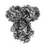
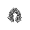
- EMDB-40762: E. coli SIR2-HerA complex (hexamer HerA bound with dodecamer Sir2) -
+ Open data

- Basic information
| Entry |  | |||||||||
|---|---|---|---|---|---|---|---|---|---|---|
| Title | E. coli SIR2-HerA complex (hexamer HerA bound with dodecamer Sir2) | |||||||||
 Map data Map data | ||||||||||
 Sample Sample |
| |||||||||
 Keywords Keywords | HerA /  SIR2 / NADase / ATPase / Anti-phage system / IMMUNE SYSTEM SIR2 / NADase / ATPase / Anti-phage system / IMMUNE SYSTEM | |||||||||
| Function / homology | Helicase HerA, central domain / Helicase HerA, central domain / SIR2-like domain / SIR2-like domain / hydrolase activity / P-loop containing nucleoside triphosphate hydrolase / SIR2-like domain-containing protein / Nucleoside triphosphate hydrolase Function and homology information Function and homology information | |||||||||
| Biological species |  Escherichia coli (E. coli) Escherichia coli (E. coli) | |||||||||
| Method | single particle reconstruction / cryo EM / Resolution: 2.83 Å | |||||||||
 Authors Authors | Shen ZF / Lin QP / Fu TM | |||||||||
| Funding support | 1 items
| |||||||||
 Citation Citation | Journal: Mol Cell / Year: 2023 Title: Assembly-mediated activation of the SIR2-HerA supramolecular complex for anti-phage defense. Authors: Zhangfei Shen / Qingpeng Lin / Xiao-Yuan Yang / Elizabeth Fosuah / Tian-Min Fu /  Abstract: SIR2-HerA, a bacterial two-protein anti-phage defense system, induces bacterial death by depleting NAD upon phage infection. Biochemical reconstitution of SIR2, HerA, and the SIR2-HerA complex ...SIR2-HerA, a bacterial two-protein anti-phage defense system, induces bacterial death by depleting NAD upon phage infection. Biochemical reconstitution of SIR2, HerA, and the SIR2-HerA complex reveals a dynamic assembly process. Unlike other ATPases, HerA can form various oligomers, ranging from dimers to nonamers. When assembled with SIR2, HerA forms a hexamer and converts SIR2 from a nuclease to an NAD hydrolase, representing an unexpected regulatory mechanism mediated by protein assembly. Furthermore, high concentrations of ATP can inhibit NAD hydrolysis by the SIR2-HerA complex. Cryo-EM structures of the SIR2-HerA complex reveal a giant supramolecular assembly up to 1 MDa, with SIR2 as a dodecamer and HerA as a hexamer, crucial for anti-phage defense. Unexpectedly, the HerA hexamer resembles a spiral staircase and exhibits helicase activities toward dual-forked DNA. Together, we reveal the supramolecular assembly of SIR2-HerA as a unique mechanism for switching enzymatic activities and bolstering anti-phage defense strategies. | |||||||||
| History |
|
- Structure visualization
| Supplemental images |
|---|

- Downloads & links
-EMDB archive
| Map data | emd_40762.map.gz | 118.2 MB | EMDB map data format | |
|---|---|---|---|---|
| Header (meta data) | emd-40762-v30.xmlemd-40762.xml | 18.6 KB 18.6 KB | Display Display | EMDB header |
| FSC (resolution estimation) | emd_40762_fsc.xml | 10.5 KB | Display | FSC data file |
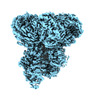
| Images | 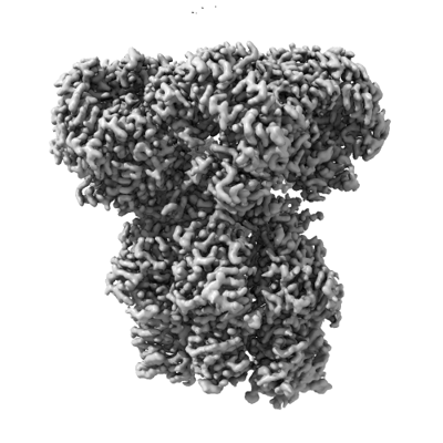 emd_40762.png emd_40762.png | 105.6 KB | ||
| Filedesc metadata | emd-40762.cif.gz | 6.1 KB | ||
| Others | emd_40762_additional_1.map.gzemd_40762_half_map_1.map.gzemd_40762_half_map_2.map.gz | 107.9 MB 115.9 MB 115.9 MB | ||
| Archive directory |  http://ftp.pdbj.org/pub/emdb/structures/EMD-40762ftp://ftp.pdbj.org/pub/emdb/structures/EMD-40762 http://ftp.pdbj.org/pub/emdb/structures/EMD-40762ftp://ftp.pdbj.org/pub/emdb/structures/EMD-40762 | HTTPS FTP |
-Related structure data
| Related structure data |  8su9MC  8subC  8suwC  8sxxC  8uaeC  8uafC C: citing same article ( M: atomic model generated by this map |
|---|---|
| Similar structure data |
-Links
| EMDB pages | EMDB (EBI/PDBe) / EMDataResource |
|---|
-Map
| File | Download / File: emd_40762.map.gz / Format: CCP4 / Size: 125 MB / Type: IMAGE STORED AS FLOATING POINT NUMBER (4 BYTES) | ||||||||||||||||||||
|---|---|---|---|---|---|---|---|---|---|---|---|---|---|---|---|---|---|---|---|---|---|
| Voxel size | X=Y=Z: 1.11 Å | ||||||||||||||||||||
| Density |
| ||||||||||||||||||||
| Symmetry | Space group: 1 | ||||||||||||||||||||
| Details | EMDB XML:
|
-Supplemental data
-Additional map: #1
| File | emd_40762_additional_1.map | ||||||||||||
|---|---|---|---|---|---|---|---|---|---|---|---|---|---|
| Projections & Slices |
| ||||||||||||
| Density Histograms |
 Z
Z Y
Y X
X


-Half map: #2
| File | emd_40762_half_map_1.map | ||||||||||||
|---|---|---|---|---|---|---|---|---|---|---|---|---|---|
| Projections & Slices |
| ||||||||||||
| Density Histograms |


-Half map: #1
| File | emd_40762_half_map_2.map | ||||||||||||
|---|---|---|---|---|---|---|---|---|---|---|---|---|---|
| Projections & Slices |
| ||||||||||||
| Density Histograms |


- Sample components
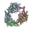
-Entire : E. coli SIR2-HerA complex
| Entire | Name: E. coli SIR2-HerA complex |
|---|---|
| Components |
|
-Supramolecule #1: E. coli SIR2-HerA complex
| Supramolecule | Name: E. coli SIR2-HerA complex / type: complex / ID: 1 / Parent: 0 / Macromolecule list: #1-#2 / Details: hexamer HerA bound with dodecamer Sir2 |
|---|---|
| Source (natural) | Organism: Escherichia coli (E. coli) |
-Macromolecule #1: SIR2-like domain-containing protein
| Macromolecule | Name: SIR2-like domain-containing protein / type: protein_or_peptide / ID: 1 / Number of copies: 12 / Enantiomer: LEVO |
|---|---|
| Source (natural) | Organism: Escherichia coli (E. coli) |
| Molecular weight | Theoretical: 46.817664 KDa |
| Recombinant expression | Organism: Escherichia coli (E. coli) |
| Sequence | String: MSIYQGGNKL NEDDFRSHVY SLCQLDNVGV LLGAGASVGC GGKTMKDVWK SFKQNYPELL GALIDKYLLV SQIDSDNNLV NVELLIDEA TKFLSVAKTR RCEDEEEEFR KILSSLYKEV TKAALLTGEQ FREKNQGKKD AFKYHKELIS KLISNRQPGQ S APAIFTTN ...String: MSIYQGGNKL NEDDFRSHVY SLCQLDNVGV LLGAGASVGC GGKTMKDVWK SFKQNYPELL GALIDKYLLV SQIDSDNNLV NVELLIDEA TKFLSVAKTR RCEDEEEEFR KILSSLYKEV TKAALLTGEQ FREKNQGKKD AFKYHKELIS KLISNRQPGQ S APAIFTTN YDLALEWAAE DLGIQLFNGF SGLHTRQFYP QNFDLAFRNV NAKGEARFGH YHAYLYKLHG SLTWYQNDSL TV NEVSASQ AYDEYINDII NKDDFYRGQH LIYPGANKYS HTIGFVYGEM FRRFGEFISK PQTALFINGF GFGDYHINRI ILG ALLNPS FHVVIYYPEL KEAITKVSKG GGSEAEKAIV TLKNMAFNQV TVVGGGSKAY FNSFVEHLPY PVLFPRDNIV DELV EAIAN LSKGEGNVPF UniProtKB: SIR2-like domain-containing protein |
-Macromolecule #2: Nucleoside triphosphate hydrolase
| Macromolecule | Name: Nucleoside triphosphate hydrolase / type: protein_or_peptide / ID: 2 / Number of copies: 6 / Enantiomer: LEVO |
|---|---|
| Source (natural) | Organism: Escherichia coli (E. coli) |
| Molecular weight | Theoretical: 68.431992 KDa |
| Recombinant expression | Organism: Escherichia coli (E. coli) |
| Sequence | String: MSLFKLTEIS AIGYVVGLEG ERIRINLHEG LQGRLASHRK GVSSVTQPGD LIGFDAGNIL VVARVTDMAF VEADKAHKAN VGTSDLADI PLRQIIAYAI GFVKRELNGY VFISEDWRLP ALGSSAVPLT SDFLNIIYSI DKEELPKAVE LGVDSRTKTV K IFASVDKL ...String: MSLFKLTEIS AIGYVVGLEG ERIRINLHEG LQGRLASHRK GVSSVTQPGD LIGFDAGNIL VVARVTDMAF VEADKAHKAN VGTSDLADI PLRQIIAYAI GFVKRELNGY VFISEDWRLP ALGSSAVPLT SDFLNIIYSI DKEELPKAVE LGVDSRTKTV K IFASVDKL LSRHLAVLGS TGYGKSNFNA LLTRKVSEKY PNSRIVIFDI NGEYAQAFTG IPNVKHTILG ESPNVDSLEK KQ QKGELYS EEYYCYKKIP YQALGFAGLI KLLRPSDKTQ LPALRNALSA INRTHFKSRN IYLEKDDGET FLLYDDCRDT NQS KLAEWL DLLRRRRLKR TNVWPPFKSL ATLVAEFGCV AADRSNGSKR DAFGFSNVLP LVKIIQQLAE DIRFKSIVNL NGGG ELADG GTHWDKAMSD EVDYFFGKEK GQENDWNVHI VNMKNLAQDH APMLLSALLE MFAEILFRRG QERSYPTVLL LEEAH HYLR DPYAEIDSQI KAYERLAKEG RKFKCSLIVS TQRPSELSPT VLAMCSNWFS LRLTNERDLQ ALRYAMESGN EQILKQ ISG LPRGDAVAFG SAFNLPVRIS INQARPGPKS SDAVFSEEWA NCTELRC UniProtKB: Nucleoside triphosphate hydrolase |
-Macromolecule #3: [(2R,3S,4R,5R)-5-(6-AMINOPURIN-9-YL)-3,4-DIHYDROXY-OXOLAN-2-YL]ME...
| Macromolecule | Name: [(2R,3S,4R,5R)-5-(6-AMINOPURIN-9-YL)-3,4-DIHYDROXY-OXOLAN-2-YL]METHYL [HYDROXY-[[(2R,3S,4R,5S)-3,4,5-TRIHYDROXYOXOLAN-2-YL]METHOXY]PHOSPHORYL] HYDROGEN PHOSPHATE type: ligand / ID: 3 / Number of copies: 12 / Formula: AR6 |
|---|---|
| Molecular weight | Theoretical: 559.316 Da |
-Macromolecule #4: ADENOSINE-5'-DIPHOSPHATE
| Macromolecule | Name: ADENOSINE-5'-DIPHOSPHATE / type: ligand / ID: 4 / Number of copies: 5 / Formula: ADP |
|---|---|
| Molecular weight | Theoretical: 427.201 Da |
| Chemical component information |  ChemComp-ADP: |
-Macromolecule #5: MAGNESIUM ION
| Macromolecule | Name: MAGNESIUM ION / type: ligand / ID: 5 / Number of copies: 5 / Formula: MG |
|---|---|
| Molecular weight | Theoretical: 24.305 Da |
-Experimental details
-Structure determination
| Method | cryo EM |
|---|---|
 Processing Processing | single particle reconstruction |
| Aggregation state | particle |
-Sample preparation
| Buffer | pH: 8 |
|---|---|
| Vitrification | Cryogen name: ETHANE |
- Electron microscopy
| Microscope | FEI TITAN KRIOS |
|---|---|
| Electron beam | Acceleration voltage: 300 kV / Electron source: FIELD EMISSION GUN |
| Electron optics | Illumination mode: FLOOD BEAM / Imaging mode: BRIGHT FIELDBright-field microscopy / Nominal defocus max: 2.0 µm / Nominal defocus min: 0.5 µm |
| Image recording | Film or detector model: GATAN K3 (6k x 4k) / Average electron dose: 50.0 e/Å2 |
| Experimental equipment |  Model: Titan Krios / Image courtesy: FEI Company |
-Image processing
| Startup model | Type of model: NONE |
|---|---|
| Initial angle assignment | Type: RANDOM ASSIGNMENT |
| Final angle assignment | Type: RANDOM ASSIGNMENT |
| Final reconstruction | Resolution.type: BY AUTHOR / Resolution: 2.83 Å / Resolution method: FSC 0.143 CUT-OFF / Number images used: 215970 |
| FSC plot (resolution estimation) |  |
