 Movie
Movie Controller

[English] 日本語
 Yorodumi
- EMDB-35664: Cryo-EM map of Euglena gracilis respirasome I+III2+IV, complex I ... -
+ Open data

- Basic information
| Entry |  | |||||||||
|---|---|---|---|---|---|---|---|---|---|---|
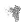
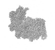
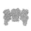
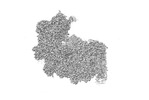
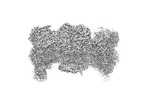
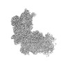
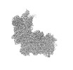
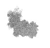
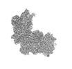
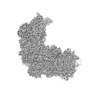
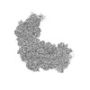
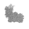
| Title | Cryo-EM map of Euglena gracilis respirasome I+III2+IV, complex I distal membrane arm focused | |||||||||
 Map data Map data | ||||||||||
 Sample Sample |
| |||||||||
 Keywords Keywords |  Electron transport chain / supercomplex / membrane protein / Euglena gracilis / ELECTRON TRANSPORT Electron transport chain / supercomplex / membrane protein / Euglena gracilis / ELECTRON TRANSPORT | |||||||||
| Biological species |  Euglena gracilis (euglena) Euglena gracilis (euglena) | |||||||||
| Method | single particle reconstruction / cryo EM / Resolution: 2.69 Å | |||||||||
 Authors Authors | Wu MC / He ZX / Tian HT / Hu YQ / Zhou L | |||||||||
| Funding support |  China, 1 items China, 1 items
| |||||||||
 Citation Citation | Journal: Nat Commun / Year: 2024 Title: Euglena's atypical respiratory chain adapts to the discoidal cristae and flexible metabolism. Authors: Zhaoxiang He / Mengchen Wu / Hongtao Tian / Liangdong Wang / Yiqi Hu / Fangzhu Han / Jiancang Zhou / Yong Wang / Long Zhou / Abstract: Euglena gracilis, a model organism of the eukaryotic supergroup Discoba harbouring also clinically important parasitic species, possesses diverse metabolic strategies and an atypical electron ...Euglena gracilis, a model organism of the eukaryotic supergroup Discoba harbouring also clinically important parasitic species, possesses diverse metabolic strategies and an atypical electron transport chain. While structures of the electron transport chain complexes and supercomplexes of most other eukaryotic clades have been reported, no similar structure is currently available for Discoba, limiting the understandings of its core metabolism and leaving a gap in the evolutionary tree of eukaryotic bioenergetics. Here, we report high-resolution cryo-EM structures of Euglena's respirasome I + III + IV and supercomplex III + IV. A previously unreported fatty acid synthesis domain locates on the tip of complex I's peripheral arm, providing a clear picture of its atypical subunit composition identified previously. Individual complexes are re-arranged in the respirasome to adapt to the non-uniform membrane curvature of the discoidal cristae. Furthermore, Euglena's conformationally rigid complex I is deactivated by restricting ubiquinone's access to its substrate tunnel. Our findings provide structural insights for therapeutic developments against euglenozoan parasite infections. | |||||||||
| History |
|
- Structure visualization
| Supplemental images |
|---|

- Downloads & links
-EMDB archive
| Map data | emd_35664.map.gz | 324.2 MB |  EMDB map data format EMDB map data format | |
|---|---|---|---|---|
| Header (meta data) | emd-35664-v30.xmlemd-35664.xml | 17.2 KB 17.2 KB | Display Display | EMDB header |
| FSC (resolution estimation) | emd_35664_fsc.xml | 14.7 KB | Display | FSC data file |
| Images | 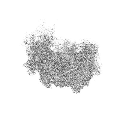 emd_35664.png emd_35664.png | 87.3 KB | ||
| Filedesc metadata | emd-35664.cif.gz | 4.6 KB | ||
| Others | emd_35664_half_map_1.map.gzemd_35664_half_map_2.map.gz | 318.9 MB 318.9 MB | ||
| Archive directory |  http://ftp.pdbj.org/pub/emdb/structures/EMD-35664ftp://ftp.pdbj.org/pub/emdb/structures/EMD-35664 http://ftp.pdbj.org/pub/emdb/structures/EMD-35664ftp://ftp.pdbj.org/pub/emdb/structures/EMD-35664 | HTTPS FTP |
-Related structure data


-Links
| EMDB pages | EMDB (EBI/PDBe) / EMDataResource |
|---|
-Map
| File | Download / File: emd_35664.map.gz / Format: CCP4 / Size: 343 MB / Type: IMAGE STORED AS FLOATING POINT NUMBER (4 BYTES) | ||||||||||||||||||||
|---|---|---|---|---|---|---|---|---|---|---|---|---|---|---|---|---|---|---|---|---|---|
| Voxel size | X=Y=Z: 1.2 Å | ||||||||||||||||||||
| Density |
| ||||||||||||||||||||
| Symmetry | Space group: 1 | ||||||||||||||||||||
| Details | EMDB XML:
|
-Supplemental data
-Half map: #1
| File | emd_35664_half_map_1.map | ||||||||||||
|---|---|---|---|---|---|---|---|---|---|---|---|---|---|
| Projections & Slices |
| ||||||||||||
| Density Histograms |
 Z
Z Y
Y X
X


-Half map: #2
| File | emd_35664_half_map_2.map | ||||||||||||
|---|---|---|---|---|---|---|---|---|---|---|---|---|---|
| Projections & Slices |
| ||||||||||||
| Density Histograms |


- Sample components
-Entire : Euglena gracilis respirasome I+III2+IV
| Entire | Name: Euglena gracilis respirasome I+III2+IV |
|---|---|
| Components |
|
-Supramolecule #1: Euglena gracilis respirasome I+III2+IV
| Supramolecule | Name: Euglena gracilis respirasome I+III2+IV / type: complex / ID: 1 / Parent: 0 |
|---|---|
| Source (natural) | Organism: Euglena gracilis (euglena) |
| Molecular weight | Theoretical: 2.5 MDa |
-Experimental details
-Structure determination
| Method | cryo EM |
|---|---|
 Processing Processing | single particle reconstruction |
| Aggregation state | particle |
-Sample preparation
| Concentration | 0.3 mg/mL | ||||||||||||||||||
|---|---|---|---|---|---|---|---|---|---|---|---|---|---|---|---|---|---|---|---|
| Buffer | pH: 7.4 Component:
Details: SEC buffer (30 mM Tris pH 7.4, 100 mM NaCl, 0.002% PMSF, 0.1% GDN (w/v), 1mM EDTA) | ||||||||||||||||||
| Grid | Model: Quantifoil R1.2/1.3 / Material: COPPER / Mesh: 300 / Support film - Material: CARBON / Support film - topology: CONTINUOUS / Support film - Film thickness: 2 / Pretreatment - Type: GLOW DISCHARGE / Pretreatment - Time: 15 sec. / Pretreatment - Atmosphere: AIR / Pretreatment - Pressure: 0.039 kPa / Details: 15mA | ||||||||||||||||||
| Vitrification | Cryogen name: ETHANE / Chamber humidity: 100 % / Chamber temperature: 277.15 K / Instrument: FEI VITROBOT MARK IV |
- Electron microscopy
| Microscope | FEI TITAN KRIOS |
|---|---|
| Electron beam | Acceleration voltage: 300 kV / Electron source: FIELD EMISSION GUN |
| Electron optics | Illumination mode: FLOOD BEAM / Imaging mode: BRIGHT FIELDBright-field microscopy / Cs: 2.7 mm / Nominal defocus max: 1.8 µm / Nominal defocus min: 0.6 µm / Nominal magnification: 105000 |
| Sample stage | Specimen holder model: FEI TITAN KRIOS AUTOGRID HOLDER / Cooling holder cryogen: NITROGEN |
| Image recording | Film or detector model: FEI FALCON IV (4k x 4k) / Digitization - Dimensions - Width: 4096 pixel / Digitization - Dimensions - Height: 4096 pixel / Number grids imaged: 1 / Number real images: 9807 / Average exposure time: 7.8 sec. / Average electron dose: 51.51 e/Å2 |
| Experimental equipment |  Model: Titan Krios / Image courtesy: FEI Company |
-Image processing
| Particle selection | Number selected: 3232325 |
|---|---|
| Startup model | Type of model: INSILICO MODEL In silico model: 3D ab-inito model reconstruction in cryosparc |
| Initial angle assignment | Type: COMMON LINE / Software - Name: cryoSPARC (ver. v3.3.2) |
| Final 3D classification | Number classes: 4 / Avg.num./class: 98936 / Software - Name: cryoSPARC (ver. v3.3.2) |
| Final angle assignment | Type: COMMON LINE / Software - Name: cryoSPARC (ver. v3.3.2) |
| Final reconstruction | Number classes used: 1 / Applied symmetry - Point group: C1 (asymmetric) / Algorithm: FOURIER SPACE / Resolution.type: BY AUTHOR / Resolution: 2.69 Å / Resolution method: FSC 0.143 CUT-OFF / Software - Name: cryoSPARC (ver. v3.3.2) / Number images used: 345048 |
| FSC plot (resolution estimation) |  |
-Atomic model buiding 1
| Refinement | Space: REAL / Protocol: RIGID BODY FIT |
|---|
