 Movie
Movie Controller


+ Open data

- Basic information
| Entry | 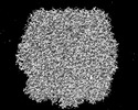 | |||||||||
|---|---|---|---|---|---|---|---|---|---|---|
| Title | Rubisco from Phaeodactylum tricornutum bound to PYCO1(452-592) | |||||||||
 Map data Map data | ||||||||||
 Sample Sample |
| |||||||||
 Keywords Keywords | Rubisco / phase separation / rubisco linker protein / condensation / pyrenoid / phaeodactylum tricornutum / PHOTOSYNTHESIS | |||||||||
| Biological species |  Phaeodactylum tricornutum (Diatom) Phaeodactylum tricornutum (Diatom) | |||||||||
| Method | single particle reconstruction / cryo EM / Resolution: 2.2 Å | |||||||||
 Authors Authors | Oh ZG / Ang WSL / Bhushan S / Mueller-Cajar O | |||||||||
| Funding support |  Singapore, 2 items Singapore, 2 items
| |||||||||
 Citation Citation | Journal: Proc Natl Acad Sci U S A / Year: 2023 Title: A linker protein from a red-type pyrenoid phase separates with Rubisco via oligomerizing sticker motifs. Authors: Zhen Guo Oh / Warren Shou Leong Ang / Cheng Wei Poh / Soak-Kuan Lai / Siu Kwan Sze / Hoi-Yeung Li / Shashi Bhushan / Tobias Wunder / Oliver Mueller-Cajar / Abstract: The slow kinetics and poor substrate specificity of the key photosynthetic CO-fixing enzyme Rubisco have prompted the repeated evolution of Rubisco-containing biomolecular condensates known as ...The slow kinetics and poor substrate specificity of the key photosynthetic CO-fixing enzyme Rubisco have prompted the repeated evolution of Rubisco-containing biomolecular condensates known as pyrenoids in the majority of eukaryotic microalgae. Diatoms dominate marine photosynthesis, but the interactions underlying their pyrenoids are unknown. Here, we identify and characterize the Rubisco linker protein PYCO1 from . PYCO1 is a tandem repeat protein containing prion-like domains that localizes to the pyrenoid. It undergoes homotypic liquid-liquid phase separation (LLPS) to form condensates that specifically partition diatom Rubisco. Saturation of PYCO1 condensates with Rubisco greatly reduces the mobility of droplet components. Cryo-electron microscopy and mutagenesis data revealed the sticker motifs required for homotypic and heterotypic phase separation. Our data indicate that the PYCO1-Rubisco network is cross-linked by PYCO1 stickers that oligomerize to bind to the small subunits lining the central solvent channel of the Rubisco holoenzyme. A second sticker motif binds to the large subunit. Pyrenoidal Rubisco condensates are highly diverse and tractable models of functional LLPS. | |||||||||
| History |
|
- Structure visualization
| Supplemental images |
|---|

- Downloads & links
-EMDB archive
| Map data | emd_35166.map.gz | 47.5 MB |  EMDB map data format EMDB map data format | |
|---|---|---|---|---|
| Header (meta data) | emd-35166-v30.xmlemd-35166.xml | 22.4 KB 22.4 KB | Display Display | EMDB header |
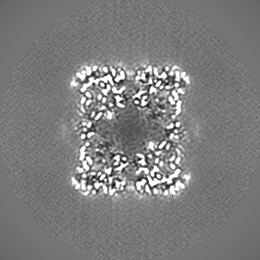
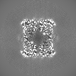
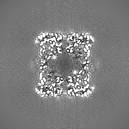
| Images | 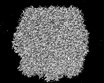 emd_35166.png emd_35166.png | 152 KB | ||
| Others | emd_35166_additional_1.map.gzemd_35166_half_map_1.map.gzemd_35166_half_map_2.map.gz | 56.4 MB 47.9 MB 47.9 MB | ||
| Archive directory |  http://ftp.pdbj.org/pub/emdb/structures/EMD-35166ftp://ftp.pdbj.org/pub/emdb/structures/EMD-35166 http://ftp.pdbj.org/pub/emdb/structures/EMD-35166ftp://ftp.pdbj.org/pub/emdb/structures/EMD-35166 | HTTPS FTP |
-Related structure data


-Links
| EMDB pages | EMDB (EBI/PDBe) / EMDataResource |
|---|
-Map
| File | Download / File: emd_35166.map.gz / Format: CCP4 / Size: 67 MB / Type: IMAGE STORED AS FLOATING POINT NUMBER (4 BYTES) | ||||||||||||||||||||
|---|---|---|---|---|---|---|---|---|---|---|---|---|---|---|---|---|---|---|---|---|---|
| Voxel size | X=Y=Z: 0.858 Å | ||||||||||||||||||||
| Density |
| ||||||||||||||||||||
| Symmetry | Space group: 1 | ||||||||||||||||||||
| Details | EMDB XML:
|
-Supplemental data
-Additional map: #1
| File | emd_35166_additional_1.map | ||||||||||||
|---|---|---|---|---|---|---|---|---|---|---|---|---|---|
| Projections & Slices |
| ||||||||||||
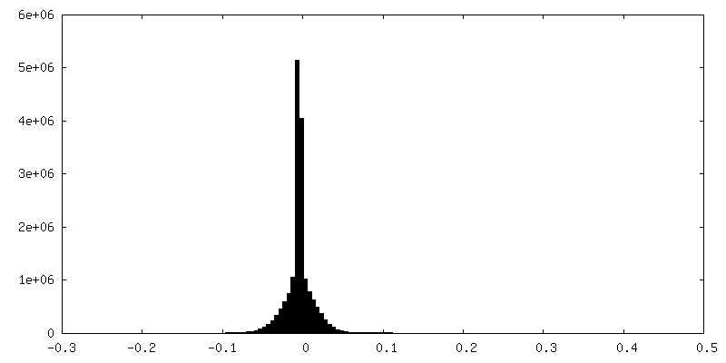
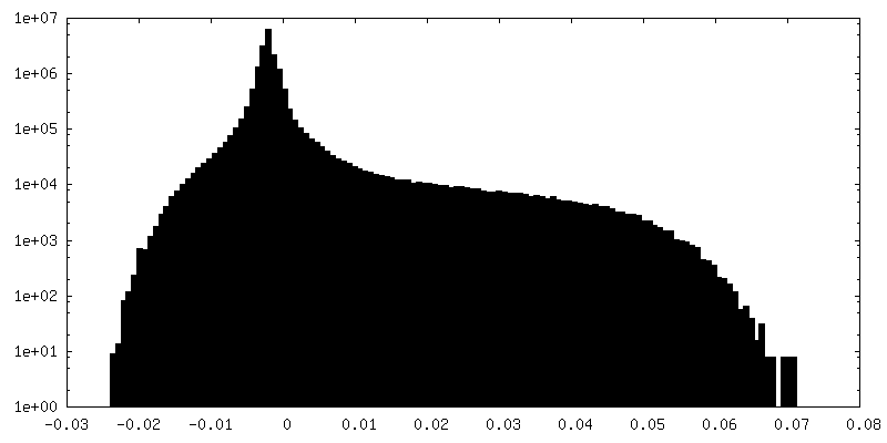
| Density Histograms |
 Z
Z Y
Y X
X


-Half map: #1
| File | emd_35166_half_map_1.map | ||||||||||||
|---|---|---|---|---|---|---|---|---|---|---|---|---|---|
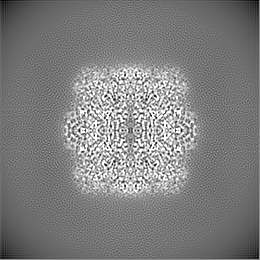
| Projections & Slices |
| ||||||||||||
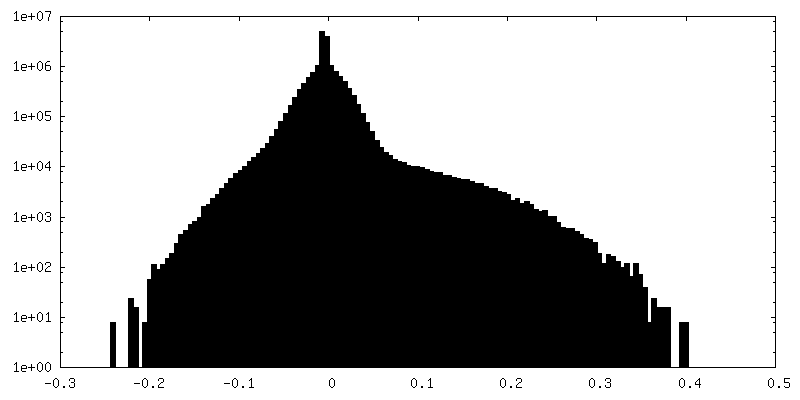
| Density Histograms |


-Half map: #2
| File | emd_35166_half_map_2.map | ||||||||||||
|---|---|---|---|---|---|---|---|---|---|---|---|---|---|
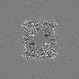
| Projections & Slices |
| ||||||||||||
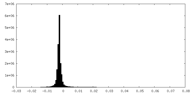
| Density Histograms |


- Sample components
-Entire : Ribulose 1,5-bisphosphate carboxylase/oxygenase from Phaeodactylu...
| Entire | Name: Ribulose 1,5-bisphosphate carboxylase/oxygenase from Phaeodactylum tricornutum |
|---|---|
| Components |
|
-Supramolecule #1: Ribulose 1,5-bisphosphate carboxylase/oxygenase from Phaeodactylu...
| Supramolecule | Name: Ribulose 1,5-bisphosphate carboxylase/oxygenase from Phaeodactylum tricornutum type: complex / ID: 1 / Parent: 0 / Macromolecule list: all |
|---|---|
| Source (natural) | Organism: Phaeodactylum tricornutum (Diatom) / Strain: Pt 1 8.6 CCMP 2561 |
| Molecular weight | Theoretical: 550 kDa/nm |
-Macromolecule #1: Ribulose 1,5-bisphosphate carboxylase/oxygenase
| Macromolecule | Name: Ribulose 1,5-bisphosphate carboxylase/oxygenase / type: protein_or_peptide / ID: 1 / Enantiomer: LEVO |
|---|---|
| Source (natural) | Organism: Phaeodactylum tricornutum (Diatom) |
| Sequence | String: MSQSVSERTR IKSDRYESGV IPYAKMGYWD AAYAVKNTDV LALFRIT(HYP)QP GVDPVEAAAA VAGESSTATW TVVWTD LLT ACDRYRAKAY RVDPVPNTTD QYFAFIAYE(CSO) DLFEEGSLAN LTASIIGNVF GFKAVSALRL EDMRIPHSYL (LYO)TFQG(HYP)ATG ...String: MSQSVSERTR IKSDRYESGV IPYAKMGYWD AAYAVKNTDV LALFRIT(HYP)QP GVDPVEAAAA VAGESSTATW TVVWTD LLT ACDRYRAKAY RVDPVPNTTD QYFAFIAYE(CSO) DLFEEGSLAN LTASIIGNVF GFKAVSALRL EDMRIPHSYL (LYO)TFQG(HYP)ATG VIVERERLNK YGIPL(HLU)GATV KPKLGLSGKN YGRVVYEGL(LYO) GGLDFL(KCX)DDE N INSQPFMR WRERFLYCME GINRASAATG ETKGSYLNIT AGTMEEVYKR AEYAKTVGSI VVMIDLVMGY TAIQSAAIWA RD NDLILHL HRAGNSTYAR QKNHGINFRV ICKWMRMCGV DHIHAGTVVG KLEGDPLMI(M3L) GFYDTLLLTH LNVNLPYGI FFEMTWASLR KCMPVASGGI HCGQMHQLVH YLGDDVVLQF GGGTIGHPDG IQAGATANRV ALEAMILARN EGADYFNSDI GPQILRNAA KTCGPLQTAL DLWKDISFNY TSTDTSDFSV TPTANV |
-Macromolecule #2: Ribulose 1,5-bisphosphate carboxylase/oxygenase small chain
| Macromolecule | Name: Ribulose 1,5-bisphosphate carboxylase/oxygenase small chain type: protein_or_peptide / ID: 2 / Enantiomer: LEVO |
|---|---|
| Source (natural) | Organism: Phaeodactylum tricornutum (Diatom) |
| Sequence | String: MRLTQGCFSF LPDLTDQQIE KQIAYCITKG WAMNVEWTDD PHPRNSYWEL WGLPLFDVKD PASVMFELRE ARKSCAAGYI RINAFNAAY GTESCVMSFI VNRPSNEPGF YLERQELEGR RIAYTTKSYS VQANPEGGRY |
-Experimental details
-Structure determination
| Method | cryo EM |
|---|---|
 Processing Processing | single particle reconstruction |
| Aggregation state | particle |
-Sample preparation
| Concentration | 0.5 mg/mL | |||||||||
|---|---|---|---|---|---|---|---|---|---|---|
| Buffer | pH: 8 Component:
Details: 20 mM Tris pH 8.0 20 mM NaCl | |||||||||
| Grid | Model: Quantifoil R2/2 / Material: COPPER / Mesh: 200 / Support film - Material: CARBON / Support film - topology: HOLEY / Support film - Film thickness: 2 / Pretreatment - Type: GLOW DISCHARGE / Pretreatment - Time: 20 sec. / Pretreatment - Atmosphere: AIR / Pretreatment - Pressure: 0.6 kPa | |||||||||
| Vitrification | Cryogen name: ETHANE / Chamber humidity: 100 % / Chamber temperature: 277 K / Instrument: FEI VITROBOT MARK IV / Details: Blotted for 2 sec with blot force of 1.. | |||||||||
| Details | 0.5 mg/mL Rubisco |
- Electron microscopy
| Microscope | FEI TITAN KRIOS |
|---|---|
| Electron beam | Acceleration voltage: 300 kV / Electron source: FIELD EMISSION GUN |
| Electron optics | C2 aperture diameter: 100.0 µm / Illumination mode: FLOOD BEAM / Imaging mode: BRIGHT FIELDBright-field microscopy / Cs: 2.7 mm / Nominal defocus max: 1.6 µm / Nominal defocus min: 0.8 µm / Nominal magnification: 165000 |
| Specialist optics | Energy filter - Name: GIF Bioquantum / Energy filter - Slit width: 20 eV / Details: Gatan EF |
| Sample stage | Specimen holder model: FEI TITAN KRIOS AUTOGRID HOLDER / Cooling holder cryogen: NITROGEN |
| Image recording | Film or detector model: GATAN K3 (6k x 4k) / Digitization - Dimensions - Width: 5760 pixel / Digitization - Dimensions - Height: 4092 pixel / Number grids imaged: 1 / Number real images: 8861 / Average exposure time: 5.0 sec. / Average electron dose: 65.0 e/Å2 Details: Images were collected in movie mode at 10 frames per second |
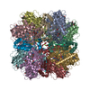
| Experimental equipment |  Model: Titan Krios / Image courtesy: FEI Company |
-Image processing
| Particle selection | Number selected: 3354751 |
|---|---|
| Startup model | Type of model: PDB ENTRY PDB model - PDB ID: Details: Rigid body docking with Rubisco from similar species. |
| Initial angle assignment | Type: COMMON LINE / Software - Name: RELION (ver. 3.1) Details: Initial model was built in situ in Relion using Initial model generation |
| Final 3D classification | Number classes: 30 / Avg.num./class: 8659 / Software - Name: RELION (ver. 3.1) / Details: Final 2D classification used 259,796 particles. |
| Final angle assignment | Type: PROJECTION MATCHING / Software - Name: RELION (ver. 3.1) |
| Final reconstruction | Number classes used: 30 / Applied symmetry - Point group: D4 (2x4 fold dihedral) / Algorithm: BACK PROJECTION / Resolution.type: BY AUTHOR / Resolution: 2.2 Å / Resolution method: FSC 0.143 CUT-OFF / Software - Name: RELION (ver. 3.1) / Number images used: 259796 |
-Atomic model buiding 1
| Details | Coot was used for model building |
|---|---|
| Refinement | Space: REAL / Protocol: RIGID BODY FIT |
