 Movie
Movie Controller

[English] 日本語
 Yorodumi
- EMDB-34266: CryoEM structure of human Pannexin1 with R217H congenital mutation. -
+ Open data

- Basic information
| Entry | 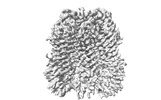 | |||||||||
|---|---|---|---|---|---|---|---|---|---|---|
| Title | CryoEM structure of human Pannexin1 with R217H congenital mutation. | |||||||||
 Map data Map data | human Pannexin 1 R217H mutant | |||||||||
 Sample Sample |
| |||||||||
 Keywords Keywords | Pannexin1 / ATP release / large-pore ion channel /  MEMBRANE PROTEIN MEMBRANE PROTEIN | |||||||||
| Function / homology |  Function and homology information Function and homology informationElectric Transmission Across Gap Junctions / leak channel activity / positive regulation of interleukin-1 alpha production / bleb / wide pore channel activity / gap junction channel activity / gap junction / positive regulation of macrophage cytokine production / oogenesis / response to ATP ...Electric Transmission Across Gap Junctions / leak channel activity / positive regulation of interleukin-1 alpha production / bleb / wide pore channel activity / gap junction channel activity / gap junction / positive regulation of macrophage cytokine production / oogenesis / response to ATP / monoatomic cation transport / The NLRP3 inflammasome / positive regulation of interleukin-1 beta production / response to ischemia / calcium channel activity / calcium ion transport / actin filament binding / cell-cell signaling / scaffold protein binding / protease binding / transmembrane transporter binding / signaling receptor binding / endoplasmic reticulum membrane / structural molecule activity / endoplasmic reticulum / protein-containing complex / membrane / identical protein binding / plasma membraneSimilarity search - Function | |||||||||
| Biological species |  Homo sapiens (human) Homo sapiens (human) | |||||||||
| Method | single particle reconstruction / cryo EM / Resolution: 3.87 Å | |||||||||
 Authors Authors | Hussain N / Penmatsa A | |||||||||
| Funding support |  India, 1 items India, 1 items
| |||||||||
 Citation Citation | Journal: To Be Published Title: Structural insights into pore dynamics of human Pannexin isoforms. Authors: Hussain N / Apotikar A / Pidathala S / Mukherjee S / Burada AP / Sikdar SK / Vinothkumar KR / Penmatsa A | |||||||||
| History |
|
- Structure visualization
| Supplemental images |
|---|

- Downloads & links
-EMDB archive
| Map data | emd_34266.map.gz | 118 MB | EMDB map data format | |
|---|---|---|---|---|
| Header (meta data) | emd-34266-v30.xmlemd-34266.xml | 19.3 KB 19.3 KB | Display Display | EMDB header |
| FSC (resolution estimation) | emd_34266_fsc.xml | 11.9 KB | Display | FSC data file |
| Images |  emd_34266.png emd_34266.png | 68.7 KB | ||
| Filedesc metadata | emd-34266.cif.gz | 6.4 KB | ||
| Others | emd_34266_half_map_1.map.gzemd_34266_half_map_2.map.gz | 115.9 MB 115.9 MB | ||
| Archive directory |  http://ftp.pdbj.org/pub/emdb/structures/EMD-34266ftp://ftp.pdbj.org/pub/emdb/structures/EMD-34266 http://ftp.pdbj.org/pub/emdb/structures/EMD-34266ftp://ftp.pdbj.org/pub/emdb/structures/EMD-34266 | HTTPS FTP |
-Related structure data
| Related structure data |  8gtsMC M: atomic model generated by this map C: citing same article ( |
|---|---|
| Similar structure data |
-Links
| EMDB pages | EMDB (EBI/PDBe) / EMDataResource |
|---|---|
| Related items in Molecule of the Month |


-Map
| File | Download / File: emd_34266.map.gz / Format: CCP4 / Size: 125 MB / Type: IMAGE STORED AS FLOATING POINT NUMBER (4 BYTES) | ||||||||||||||||||||||||||||||||||||
|---|---|---|---|---|---|---|---|---|---|---|---|---|---|---|---|---|---|---|---|---|---|---|---|---|---|---|---|---|---|---|---|---|---|---|---|---|---|
| Annotation | human Pannexin 1 R217H mutant | ||||||||||||||||||||||||||||||||||||
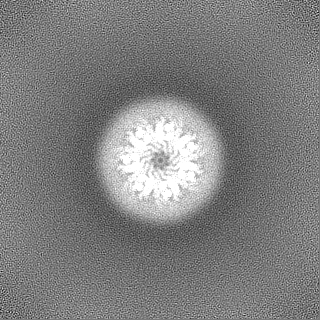
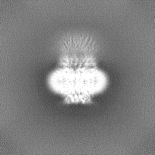
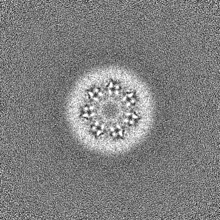
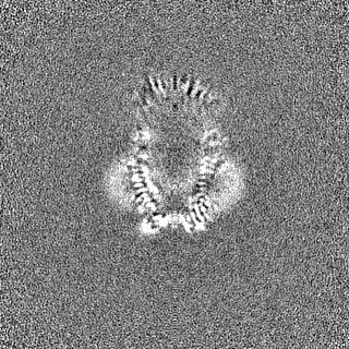
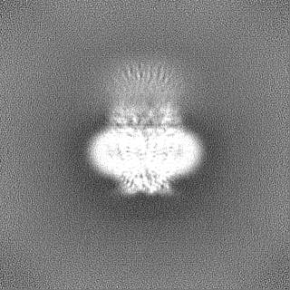
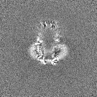
| Projections & slices | Image control
Images are generated by Spider. | ||||||||||||||||||||||||||||||||||||
| Voxel size | X=Y=Z: 1.07 Å | ||||||||||||||||||||||||||||||||||||
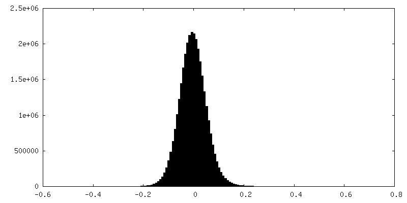
| Density |
| ||||||||||||||||||||||||||||||||||||
| Symmetry | Space group: 1 | ||||||||||||||||||||||||||||||||||||
| Details | EMDB XML:
|
 Z (Sec.)
 Y (Row.)
 X (Col.)


-Supplemental data
-Half map: half map B
| File | emd_34266_half_map_1.map | ||||||||||||
|---|---|---|---|---|---|---|---|---|---|---|---|---|---|
| Annotation | half map B | ||||||||||||
| Projections & Slices |
| ||||||||||||
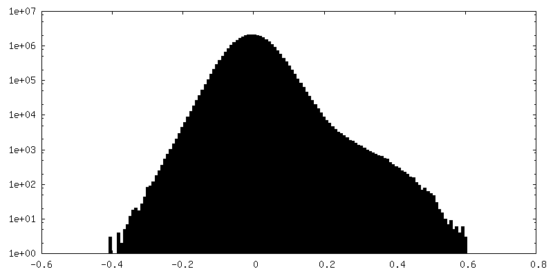
| Density Histograms |


-Half map: half map A
| File | emd_34266_half_map_2.map | ||||||||||||
|---|---|---|---|---|---|---|---|---|---|---|---|---|---|
| Annotation | half map A | ||||||||||||
| Projections & Slices |
| ||||||||||||
| Density Histograms |


- Sample components
-Entire : Pannexin 1 (R217H)
| Entire | Name: Pannexin 1 (R217H) |
|---|---|
| Components |
|
-Supramolecule #1: Pannexin 1 (R217H)
| Supramolecule | Name: Pannexin 1 (R217H) / type: organelle_or_cellular_component / ID: 1 / Parent: 0 / Macromolecule list: all Details: human isoform 1 of Pannexin. Expressed in plasma membranes involved in ATP release |
|---|---|
| Source (natural) | Organism: Homo sapiens (human) |
| Molecular weight | Theoretical: 48 kDa/nm |
-Macromolecule #1: Pannexin-1
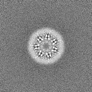
| Macromolecule | Name: Pannexin-1 / type: protein_or_peptide / ID: 1 / Number of copies: 7 / Enantiomer: LEVO |
|---|---|
| Source (natural) | Organism: Homo sapiens (human) |
| Molecular weight | Theoretical: 48.074816 KDa |
| Recombinant expression | Organism: Homo sapiens (human) |
| Sequence | String: MAIAQLATEY VFSDFLLKEP TEPKFKGLRL ELAVDKMVTC IAVGLPLLLI SLAFAQEISI GTQISCFSPS SFSWRQAAFV DSYCWAAVQ QKNSLQSESG NLPLWLHKFF PYILLLFAIL LYLPPLFWRF AAAPHICSDL KFIMEELDKV YNRAIKAAKS A RDLDMRDG ...String: MAIAQLATEY VFSDFLLKEP TEPKFKGLRL ELAVDKMVTC IAVGLPLLLI SLAFAQEISI GTQISCFSPS SFSWRQAAFV DSYCWAAVQ QKNSLQSESG NLPLWLHKFF PYILLLFAIL LYLPPLFWRF AAAPHICSDL KFIMEELDKV YNRAIKAAKS A RDLDMRDG ACSVPGVTEN LGQSLWEVSE SHFKYPIVEQ YLKTKKNSNN LIIKYISCHL LTLIIILLAC IYLGYYFSLS SL SDEFVCS IKSGILRNDS TVPDQFQCKL IAVGIFQLLS VINLVVYVLL APVVVYTLFV PFRQKTDVLK VYEILPTFDV LHF KSEGYN DLSLYNLFLE ENISEVKSYK CLKVLENIKS SGQGIDPMLL LTNLGMIKMD VVDGKTPMSA EMREEQGNQT AELQ GMNID SETKANNGEK NARQRLLDSS C UniProtKB: Pannexin-1 |
-Experimental details
-Structure determination
| Method | cryo EM |
|---|---|
 Processing Processing | single particle reconstruction |
| Aggregation state | particle |
-Sample preparation
| Concentration | 2.5 mg/mL | |||||||||||||||
|---|---|---|---|---|---|---|---|---|---|---|---|---|---|---|---|---|
| Buffer | pH: 8 Component:
Details: Fresh solution containing detergent was prepared for every prep. | |||||||||||||||
| Grid | Model: Quantifoil R1.2/1.3 / Material: GOLD / Mesh: 300 / Support film - Material: CARBON / Support film - topology: HOLEY / Pretreatment - Type: GLOW DISCHARGE / Pretreatment - Time: 60 sec. / Pretreatment - Atmosphere: AIR / Details: 25 milli amps for 60s | |||||||||||||||
| Vitrification | Cryogen name: ETHANE / Chamber humidity: 100 % / Chamber temperature: 288 K / Instrument: FEI VITROBOT MARK IV Details: sequentially blotted for 2 and 3.5 seconds with a wait of 10 s.. | |||||||||||||||
| Details | Sample is homoheptamer purified to homogeneity. |
- Electron microscopy
| Microscope | FEI TITAN KRIOS |
|---|---|
| Electron beam | Acceleration voltage: 300 kV / Electron source: FIELD EMISSION GUN |
| Electron optics | Illumination mode: OTHER / Imaging mode: OTHER / Cs: 2.7 mm / Nominal defocus max: 3.3000000000000003 µm / Nominal defocus min: 1.8 µm / Nominal magnification: 130000 |
| Specialist optics | Phase plate: OTHER / Spherical aberration corrector: None / Chromatic aberration corrector: None / Energy filter - Name: GIF Bioquantum / Energy filter - Slit width: 20 eV / Details: Bioquantum with K2 camera |
| Sample stage | Specimen holder model: FEI TITAN KRIOS AUTOGRID HOLDER / Cooling holder cryogen: NITROGEN |
| Temperature | Min: 100.0 K / Max: 100.0 K |
| Image recording | Film or detector model: GATAN K2 QUANTUM (4k x 4k) / Detector mode: COUNTING / Digitization - Frames/image: 1-40 / Number grids imaged: 1 / Number real images: 1776 / Average electron dose: 41.12 e/Å2 |
| Experimental equipment |  Model: Titan Krios / Image courtesy: FEI Company |
-Image processing
| Particle selection | Number selected: 781859 / Details: selected through particle picking |
|---|---|
| Startup model | Type of model: PDB ENTRY PDB model - PDB ID: |
| Initial angle assignment | Type: NOT APPLICABLE |
| Final 3D classification | Number classes: 5 / Avg.num./class: 8174 / Software - Name: cryoSPARC / Software - details: 2D classification |
| Final angle assignment | Type: NOT APPLICABLE |
| Final reconstruction | Number classes used: 5 / Applied symmetry - Point group: C7 (7 fold cyclic) / Resolution.type: BY AUTHOR / Resolution: 3.87 Å / Resolution method: FSC 0.143 CUT-OFF / Software - Name: cryoSPARC / Software - details: non uniform refinement / Number images used: 40873 |
| Details | Images were screened for ice thickness |
| FSC plot (resolution estimation) | 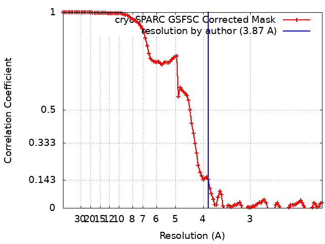 |

-Atomic model buiding 1
| Initial model | PDB ID: Chain - Chain ID: A / Chain - Source name: PDB / Chain - Initial model type: experimental model |
|---|---|
| Refinement | Space: REAL / Protocol: AB INITIO MODEL / Overall B value: 156 / Target criteria: Correlation coeficient |
| Output model | PDB-8gts: |
