- EMDB-30822: Structure of Wild-type PSI tetramer from Cyanophora paradoxa -
+
Open data
ID or keywords:
Loading...
-
Basic information
Entry
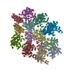
Database: EMDB / ID: EMD-30822
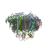
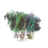
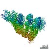
Title
Structure of Wild-type PSI tetramer from Cyanophora paradoxa
Map data
Sample
Complex: PSI tetramer
Function / homology
Function and homology information
cyanelle thylakoid membrane / thylakoid membrane / photosystem I reaction center / photosystem I / photosynthetic electron transport in photosystem I / photosystem I / plastid / chlorophyll binding / photosynthesis / 4 iron, 4 sulfur cluster binding ...cyanelle thylakoid membrane / thylakoid membrane / photosystem I reaction center / photosystem I / photosynthetic electron transport in photosystem I / photosystem I / plastid / chlorophyll binding / photosynthesis / 4 iron, 4 sulfur cluster binding / electron transfer activity / magnesium ion binding / metal ion binding Similarity search - Function
Photosystem I PsaM, reaction centre superfamily / Photosystem I PsaM, reaction centre / Photosystem I protein M (PsaM) / Photosystem I reaction centre subunit VIII / Photosystem I reaction centre subunit VIII / Photosystem I reaction centre subunit VIII superfamily / Photosystem I PsaF, reaction centre subunit III / Photosystem I PsaF, reaction centre subunit III superfamily / Photosystem I reaction centre subunit III / Photosystem I PsaJ, reaction centre subunit IX ...Photosystem I PsaM, reaction centre superfamily / Photosystem I PsaM, reaction centre / Photosystem I protein M (PsaM) / Photosystem I reaction centre subunit VIII / Photosystem I reaction centre subunit VIII / Photosystem I reaction centre subunit VIII superfamily / Photosystem I PsaF, reaction centre subunit III / Photosystem I PsaF, reaction centre subunit III superfamily / Photosystem I reaction centre subunit III / Photosystem I PsaJ, reaction centre subunit IX / Photosystem I PsaD / Photosystem I PsaJ, reaction centre subunit IX superfamily / Photosystem I, reaction centre subunit PsaD superfamily / Photosystem I reaction centre subunit IX / PsaJ / PsaD / Photosystem I PsaE, reaction centre subunit IV / Photosystem I reaction centre subunit IV / PsaE / Photosystem I protein PsaC / Photosystem I PsaA / Photosystem I PsaB / Photosystem I PsaA/PsaB, conserved site / Photosystem I psaA and psaB proteins signature. / Photosystem I PsaA/PsaB / Photosystem I PsaA/PsaB superfamily / Photosystem I psaA/psaB protein / Electron transport accessory-like domain superfamily / 4Fe-4S dicluster domain / 4Fe-4S ferredoxin, iron-sulphur binding, conserved site / 4Fe-4S ferredoxin-type iron-sulfur binding region signature. / 4Fe-4S ferredoxin-type iron-sulfur binding domain profile. / 4Fe-4S ferredoxin-type, iron-sulphur binding domain Similarity search - Domain/homology
Photosystem I P700 chlorophyll a apoprotein A1 / Photosystem I iron-sulfur center / Photosystem I P700 chlorophyll a apoprotein A2 / Photosystem I reaction center subunit IV / Photosystem I reaction center subunit III / Photosystem I reaction center subunit VIII / Photosystem I reaction center subunit IX / Photosystem I reaction center subunit XII / Photosystem I reaction center subunit II, cyanelle Similarity search - Component
Biological species
Cyanophora paradoxa (eukaryote)
Method
single particle reconstruction / cryo EM / Resolution: 4.0 Å
In the structure databanks used in Yorodumi, some data are registered as the other names, "COVID-19 virus" and "2019-nCoV". Here are the details of the virus and the list of structure data.
Jan 31, 2019. EMDB accession codes are about to change! (news from PDBe EMDB page)
EMDB accession codes are about to change! (news from PDBe EMDB page)
The allocation of 4 digits for EMDB accession codes will soon come to an end. Whilst these codes will remain in use, new EMDB accession codes will include an additional digit and will expand incrementally as the available range of codes is exhausted. The current 4-digit format prefixed with “EMD-” (i.e. EMD-XXXX) will advance to a 5-digit format (i.e. EMD-XXXXX), and so on. It is currently estimated that the 4-digit codes will be depleted around Spring 2019, at which point the 5-digit format will come into force.
The EM Navigator/Yorodumi systems omit the EMD- prefix.
Related info.:Q: What is EMD? / ID/Accession-code notation in Yorodumi/EM Navigator
Yorodumi is a browser for structure data from EMDB, PDB, SASBDB, etc.
This page is also the successor to EM Navigator detail page, and also detail information page/front-end page for Omokage search.
The word "yorodu" (or yorozu) is an old Japanese word meaning "ten thousand". "mi" (miru) is to see.
Related info.:EMDB / PDB / SASBDB / Comparison of 3 databanks / Yorodumi Search / Aug 31, 2016. New EM Navigator & Yorodumi / Yorodumi Papers / Jmol/JSmol / Function and homology information / Changes in new EM Navigator and Yorodumi
 Movie
Movie Controller


 Open data
 Basic information
 Map data
 Sample
 Function and homology information
 thylakoid membrane /
 Authors
 Citation
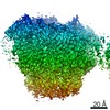
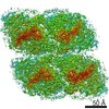
 Structure visualization
 Downloads & links
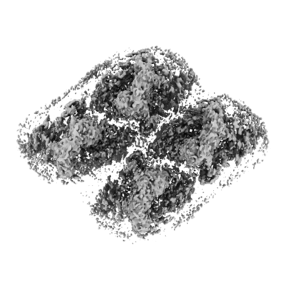 emd_30822.png
 http://ftp.pdbj.org/pub/emdb/structures/EMD-30822
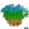


 Sample components
 Processing
 Electron microscopy

