 Movie
Movie Controller


+ Open data

- Basic information
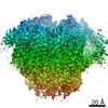
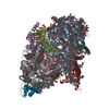
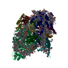
| Entry | Database: EMDB / ID: EMD-30821 | |||||||||
|---|---|---|---|---|---|---|---|---|---|---|
| Title | Structure of Wild-type PSI monomer2 from Cyanophora paradoxa | |||||||||
 Map data Map data | ||||||||||
 Sample Sample |
| |||||||||
| Function / homology |  Function and homology information Function and homology informationcyanelle thylakoid membrane /  thylakoid membrane / photosystem I reaction center / photosystem I / photosynthetic electron transport in photosystem I / photosystem I / plastid / chlorophyll binding / photosynthesis / 4 iron, 4 sulfur cluster binding ...cyanelle thylakoid membrane / thylakoid membrane / photosystem I reaction center / photosystem I / photosynthetic electron transport in photosystem I / photosystem I / plastid / chlorophyll binding / photosynthesis / 4 iron, 4 sulfur cluster binding / electron transfer activity / magnesium ion binding / metal ion binding thylakoid membrane / photosystem I reaction center / photosystem I / photosynthetic electron transport in photosystem I / photosystem I / plastid / chlorophyll binding / photosynthesis / 4 iron, 4 sulfur cluster binding ...cyanelle thylakoid membrane / thylakoid membrane / photosystem I reaction center / photosystem I / photosynthetic electron transport in photosystem I / photosystem I / plastid / chlorophyll binding / photosynthesis / 4 iron, 4 sulfur cluster binding / electron transfer activity / magnesium ion binding / metal ion bindingSimilarity search - Function | |||||||||
| Biological species |  Cyanophora paradoxa (eukaryote) Cyanophora paradoxa (eukaryote) | |||||||||
| Method | single particle reconstruction / cryo EM / Resolution: 3.2 Å | |||||||||
 Authors Authors | Kato K / Nagao R / Akita F / Miyazaki N / Shen JR | |||||||||
 Citation Citation | Journal: Biorxiv / Year: 2022 Title: Structural insights into an evolutionary turning-point of photosystem I from prokaryotes to eukaryotes Authors: Kato K / Nagao R / Ueno Y / Yokono M / Suzuki T / Jiang TY / Dohmae N / Akita F / Akimoto S / Miyazaki N / Shen JR | |||||||||
| History |
|
- Structure visualization
| Movie |
Movie viewer |
|---|---|
| Structure viewer | EM map: SurfViewMolmilJmol/JSmol |
| Supplemental images |

- Downloads & links
-EMDB archive
| Map data | emd_30821.map.gz | 28.4 MB | EMDB map data format | |
|---|---|---|---|---|
| Header (meta data) | emd-30821-v30.xmlemd-30821.xml | 23.5 KB 23.5 KB | Display Display | EMDB header |
| FSC (resolution estimation) | emd_30821_fsc.xml | 14.2 KB | Display | FSC data file |
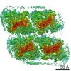
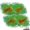
| Images | 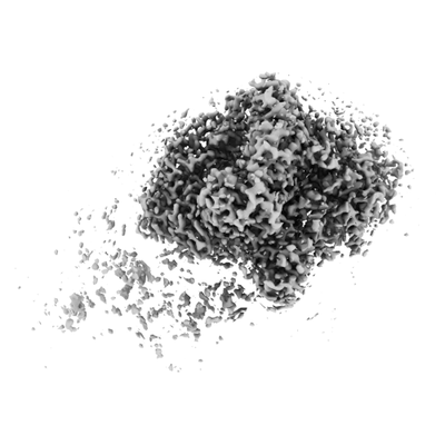 emd_30821.png emd_30821.png | 45.3 KB | ||
| Archive directory |  http://ftp.pdbj.org/pub/emdb/structures/EMD-30821ftp://ftp.pdbj.org/pub/emdb/structures/EMD-30821 http://ftp.pdbj.org/pub/emdb/structures/EMD-30821ftp://ftp.pdbj.org/pub/emdb/structures/EMD-30821 | HTTPS FTP |
-Related structure data
| Related structure data | 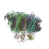 7dr1MC 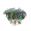 7dr0C 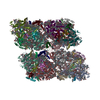 7dr2C M: atomic model generated by this map C: citing same article ( |
|---|---|
| Similar structure data |


-Links
| EMDB pages | EMDB (EBI/PDBe) / EMDataResource |
|---|---|
| Related items in Molecule of the Month |


-Map
| File | Download / File: emd_30821.map.gz / Format: CCP4 / Size: 244.1 MB / Type: IMAGE STORED AS FLOATING POINT NUMBER (4 BYTES) | ||||||||||||||||||||||||||||||||||||||||||||||||||||||||||||||||||||
|---|---|---|---|---|---|---|---|---|---|---|---|---|---|---|---|---|---|---|---|---|---|---|---|---|---|---|---|---|---|---|---|---|---|---|---|---|---|---|---|---|---|---|---|---|---|---|---|---|---|---|---|---|---|---|---|---|---|---|---|---|---|---|---|---|---|---|---|---|---|
| Voxel size | X=Y=Z: 1.093 Å | ||||||||||||||||||||||||||||||||||||||||||||||||||||||||||||||||||||
| Density |
| ||||||||||||||||||||||||||||||||||||||||||||||||||||||||||||||||||||
| Symmetry | Space group: 1 | ||||||||||||||||||||||||||||||||||||||||||||||||||||||||||||||||||||
| Details | EMDB XML:
CCP4 map header:
| ||||||||||||||||||||||||||||||||||||||||||||||||||||||||||||||||||||
-Supplemental data
- Sample components
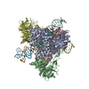
+Entire : PSI monomer2
+Supramolecule #1: PSI monomer2
+Macromolecule #1: Photosystem I P700 chlorophyll a apoprotein A1
+Macromolecule #2: Photosystem I P700 chlorophyll a apoprotein A2
+Macromolecule #3: Photosystem I iron-sulfur center
+Macromolecule #4: Photosystem I reaction center subunit II, cyanelle
+Macromolecule #5: Photosystem I reaction center subunit IV
+Macromolecule #6: Photosystem I reaction center subunit III
+Macromolecule #7: Photosystem I reaction center subunit VIII
+Macromolecule #8: Photosystem I reaction center subunit IX
+Macromolecule #9: Photosystem I reaction center subunit XI
+Macromolecule #10: Photosystem I reaction center subunit XII
+Macromolecule #11: CHLOROPHYLL A ISOMER

+Macromolecule #12: CHLOROPHYLL A

+Macromolecule #13: PHYLLOQUINONE

+Macromolecule #14: IRON/SULFUR CLUSTER

+Macromolecule #15: BETA-CAROTENE

+Macromolecule #16: 1,2-DIPALMITOYL-PHOSPHATIDYL-GLYCEROLE

+Macromolecule #17: 1,2-DISTEAROYL-MONOGALACTOSYL-DIGLYCERIDE

-Experimental details
-Structure determination
| Method | cryo EM |
|---|---|
 Processing Processing | single particle reconstruction |
| Aggregation state | particle |
-Sample preparation
| Concentration | 0.007 mg/mL | ||||||
|---|---|---|---|---|---|---|---|
| Buffer | pH: 6.5 / Component:
| ||||||
| Grid | Model: Quantifoil R2/1 / Material: COPPER / Mesh: 300 / Support film - Material: CARBON / Support film - topology: HOLEY ARRAY / Pretreatment - Type: GLOW DISCHARGE | ||||||
| Vitrification | Cryogen name: ETHANE / Chamber humidity: 100 % / Chamber temperature: 277 K / Instrument: FEI VITROBOT MARK IV |
- Electron microscopy
| Microscope | FEI TALOS ARCTICA |
|---|---|
| Electron beam | Acceleration voltage: 200 kV / Electron source: FIELD EMISSION GUN |
| Electron optics | Illumination mode: FLOOD BEAM / Imaging mode: BRIGHT FIELDBright-field microscopy |
| Image recording | Film or detector model: FEI FALCON III (4k x 4k) / Average electron dose: 50.0 e/Å2 |
| Experimental equipment |  Model: Talos Arctica / Image courtesy: FEI Company |
-Image processing
| Particle selection | Number selected: 1603082 |
|---|---|
| CTF correction | Software - Name: Gctf (ver. 1.18) |
| Startup model | Type of model: OTHER / Details: De novo generation |
| Initial angle assignment | Type: MAXIMUM LIKELIHOOD / Software - Name: RELION (ver. 3) |
| Final 3D classification | Software - Name: RELION (ver. 3) |
| Final angle assignment | Type: MAXIMUM LIKELIHOOD / Software - Name: RELION (ver. 3) |
| Final reconstruction | Applied symmetry - Point group: C1 (asymmetric) / Algorithm: FOURIER SPACE / Resolution.type: BY AUTHOR / Resolution: 3.2 Å / Resolution method: FSC 0.143 CUT-OFF / Software - Name: RELION (ver. 3) / Number images used: 110380 |
| FSC plot (resolution estimation) |  |
-Atomic model buiding 1
| Refinement | Space: REAL / Protocol: FLEXIBLE FIT / Target criteria: Correlation coefficient |
|---|---|
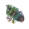
| Output model | PDB-7dr1: |
