 Movie
Movie Controller


+ Open data

- Basic information
| Entry | Database: EMDB / ID: EMD-30375 | ||||||||||||
|---|---|---|---|---|---|---|---|---|---|---|---|---|---|
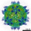
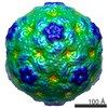
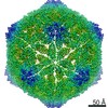
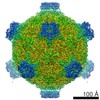
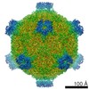
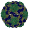
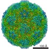
| Title | Cryo-EM Structure of Apple Latent Spherical Virus (ALSV) | ||||||||||||
 Map data Map data | |||||||||||||
 Sample Sample |
| ||||||||||||
 Keywords Keywords |  Cheravirus / capsid stabilization / genome release / single particle cryo-EM / STRUCTURAL PROTEIN Cheravirus / capsid stabilization / genome release / single particle cryo-EM / STRUCTURAL PROTEIN | ||||||||||||
| Function / homology | Viral coat protein subunit / 108K polyprotein Function and homology information Function and homology information | ||||||||||||
| Biological species |  Apple latent spherical virus Apple latent spherical virus | ||||||||||||
| Method | single particle reconstruction / cryo EM / Resolution: 2.87 Å | ||||||||||||
 Authors Authors | Naitow H / Hamaguchi T | ||||||||||||
| Funding support |  Japan, 3 items Japan, 3 items
| ||||||||||||
 Citation Citation | Journal: Commun Biol / Year: 2020 Title: Apple latent spherical virus structure with stable capsid frame supports quasi-stable protrusions expediting genome release. Authors: Hisashi Naitow / Tasuku Hamaguchi / Saori Maki-Yonekura / Masamichi Isogai / Nobuyuki Yoshikawa / Koji Yonekura / Abstract: Picorna-like plant viruses are non-enveloped RNA spherical viruses of ~30 nm. Part of the survival of these viruses depends on their capsid being stable enough to harbour the viral genome and yet ...Picorna-like plant viruses are non-enveloped RNA spherical viruses of ~30 nm. Part of the survival of these viruses depends on their capsid being stable enough to harbour the viral genome and yet malleable enough to allow its release. However, molecular mechanisms remain obscure. Here, we report a structure of a picorna-like plant virus, apple latent spherical virus, at 2.87 Å resolution by single-particle cryo-electron microscopy (cryo-EM) with a cold-field emission beam. The cryo-EM map reveals a unique structure composed of three capsid proteins Vp25, Vp20, and Vp24. Strikingly Vp25 has a long N-terminal extension, which substantially stabilises the capsid frame of Vp25 and Vp20 subunits. Cryo-EM images also resolve RNA genome leaking from a pentameric protrusion of Vp24 subunits. The structures and observations suggest that genome release occurs through occasional opening of the Vp24 subunits, possibly suppressed to a low frequency by the rigid frame of the other subunits. | ||||||||||||
| History |
|
- Structure visualization
| Movie |
Movie viewer |
|---|---|
| Structure viewer | EM map: SurfViewMolmilJmol/JSmol |
| Supplemental images |

- Downloads & links
-EMDB archive
| Map data | emd_30375.map.gz | 479.6 MB | EMDB map data format | |
|---|---|---|---|---|
| Header (meta data) | emd-30375-v30.xmlemd-30375.xml | 13.7 KB 13.7 KB | Display Display | EMDB header |
| Images | 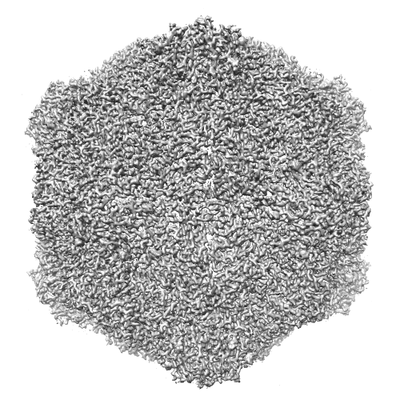 emd_30375.png emd_30375.png | 95.6 KB | ||
| Filedesc metadata | emd-30375.cif.gz | 5.6 KB | ||
| Archive directory |  http://ftp.pdbj.org/pub/emdb/structures/EMD-30375ftp://ftp.pdbj.org/pub/emdb/structures/EMD-30375 http://ftp.pdbj.org/pub/emdb/structures/EMD-30375ftp://ftp.pdbj.org/pub/emdb/structures/EMD-30375 | HTTPS FTP |
-Related structure data
| Related structure data |  7chkMC M: atomic model generated by this map C: citing same article ( |
|---|---|
| Similar structure data |


-Links
| EMDB pages | EMDB (EBI/PDBe) / EMDataResource |
|---|
-Map
| File | Download / File: emd_30375.map.gz / Format: CCP4 / Size: 512 MB / Type: IMAGE STORED AS FLOATING POINT NUMBER (4 BYTES) | ||||||||||||||||||||||||||||||||||||||||||||||||||||||||||||
|---|---|---|---|---|---|---|---|---|---|---|---|---|---|---|---|---|---|---|---|---|---|---|---|---|---|---|---|---|---|---|---|---|---|---|---|---|---|---|---|---|---|---|---|---|---|---|---|---|---|---|---|---|---|---|---|---|---|---|---|---|---|
| Voxel size | X=Y=Z: 1.24 Å | ||||||||||||||||||||||||||||||||||||||||||||||||||||||||||||
| Density |
| ||||||||||||||||||||||||||||||||||||||||||||||||||||||||||||
| Symmetry | Space group: 1 | ||||||||||||||||||||||||||||||||||||||||||||||||||||||||||||
| Details | EMDB XML:
CCP4 map header:
| ||||||||||||||||||||||||||||||||||||||||||||||||||||||||||||
-Supplemental data
- Sample components
-Entire : Apple latent spherical virus
| Entire | Name: Apple latent spherical virus |
|---|---|
| Components |
|
-Supramolecule #1: Apple latent spherical virus
| Supramolecule | Name: Apple latent spherical virus / type: virus / ID: 1 / Parent: 0 / Macromolecule list: all Details: ALSV was purified and isolated from infected Chenopodium quinoa leaf. NCBI-ID: 101688 / Sci species name: Apple latent spherical virus / Virus type: VIRION / Virus isolate: SPECIES / Virus enveloped: No / Virus empty: No |
|---|---|
| Host (natural) | Organism:  Malus domestica (apple) Malus domestica (apple) |
| Virus shell | Shell ID: 1 / Diameter: 300.0 Å / T number (triangulation number): 3 |
-Macromolecule #1: VP20 protein
| Macromolecule | Name: VP20 protein / type: protein_or_peptide / ID: 1 / Number of copies: 1 / Enantiomer: LEVO |
|---|---|
| Source (natural) | Organism: Apple latent spherical virus |
| Molecular weight | Theoretical: 19.957994 KDa |
| Sequence | String: GACLSIPNFP VHITGKTQQL HVGPKPSIAR FSFNPFDLGT VFNRFQSLCA HLEGYSGDLI VNWLVTCSAL TNARLYIIPV YDNYSFEKF SEEKLIQCKY EFKQISLVRK GTVHIPFVNW FGSYSRTRFP KLLFYFPNGV SGPSGEKIHV TVQLDRILNF S GLGHRLFK EIGPLVGE UniProtKB: 108K polyprotein |
-Macromolecule #2: VP24 protein
| Macromolecule | Name: VP24 protein / type: protein_or_peptide / ID: 2 / Number of copies: 1 / Enantiomer: LEVO |
|---|---|
| Source (natural) | Organism: Apple latent spherical virus |
| Molecular weight | Theoretical: 21.575467 KDa |
| Sequence | String: GSDPFSFLLN YSHCGTLVES SLNKGGMWCV PVSPVNLAAY TLQGEALVFN DAFVSKTHNW LHFMASTTAY WRGTLHYQMR VTYKDRNAA CRNLVAFYTT NNESLFGFNN KPVGDTGISS VMGDSFSVDI TVPFLIPTCY LQTIRGKFDY LNSCNGCIYF H LPTKSATS ...String: GSDPFSFLLN YSHCGTLVES SLNKGGMWCV PVSPVNLAAY TLQGEALVFN DAFVSKTHNW LHFMASTTAY WRGTLHYQMR VTYKDRNAA CRNLVAFYTT NNESLFGFNN KPVGDTGISS VMGDSFSVDI TVPFLIPTCY LQTIRGKFDY LNSCNGCIYF H LPTKSATS VQLWVRPGQD FDFARFRLLK AGYT UniProtKB: 108K polyprotein |
-Macromolecule #3: VP25 protein
| Macromolecule | Name: VP25 protein / type: protein_or_peptide / ID: 3 / Number of copies: 1 / Enantiomer: LEVO |
|---|---|
| Source (natural) | Organism: Apple latent spherical virus |
| Molecular weight | Theoretical: 24.098459 KDa |
| Sequence | String: GPDFTKIIWP TVVERNFSNP QSEITTTLQE LYGDTFETVS ICPPQSYGGE LLKGKIFFSS TPEFTREDLV EGKILASFKL DEVLSGLGM GAMLMTQIMS GHATIRVSAK VMLSKFCSFA LKLVYDELMQ LNSDTTDFGK ISVLPGAIFS TQEEEFSFDF E LFSPGVHL ...String: GPDFTKIIWP TVVERNFSNP QSEITTTLQE LYGDTFETVS ICPPQSYGGE LLKGKIFFSS TPEFTREDLV EGKILASFKL DEVLSGLGM GAMLMTQIMS GHATIRVSAK VMLSKFCSFA LKLVYDELMQ LNSDTTDFGK ISVLPGAIFS TQEEEFSFDF E LFSPGVHL KFDNNKLLGK VHLAALSAPN LTENMPESFS CTFNFSIVDV KTTFYNIGQ UniProtKB: 108K polyprotein |
-Experimental details
-Structure determination
| Method | cryo EM |
|---|---|
 Processing Processing | single particle reconstruction |
| Aggregation state | particle |
-Sample preparation
| Concentration | 1 mg/mL | ||||||||
|---|---|---|---|---|---|---|---|---|---|
| Buffer | pH: 7.8 Component:
| ||||||||
| Sugar embedding | Material: ice | ||||||||
| Grid | Model: Quantifoil / Material: COPPER / Support film - Material: CARBON / Support film - topology: HOLEY | ||||||||
| Vitrification | Cryogen name: ETHANE | ||||||||
| Details | In practice, the sample concentration is 1-2 mg/ml. |
- Electron microscopy
| Microscope | JEOL CRYO ARM 300 |
|---|---|
| Electron beam | Acceleration voltage: 300 kV / Electron source: FIELD EMISSION GUN |
| Electron optics | Illumination mode: FLOOD BEAM / Imaging mode: BRIGHT FIELDBright-field microscopy / Nominal defocus max: 2.0 µm / Nominal defocus min: 0.5 µm / Nominal magnification: 40000 |
| Sample stage | Specimen holder model: JEOL / Cooling holder cryogen: NITROGEN |
| Image recording | Film or detector model: GATAN K2 SUMMIT (4k x 4k) / Detector mode: SUPER-RESOLUTION / Average electron dose: 8.5 e/Å2 |
-Image processing
| Startup model | Type of model: OTHER / Details: Ab initio |
|---|---|
| Initial angle assignment | Type: MAXIMUM LIKELIHOOD |
| Final angle assignment | Type: MAXIMUM LIKELIHOOD |
| Final reconstruction | Resolution.type: BY AUTHOR / Resolution: 2.87 Å / Resolution method: FSC 0.143 CUT-OFF / Number images used: 8018 |
-Atomic model buiding 1
| Refinement | Space: REAL |
|---|---|
| Output model | PDB-7chk: |
