 Movie
Movie Controller


+ Open data

- Basic information
| Entry | 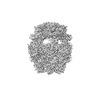 | |||||||||||||||
|---|---|---|---|---|---|---|---|---|---|---|---|---|---|---|---|---|
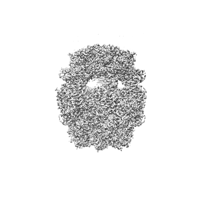
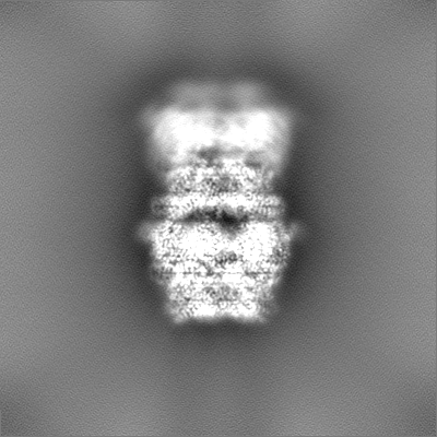
| Title | Structure of RdrA from Streptococcus suis RADAR defense system | |||||||||||||||
 Map data Map data | ||||||||||||||||
 Sample Sample |
| |||||||||||||||
| Function / homology | KAP family P-loop domain / KAP family P-loop domain / P-loop containing nucleoside triphosphate hydrolase / KAP NTPase domain-containing protein Function and homology information Function and homology information | |||||||||||||||
| Biological species |   Streptococcus suis (bacteria) Streptococcus suis (bacteria) | |||||||||||||||
| Method | single particle reconstruction / cryo EM / Resolution: 2.5 Å | |||||||||||||||
 Authors Authors | Duncan-Lowey B / Johnson AG / Rawson S / Mayer ML / Kranzusch PJ | |||||||||||||||
| Funding support |  United States, 4 items United States, 4 items
| |||||||||||||||
 Citation Citation | Journal: Cell / Year: 2023 Title: Cryo-EM structure of the RADAR supramolecular anti-phage defense complex. Authors: Brianna Duncan-Lowey / Nitzan Tal / Alex G Johnson / Shaun Rawson / Megan L Mayer / Shany Doron / Adi Millman / Sarah Melamed / Taya Fedorenko / Assaf Kacen / Alexander Brandis / Tevie ...Authors: Brianna Duncan-Lowey / Nitzan Tal / Alex G Johnson / Shaun Rawson / Megan L Mayer / Shany Doron / Adi Millman / Sarah Melamed / Taya Fedorenko / Assaf Kacen / Alexander Brandis / Tevie Mehlman / Gil Amitai / Rotem Sorek / Philip J Kranzusch /  Abstract: RADAR is a two-protein bacterial defense system that was reported to defend against phage by "editing" messenger RNA. Here, we determine cryo-EM structures of the RADAR defense complex, revealing ...RADAR is a two-protein bacterial defense system that was reported to defend against phage by "editing" messenger RNA. Here, we determine cryo-EM structures of the RADAR defense complex, revealing RdrA as a heptameric, two-layered AAA+ ATPase and RdrB as a dodecameric, hollow complex with twelve surface-exposed deaminase active sites. RdrA and RdrB join to form a giant assembly up to 10 MDa, with RdrA docked as a funnel over the RdrB active site. Surprisingly, our structures reveal an RdrB active site that targets mononucleotides. We show that RdrB catalyzes ATP-to-ITP conversion in vitro and induces the massive accumulation of inosine mononucleotides during phage infection in vivo, limiting phage replication. Our results define ATP mononucleotide deamination as a determinant of RADAR immunity and reveal supramolecular assembly of a nucleotide-modifying machine as a mechanism of anti-phage defense. | |||||||||||||||
| History |
|
- Structure visualization
| Supplemental images |
|---|
- Downloads & links
-EMDB archive
| Map data | emd_29326.map.gz | 230.1 MB | EMDB map data format | |
|---|---|---|---|---|
| Header (meta data) | emd-29326-v30.xmlemd-29326.xml | 15.2 KB 15.2 KB | Display Display | EMDB header |
| Images |  emd_29326.png emd_29326.png | 75.4 KB | ||
| Others | emd_29326_half_map_1.map.gzemd_29326_half_map_2.map.gz | 226.4 MB 226.4 MB | ||
| Archive directory |  http://ftp.pdbj.org/pub/emdb/structures/EMD-29326ftp://ftp.pdbj.org/pub/emdb/structures/EMD-29326 http://ftp.pdbj.org/pub/emdb/structures/EMD-29326ftp://ftp.pdbj.org/pub/emdb/structures/EMD-29326 | HTTPS FTP |
-Related structure data
| Related structure data | 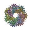 8fnuMC 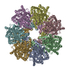 8fntC 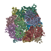 8fnvC 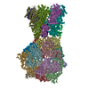 8fnwC M: atomic model generated by this map C: citing same article ( |
|---|---|
| Similar structure data |
-Links
| EMDB pages | EMDB (EBI/PDBe) / EMDataResource |
|---|
-Map
| File | Download / File: emd_29326.map.gz / Format: CCP4 / Size: 244.1 MB / Type: IMAGE STORED AS FLOATING POINT NUMBER (4 BYTES) | ||||||||||||||||||||
|---|---|---|---|---|---|---|---|---|---|---|---|---|---|---|---|---|---|---|---|---|---|
| Voxel size | X=Y=Z: 1.1 Å | ||||||||||||||||||||
| Density |
| ||||||||||||||||||||
| Symmetry | Space group: 1 | ||||||||||||||||||||
| Details | EMDB XML:
|
-Supplemental data
-Half map: #2
| File | emd_29326_half_map_1.map | ||||||||||||
|---|---|---|---|---|---|---|---|---|---|---|---|---|---|
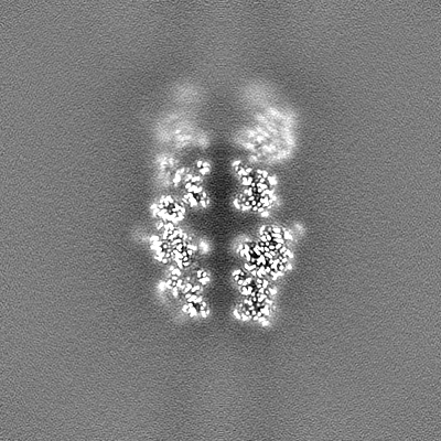
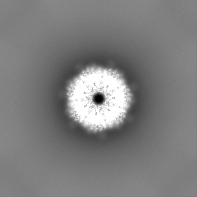
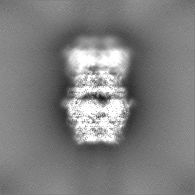
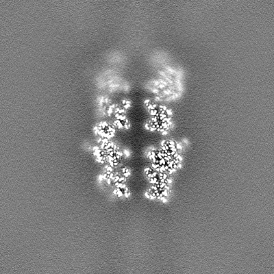
| Projections & Slices |
| ||||||||||||
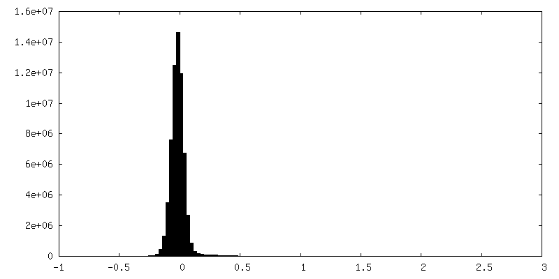
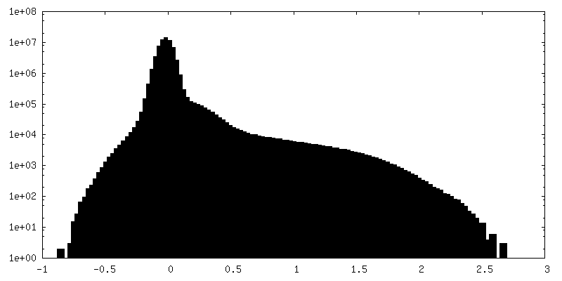
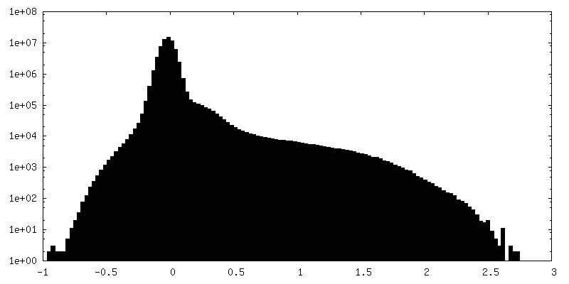
| Density Histograms |
 Z
Z Y
Y X
X


-Half map: #1
| File | emd_29326_half_map_2.map | ||||||||||||
|---|---|---|---|---|---|---|---|---|---|---|---|---|---|
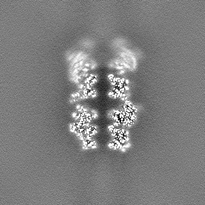
| Projections & Slices |
| ||||||||||||
| Density Histograms |


- Sample components
-Entire : RdrA heptameric complex
| Entire | Name: RdrA heptameric complex |
|---|---|
| Components |
|
-Supramolecule #1: RdrA heptameric complex
| Supramolecule | Name: RdrA heptameric complex / type: complex / ID: 1 / Chimera: Yes / Parent: 0 / Macromolecule list: all |
|---|---|
| Source (natural) | Organism: Streptococcus suis (bacteria) |
-Macromolecule #1: KAP NTPase domain-containing protein
| Macromolecule | Name: KAP NTPase domain-containing protein / type: protein_or_peptide / ID: 1 / Number of copies: 7 / Enantiomer: LEVO |
|---|---|
| Source (natural) | Organism: Streptococcus suis (bacteria) |
| Molecular weight | Theoretical: 106.413305 KDa |
| Recombinant expression | Organism: Escherichia coli (E. coli) |
| Sequence | String: MTKINWEKYK KVIKKEFSEK EETEEVKNYI FSSQLDKVNL ILEHMDTVGG IHSRNIAITG DRGTGKTSFI ETLKLVLEKQ NYYVFDIVS PTVLSSHLNI LEIVISSIYR EIDQFIDSHD VHDRGRLIQH LKKVMNAIAV EKKQSDYFKQ SKPEIEMLTD L SHRTFLDE ...String: MTKINWEKYK KVIKKEFSEK EETEEVKNYI FSSQLDKVNL ILEHMDTVGG IHSRNIAITG DRGTGKTSFI ETLKLVLEKQ NYYVFDIVS PTVLSSHLNI LEIVISSIYR EIDQFIDSHD VHDRGRLIQH LKKVMNAIAV EKKQSDYFKQ SKPEIEMLTD L SHRTFLDE EIKELFCYFK KVLNNRQDSC KEVIKDLVLI IDDLDLVENN LVYDLLRDIQ HYLDSQLIVI FAYKEGQLEQ SM FEHLAKG NEALLNHGVI DSNAIFGQIE RFLTKLVPLS NRIPLFKQDE LLNKTIGEFL ASLDPSYGVG ENLEFITKDS EKN KNNLTI REWFYESIFY RTNLKLDPID IREEASRLMP KTLREMVQLC EELHSMQVIT RSMDKLAGVE GLRKNIGAFR RYIG YKNST YFNLATMEFF QKWELAESHQ ANYLAYHFLM SYYQESFEQN QKLGYPLNLS KSGYPLTLRT MEPYNITLGD IYALM EELK YTEGISADTY YIVYILKVYY SLRLSELLYN VVLHHKLFVH VKEEATTFYM ATSTEELQIE KNHEQEATKL TDKEYR EHI MTAIEKVPAL QAYLELVNAQ FMPQNFNYDR SGSRDDDFYL ISWLKDDDLP EYSRLFKSLF LNSEVAAKGQ IQRNIGK RE SVFRYRNLYS YLPLQLTSAT FYKIDFLAFA IKADLLMYNV VRFVEEEGDT IPYFMSNMFH IDVFVRHNYN ENNNKGKF A YIAKQIVFGL WQGSNQRAHD LKHWYKSFDT VFGTKIEALH LLVDIAEQIK ISDKQTTDVS ALSEEQKRDE QAKKVAEKL AAIYHHIGMS RILSRLHQLP FIAEIKSNKE LLQHFSEAIV KLEKYASDTI NVGNLSQFRE SLKKIGQTYP SIQVLVDKLH RKQKLYVEF IQDFIETVNK LGEADESN |
-Experimental details
-Structure determination
| Method | cryo EM |
|---|---|
 Processing Processing | single particle reconstruction |
| Aggregation state | particle |
-Sample preparation
| Buffer | pH: 7.5 Component:
| ||||||||||||
|---|---|---|---|---|---|---|---|---|---|---|---|---|---|
| Vitrification | Cryogen name: ETHANE / Chamber humidity: 100 % |
- Electron microscopy
| Microscope | FEI TALOS ARCTICA |
|---|---|
| Electron beam | Acceleration voltage: 200 kV / Electron source: FIELD EMISSION GUN |
| Electron optics | Illumination mode: OTHER / Imaging mode: OTHER / Nominal defocus max: 2.0 µm / Nominal defocus min: 0.7000000000000001 µm |
| Image recording | Film or detector model: GATAN K3 (6k x 4k) / Average electron dose: 42.02 e/Å2 |
| Experimental equipment |  Model: Talos Arctica / Image courtesy: FEI Company |
-Image processing
| Initial angle assignment | Type: OTHER |
|---|---|
| Final angle assignment | Type: OTHER |
| Final reconstruction | Resolution.type: BY AUTHOR / Resolution: 2.5 Å / Resolution method: FSC 0.143 CUT-OFF / Number images used: 193305 |
