 Movie
Movie Controller

[English] 日本語
 Yorodumi
- EMDB-28441: Cryo-ET 3D reconstruction of an individual tetra-nucleosome parti... -
+ Open data

- Basic information
| Entry |  | |||||||||
|---|---|---|---|---|---|---|---|---|---|---|
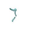
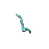
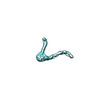
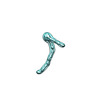
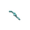
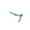
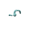
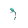
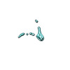
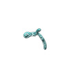
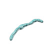
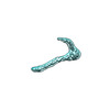
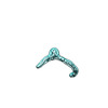
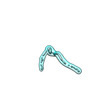
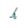
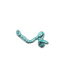
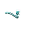
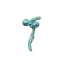
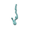
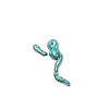
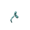
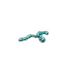
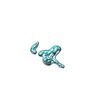
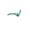
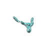
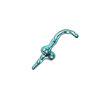
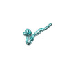
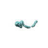
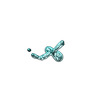
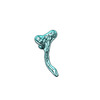
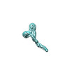
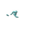
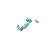
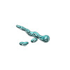
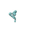
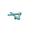
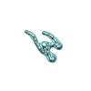
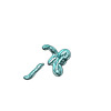
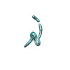
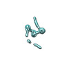
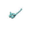
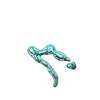
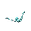
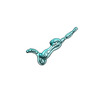
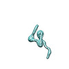
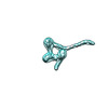
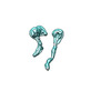
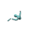
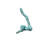
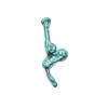
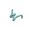
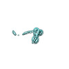
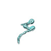
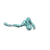
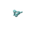
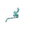
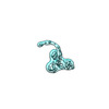
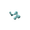
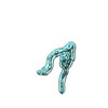
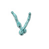
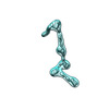
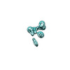
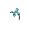
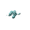
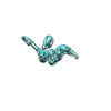
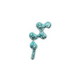
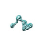
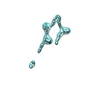
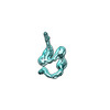
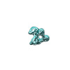
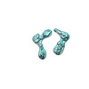
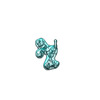
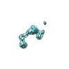
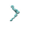
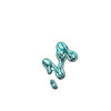
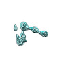
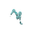
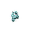
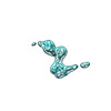
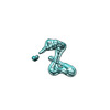
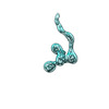
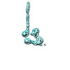
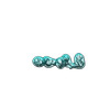
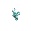
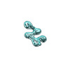
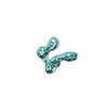
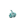
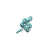
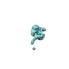
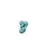
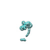
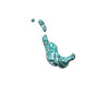
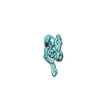
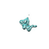
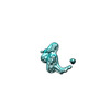
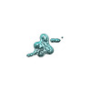
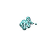
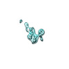
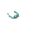
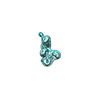
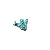
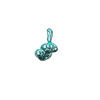
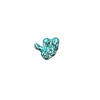
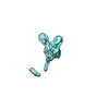
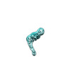
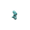
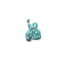
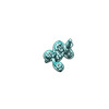
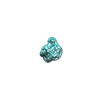
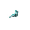
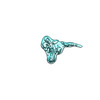
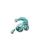
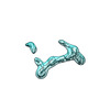
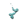
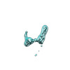
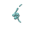
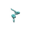
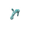
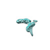
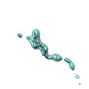
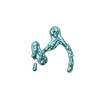
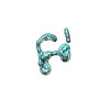
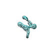
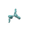
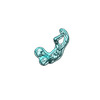
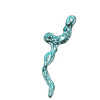
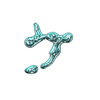
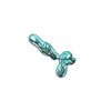
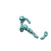
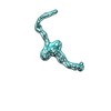
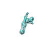
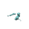
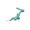
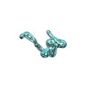
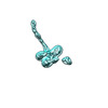
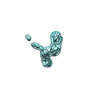
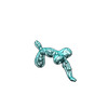
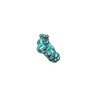
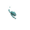
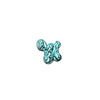
| Title | Cryo-ET 3D reconstruction of an individual tetra-nucleosome particle in 5 mM NaCl and 20 mM HEPES buffer --- Particle #150 | |||||||||
 Map data Map data | Cryo-ET 3D reconstruction of an individual tetra-nucleosome particle in 5 mM NaCl and 20 mM HEPES buffer --- Particle #150 | |||||||||
 Sample Sample |
| |||||||||
 Keywords Keywords | Nucleosome Array /  DNA BINDING PROTEIN DNA BINDING PROTEIN | |||||||||
| Biological species | Xenopus laevis (African clawed frog) | |||||||||
| Method | electron tomography / cryo EM | |||||||||
 Authors Authors | Zhang M / Celis CD / Liu JF / Bustamante C / Ren G | |||||||||
| Funding support |  United States, 1 items United States, 1 items
| |||||||||
 Citation Citation | Journal: To Be Published Title: Individual-molecule 3D structures reveal nucleosome array dynamics and regulations Authors: Zhang M / Celis CD / Liu JF / Bustamante C / Ren G | |||||||||
| History |
|
- Structure visualization
| Supplemental images |
|---|

- Downloads & links
-EMDB archive
| Map data | emd_28441.map.gz | 27.8 MB |  EMDB map data format EMDB map data format | |
|---|---|---|---|---|
| Header (meta data) | emd-28441-v30.xmlemd-28441.xml | 7.4 KB 7.4 KB | Display Display | EMDB header |
| Images |  emd_28441.png emd_28441.png | 20.5 KB | ||
| Filedesc metadata | emd-28441.cif.gz | 3.5 KB | ||
| Archive directory |  http://ftp.pdbj.org/pub/emdb/structures/EMD-28441ftp://ftp.pdbj.org/pub/emdb/structures/EMD-28441 http://ftp.pdbj.org/pub/emdb/structures/EMD-28441ftp://ftp.pdbj.org/pub/emdb/structures/EMD-28441 | HTTPS FTP |
-Related structure data
| Related structure data | C: citing same article ( |
|---|


-Links
| EMDB pages | EMDB (EBI/PDBe) / EMDataResource |
|---|
-Map
| File | Download / File: emd_28441.map.gz / Format: CCP4 / Size: 30.5 MB / Type: IMAGE STORED AS FLOATING POINT NUMBER (4 BYTES) | ||||||||||||||||||||
|---|---|---|---|---|---|---|---|---|---|---|---|---|---|---|---|---|---|---|---|---|---|
| Annotation | Cryo-ET 3D reconstruction of an individual tetra-nucleosome particle in 5 mM NaCl and 20 mM HEPES buffer --- Particle #150 | ||||||||||||||||||||
| Voxel size | X=Y=Z: 7.4 Å | ||||||||||||||||||||
| Density |
| ||||||||||||||||||||
| Symmetry | Space group: 1 | ||||||||||||||||||||
| Details | EMDB XML:
|
-Supplemental data
- Sample components
-Entire : Tetranucleosome array
| Entire | Name: Tetranucleosome array |
|---|---|
| Components |
|
-Supramolecule #1: Tetranucleosome array
| Supramolecule | Name: Tetranucleosome array / type: complex / ID: 1 / Parent: 0 |
|---|---|
| Source (natural) | Organism: Xenopus laevis (African clawed frog) |
-Experimental details
-Structure determination
| Method | cryo EM |
|---|---|
 Processing Processing | electron tomography |
| Aggregation state | particle |
-Sample preparation
| Buffer | pH: 7.5 / Details: 5 mM NaCl 20 mM HEPES 1 mM DTT 1 mM EDTA |
|---|---|
| Vitrification | Cryogen name: ETHANE / Chamber humidity: 99 % / Chamber temperature: 277 K |
| Sectioning | Other: NO SECTIONING |
- Electron microscopy
| Microscope | FEI TITAN KRIOS |
|---|---|
| Electron beam | Acceleration voltage: 300 kV / Electron source: FIELD EMISSION GUN |
| Electron optics | Illumination mode: FLOOD BEAM / Imaging mode: BRIGHT FIELDBright-field microscopy / Nominal defocus max: 3.5 µm / Nominal defocus min: 2.5 µm |
| Image recording | Film or detector model: GATAN K2 QUANTUM (4k x 4k) / Average exposure time: 2.0 sec. / Average electron dose: 5.4 e/Å2 |
| Experimental equipment |  Model: Titan Krios / Image courtesy: FEI Company |
-Image processing
| Final reconstruction | Software - Name: SPIDER / Number images used: 35 |
|---|
-Atomic model buiding 1
| Refinement | Protocol: FLEXIBLE FIT |
|---|
