 Movie
Movie Controller

[English] 日本語
 Yorodumi
- EMDB-26810: KDM2B-nucleosome complex stabilized by H3K36C-UNC8015 covalent co... -
+ Open data

- Basic information
| Entry |  | |||||||||||||||
|---|---|---|---|---|---|---|---|---|---|---|---|---|---|---|---|---|
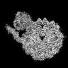
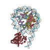
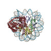
| Title | KDM2B-nucleosome complex stabilized by H3K36C-UNC8015 covalent conjugate | |||||||||||||||
 Map data Map data | KDM2B-nucleosome complex stabilized by H3K36C-UNC8015 covalent conjugate | |||||||||||||||
 Sample Sample |
| |||||||||||||||
| Biological species |   Homo sapiens (human) / synthetic construct (others) Homo sapiens (human) / synthetic construct (others) | |||||||||||||||
| Method | single particle reconstruction / cryo EM / Resolution: 3.6 Å | |||||||||||||||
 Authors Authors | Spangler CJ / Skrajna A / Foley CA / Budziszewski GR / Azzam DN / James LI / Frye SV / McGinty RK | |||||||||||||||
| Funding support |  United States, 4 items United States, 4 items
| |||||||||||||||
 Citation Citation | Journal: Nat Chem Biol / Year: 2023 Title: Structural basis of paralog-specific KDM2A/B nucleosome recognition. Authors: Cathy J Spangler / Aleksandra Skrajna / Caroline A Foley / Anh Nguyen / Gabrielle R Budziszewski / Dalal N Azzam / Eyla C Arteaga / Holly C Simmons / Charlotte B Smith / Nathaniel A Wesley / ...Authors: Cathy J Spangler / Aleksandra Skrajna / Caroline A Foley / Anh Nguyen / Gabrielle R Budziszewski / Dalal N Azzam / Eyla C Arteaga / Holly C Simmons / Charlotte B Smith / Nathaniel A Wesley / Emily M Wilkerson / Jeanne-Marie E McPherson / Dmitri Kireev / Lindsey I James / Stephen V Frye / Dennis Goldfarb / Robert K McGinty / Abstract: The nucleosome acidic patch is a major interaction hub for chromatin, providing a platform for enzymes to dock and orient for nucleosome-targeted activities. To define the molecular basis of acidic ...The nucleosome acidic patch is a major interaction hub for chromatin, providing a platform for enzymes to dock and orient for nucleosome-targeted activities. To define the molecular basis of acidic patch recognition proteome wide, we performed an amino acid resolution acidic patch interactome screen. We discovered that the histone H3 lysine 36 (H3K36) demethylase KDM2A, but not its closely related paralog, KDM2B, requires the acidic patch for nucleosome binding. Despite fundamental roles in transcriptional repression in health and disease, the molecular mechanisms governing nucleosome substrate specificity of KDM2A/B, or any related JumonjiC (JmjC) domain lysine demethylase, remain unclear. We used a covalent conjugate between H3K36 and a demethylase inhibitor to solve cryogenic electron microscopy structures of KDM2A and KDM2B trapped in action on a nucleosome substrate. Our structures show that KDM2-nucleosome binding is paralog specific and facilitated by dynamic nucleosomal DNA unwrapping and histone charge shielding that mobilize the H3K36 sequence for demethylation. | |||||||||||||||
| History |
|
- Structure visualization
| Supplemental images |
|---|

- Downloads & links
-EMDB archive
| Map data | emd_26810.map.gz | 9.4 MB |  EMDB map data format EMDB map data format | |
|---|---|---|---|---|
| Header (meta data) | emd-26810-v30.xmlemd-26810.xml | 23 KB 23 KB | Display Display | EMDB header |
| FSC (resolution estimation) | emd_26810_fsc.xml | 11.6 KB | Display | FSC data file |
| Images |  emd_26810.png emd_26810.png | 119.8 KB | ||
| Others | emd_26810_half_map_1.map.gzemd_26810_half_map_2.map.gz | 41.3 MB 41.3 MB | ||
| Archive directory |  http://ftp.pdbj.org/pub/emdb/structures/EMD-26810ftp://ftp.pdbj.org/pub/emdb/structures/EMD-26810 http://ftp.pdbj.org/pub/emdb/structures/EMD-26810ftp://ftp.pdbj.org/pub/emdb/structures/EMD-26810 | HTTPS FTP |
-Related structure data

-Links
| EMDB pages | EMDB (EBI/PDBe) / EMDataResource |
|---|
-Map
| File | Download / File: emd_26810.map.gz / Format: CCP4 / Size: 137.1 MB / Type: IMAGE STORED AS FLOATING POINT NUMBER (4 BYTES) | ||||||||||||||||||||
|---|---|---|---|---|---|---|---|---|---|---|---|---|---|---|---|---|---|---|---|---|---|
| Annotation | KDM2B-nucleosome complex stabilized by H3K36C-UNC8015 covalent conjugate | ||||||||||||||||||||
| Voxel size | X=Y=Z: 0.91 Å | ||||||||||||||||||||
| Density |
| ||||||||||||||||||||
| Symmetry | Space group: 1 | ||||||||||||||||||||
| Details | EMDB XML:
|
-Supplemental data
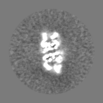
-Half map: KDM2B-nucleosome complex stabilized by H3K36C-UNC8015 covalent conjugate half...
| File | emd_26810_half_map_1.map | ||||||||||||
|---|---|---|---|---|---|---|---|---|---|---|---|---|---|
| Annotation | KDM2B-nucleosome complex stabilized by H3K36C-UNC8015 covalent conjugate half map 2 | ||||||||||||
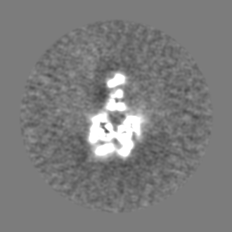
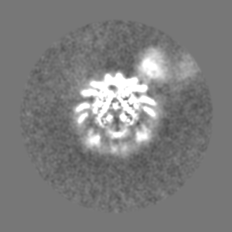
| Projections & Slices |
| ||||||||||||
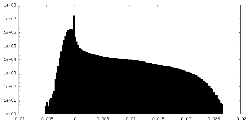
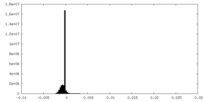
| Density Histograms |
 Z
Z Y
Y X
X


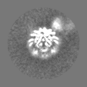
-Half map: KDM2B-nucleosome complex stabilized by H3K36C-UNC8015 covalent conjugate half...
| File | emd_26810_half_map_2.map | ||||||||||||
|---|---|---|---|---|---|---|---|---|---|---|---|---|---|
| Annotation | KDM2B-nucleosome complex stabilized by H3K36C-UNC8015 covalent conjugate half map 1 | ||||||||||||
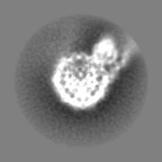
| Projections & Slices |
| ||||||||||||
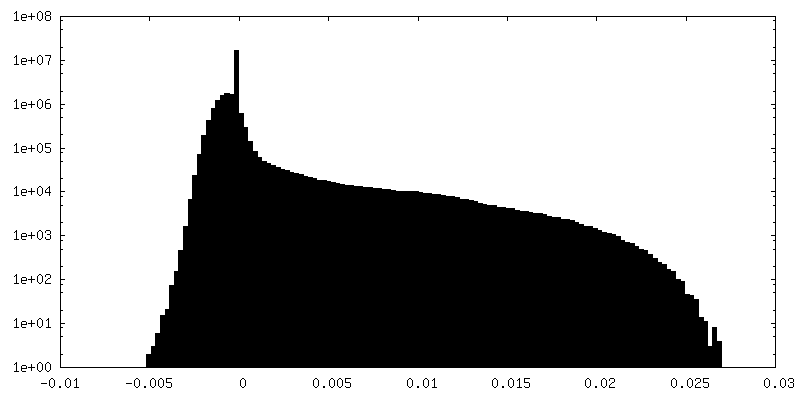
| Density Histograms |


- Sample components
-Entire : KDM2B-nucleosome complex stabilized by H3K36C-UNC8015 covalent co...
| Entire | Name: KDM2B-nucleosome complex stabilized by H3K36C-UNC8015 covalent conjugate |
|---|---|
| Components |
|
-Supramolecule #1: KDM2B-nucleosome complex stabilized by H3K36C-UNC8015 covalent co...
| Supramolecule | Name: KDM2B-nucleosome complex stabilized by H3K36C-UNC8015 covalent conjugate type: complex / ID: 1 / Chimera: Yes / Parent: 0 / Macromolecule list: #1-#7 |
|---|---|
| Source (natural) | Organism: Homo sapiens (human) |
| Molecular weight | Theoretical: 320 KDa |
-Macromolecule #1: human histone H3
| Macromolecule | Name: human histone H3 / type: protein_or_peptide / ID: 1 / Enantiomer: LEVO |
|---|---|
| Source (natural) | Organism: Homo sapiens (human) |
| Recombinant expression | Organism:  Escherichia coli BL21(DE3) (bacteria) Escherichia coli BL21(DE3) (bacteria) |
| Sequence | String: ARTKQTARKS TGGKAPRKQL ATKAARKSAP ATGGVCKPHR YRPGTVALRE IRRYQKSTEL LIRKLPFQRL VREIAQDFKT DLRFQSSAVM ALQEASEAYL VGLFEDTNLA AIHAKRVTIM PKDIQLARRI RGERA |
-Macromolecule #2: human histone H4
| Macromolecule | Name: human histone H4 / type: protein_or_peptide / ID: 2 / Enantiomer: LEVO |
|---|---|
| Source (natural) | Organism: Homo sapiens (human) |
| Recombinant expression | Organism: Escherichia coli BL21(DE3) (bacteria) |
| Sequence | String: SGRGKGGKGL GKGGAKRHRK VLRDNIQGIT KPAIRRLARR GGVKRISGLI YEETRGVLKV FLENVIRDAV TYTEHAKRKT VTAMDVVYAL KRQGRTLYGF GG |
-Macromolecule #3: human histone H2A
| Macromolecule | Name: human histone H2A / type: protein_or_peptide / ID: 3 / Enantiomer: LEVO |
|---|---|
| Source (natural) | Organism: Homo sapiens (human) |
| Recombinant expression | Organism: Escherichia coli BL21(DE3) (bacteria) |
| Sequence | String: SGRGKQGGKA RAKAKTRSSR AGLQFPVGRV HRLLRKGNYA ERVGAGAPVY LAAVLEYLTA EILELAGNAA RDNKKTRIIP RHLQLAIRND EELNKLLGKV TIAQGGVLPN IQAVLLPKKT ESHHKAKGK |
-Macromolecule #4: human histone H2B
| Macromolecule | Name: human histone H2B / type: protein_or_peptide / ID: 4 / Enantiomer: LEVO |
|---|---|
| Source (natural) | Organism: Homo sapiens (human) |
| Recombinant expression | Organism: Escherichia coli BL21(DE3) (bacteria) |
| Sequence | String: PEPAKSAPAP KKGSKKAVTK AQKKDGKKRK RSRKESYSVY VYKVLKQVHP DTGISSKAMG IMNSFVNDIF ERIAGEASRL AHYNKRSTIT SREIQTAVRL LLPGELAKHA VSEGTKAVTK YTSSK |
-Macromolecule #7: Lysine-specific demethylase 2B (KDM2B)
| Macromolecule | Name: Lysine-specific demethylase 2B (KDM2B) / type: protein_or_peptide / ID: 7 / Enantiomer: LEVO |
|---|---|
| Source (natural) | Organism: Homo sapiens (human) |
| Recombinant expression | Organism: Escherichia coli BL21(DE3) (bacteria) |
| Sequence | String: GSEPEEERIR YSQRLRGTMR RRYEDDGISD DEIEGKRTFD LEEKLHTNKY NANFVTFMEG KDFNVEYIQR GGLRDPLIFK NSDGLGIKMP DPDFTVNDVK MCVGSRRMVD VMDVNTQKGI EMTMAQWTRY YETPEEEREK LYNVISLEFS HTRLENMVQR PSTVDFIDWV ...String: GSEPEEERIR YSQRLRGTMR RRYEDDGISD DEIEGKRTFD LEEKLHTNKY NANFVTFMEG KDFNVEYIQR GGLRDPLIFK NSDGLGIKMP DPDFTVNDVK MCVGSRRMVD VMDVNTQKGI EMTMAQWTRY YETPEEEREK LYNVISLEFS HTRLENMVQR PSTVDFIDWV DNMWPRHLKE SQTESTNAIL EMQYPKVQKY CLMSVRGCYT DFHVDFGGTS VWYHIHQGGK VFWLIPPTAH NLELYENWLL SGKQGDIFLG DRVSDCQRIE LKQGYTFVIP SGWIHAVYTP TDTLVFGGNF LHSFNIPMQL KIYNIEDRTR VPNKFRYPFY YEMCWYVLER YVYCITNRSH LTKEFQKESL SMDLELNGLE SGNGDEEAVD REPRRLSSRR SVLTSPVANG VNLDYDGLGK TCRSLPSLKK TLAGDSSSDC SRGSHNGQVW DPQCAPRKDR QVHLTHFELE GLRCLVDKLE SLPLHKKCVP TGIEDEDALI ADVKILLEEL ANSDPKLALT GVPIVQWPKR DKLKFPTRPK VRVPTIPITK PHTMKPAPRL TPVRPAAASP IVSGARRRRV RCRKCKACVQ GECGVCHYCR DMKKFGGPGR MKQSCVLRQC LAPRLPHSVT CSLCGEVDQN EETQDFEKKL MECCICNEIV HPGCLQMDGE GLLNEELPNC WECPKCYQED SSEKAQHHHH HH |
-Macromolecule #5: DNA 185-MER
| Macromolecule | Name: DNA 185-MER / type: dna / ID: 5 / Classification: DNA |
|---|---|
| Source (natural) | Organism: synthetic construct (others) |
| Sequence | String: ATCCCTATAC GCGGCCGCCC TGGAGAATCC CGGTGCCGAG GCCGCTCAAT TGGTCGTAGA CAGCTCTAGC ACCGCTTAAA CGCACGTACG CGCTGTCCCC CGCGTTTTAA CCGCCAAGGG GATTACTCCC TAGTCTCCAG GCACGTGTCA GATATATACA TCCTGTGCAT GTATTGAACA GCGAT |
-Macromolecule #6: DNA 185-MER
| Macromolecule | Name: DNA 185-MER / type: dna / ID: 6 / Classification: DNA |
|---|---|
| Source (natural) | Organism: synthetic construct (others) |
| Sequence | String: ATCGCTGTTC AATACATGCA CAGGATGTAT ATATCTGACA CGTGCCTGGA GACTAGGGAG TAATCCCCTT GGCGGTTAAA ACGCGGGGGA CAGCGCGTAC GTGCGTTTAA GCGGTGCTAG AGCTGTCTAC GACCAATTGA GCGGCCTCGG CACCGGGATT CTCCAGGGCG GCCGCGTATA GGGAT |
-Experimental details
-Structure determination
| Method | cryo EM |
|---|---|
 Processing Processing | single particle reconstruction |
| Aggregation state | particle |
-Sample preparation
| Concentration | 1 mg/mL | |||||||||
|---|---|---|---|---|---|---|---|---|---|---|
| Buffer | pH: 7.5 Component:
| |||||||||
| Vitrification | Cryogen name: ETHANE-PROPANE / Chamber humidity: 100 % / Chamber temperature: 277 K / Instrument: FEI VITROBOT MARK IV |
- Electron microscopy
| Microscope | FEI TALOS ARCTICA |
|---|---|
| Electron beam | Acceleration voltage: 200 kV / Electron source: FIELD EMISSION GUN |
| Electron optics | Illumination mode: FLOOD BEAM / Imaging mode: BRIGHT FIELDBright-field microscopy / Nominal defocus max: 3.0 µm / Nominal defocus min: 0.5 µm / Nominal magnification: 45000 |
| Sample stage | Specimen holder model: FEI TITAN KRIOS AUTOGRID HOLDER |
| Image recording | Film or detector model: GATAN K3 BIOQUANTUM (6k x 4k) / Number grids imaged: 1 / Number real images: 15671 / Average electron dose: 45.0 e/Å2 |
| Experimental equipment |  Model: Talos Arctica / Image courtesy: FEI Company |
-Image processing
| Particle selection | Number selected: 3435247 |
|---|---|
| Startup model | Type of model: PDB ENTRY PDB model - PDB ID: |
| Initial angle assignment | Type: MAXIMUM LIKELIHOOD / Software - Name: RELION (ver. 3.1) |
| Final angle assignment | Type: MAXIMUM LIKELIHOOD / Software - Name: RELION (ver. 3.1) |
| Final reconstruction | Resolution.type: BY AUTHOR / Resolution: 3.6 Å / Resolution method: FSC 0.143 CUT-OFF / Software - Name: RELION (ver. 3.1) / Number images used: 47729 |
| FSC plot (resolution estimation) |  |
-Atomic model buiding 1
| Initial model | PDB ID: |
|---|---|
| Refinement | Space: REAL |
