National Institutes of Health/National Institute Of Allergy and Infectious Diseases (NIH/NIAID)
U54 AI150472
United States
National Institutes of Health/National Institute Of Allergy and Infectious Diseases (NIH/NIAID)
R01 AI136680
United States
The Francis Crick Institute
FC10061
United Kingdom
National Institutes of Health/National Institute Of Allergy and Infectious Diseases (NIH/NIAID)
P50AI150481
United States
Citation
Journal: Nat Commun / Year: 2022 Title: Multivalent interactions essential for lentiviral integrase function. Authors: Allison Ballandras-Colas / Vidya Chivukula / Dominika T Gruszka / Zelin Shan / Parmit K Singh / Valerie E Pye / Rebecca K McLean / Gregory J Bedwell / Wen Li / Andrea Nans / Nicola J Cook / ...Authors: Allison Ballandras-Colas / Vidya Chivukula / Dominika T Gruszka / Zelin Shan / Parmit K Singh / Valerie E Pye / Rebecca K McLean / Gregory J Bedwell / Wen Li / Andrea Nans / Nicola J Cook / Hind J Fadel / Eric M Poeschla / David J Griffiths / Javier Vargas / Ian A Taylor / Dmitry Lyumkis / Hasan Yardimci / Alan N Engelman / Peter Cherepanov / Abstract: A multimer of retroviral integrase (IN) synapses viral DNA ends within a stable intasome nucleoprotein complex for integration into a host cell genome. Reconstitution of the intasome from the maedi- ...A multimer of retroviral integrase (IN) synapses viral DNA ends within a stable intasome nucleoprotein complex for integration into a host cell genome. Reconstitution of the intasome from the maedi-visna virus (MVV), an ovine lentivirus, revealed a large assembly containing sixteen IN subunits. Herein, we report cryo-EM structures of the lentiviral intasome prior to engagement of target DNA and following strand transfer, refined at 3.4 and 3.5 Å resolution, respectively. The structures elucidate details of the protein-protein and protein-DNA interfaces involved in lentiviral intasome formation. We show that the homomeric interfaces involved in IN hexadecamer formation and the α-helical configuration of the linker connecting the C-terminal and catalytic core domains are critical for MVV IN strand transfer activity in vitro and for virus infectivity. Single-molecule microscopy in conjunction with photobleaching reveals that the MVV intasome can bind a variable number, up to sixteen molecules, of the lentivirus-specific host factor LEDGF/p75. Concordantly, ablation of endogenous LEDGF/p75 results in gross redistribution of MVV integration sites in human and ovine cells. Our data confirm the importance of the expanded architecture observed in cryo-EM studies of lentiviral intasomes and suggest that this organization underlies multivalent interactions with chromatin for integration targeting to active genes.
Name: ZINC ION / type: ligand / ID: 4 / Number of copies: 12 / Formula: ZN
Molecular weight
Theoretical: 65.409 Da
-
Macromolecule #5: CALCIUM ION
Macromolecule
Name: CALCIUM ION / type: ligand / ID: 5 / Number of copies: 2 / Formula: CA
Molecular weight
Theoretical: 40.078 Da
-
Experimental details
-
Structure determination
Method
cryo EM
Processing
single particle reconstruction
Aggregation state
particle
-
Sample preparation
Buffer
pH: 6.5 Component:
Concentration
Name
Formula
25.0 mM
Tris-HClTris
350.0 mM
sodium chloride
NaClSodium chloride
1.0 mM
TCEP
3.0 mM
Calcium chloride
CaCl2
Grid
Model: Quantifoil R1.2/1.3 / Material: GOLD / Mesh: 300 / Support film - Material: GOLD / Support film - topology: HOLEY / Pretreatment - Type: PLASMA CLEANING / Pretreatment - Time: 7 sec.
Vitrification
Cryogen name: ETHANE / Instrument: HOMEMADE PLUNGER Details: Cryo-EM grids were prepared by freezing using a manual plunger in cold room at 4C.
Details
MVV CSC intasomes, assembled and purified as previously described, were applied onto R1.2/1.3 gold UltrAufoil grids, Au 300 mesh (Quantifoil). Cryo-EM grids were prepared by manually freezing using a manual plunger in cold room at 4C and stored in liquid nitrogen for future data acquisition.
-
Electron microscopy
Microscope
FEI TALOS ARCTICA
Electron beam
Acceleration voltage: 200 kV / Electron source: FIELD EMISSION GUN
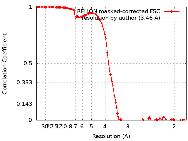
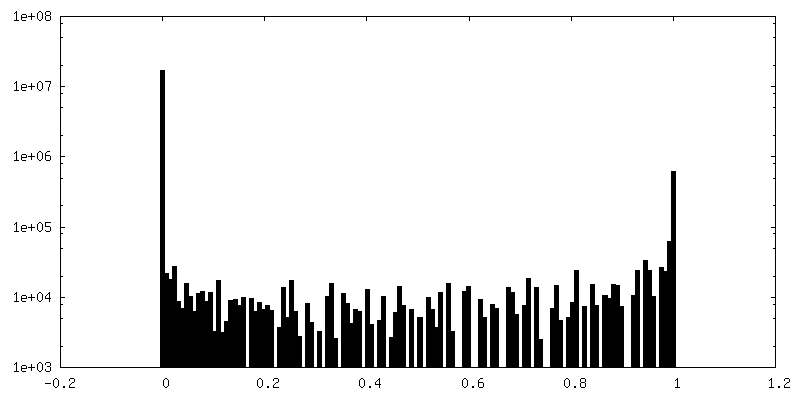
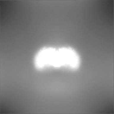
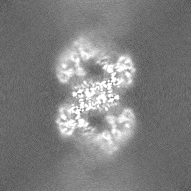
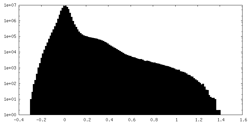
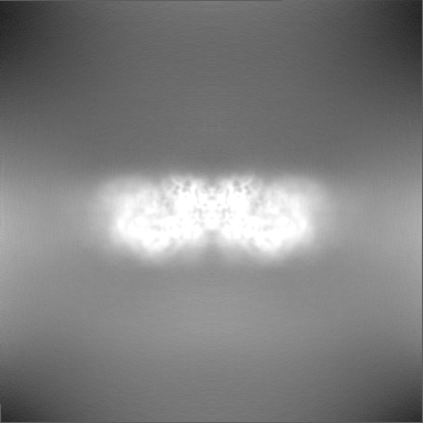
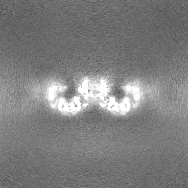
Applied symmetry - Point group: C2 (2 fold cyclic) / Algorithm: FOURIER SPACE / Resolution.type: BY AUTHOR / Resolution: 3.46 Å / Resolution method: FSC 0.143 CUT-OFF / Software - Name: cryoSPARC (ver. 3.0) / Number images used: 147860
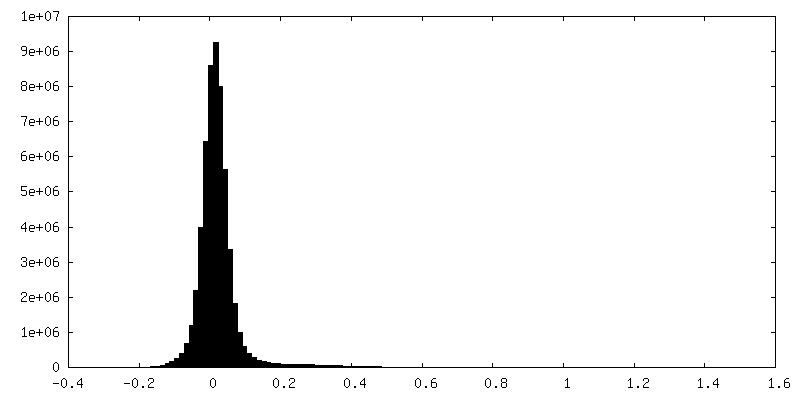
FSC plot (resolution estimation)
-
Atomic model buiding 1
Details
The STC model refined in study, with the tDNA removed, was docked into the CSC cryoEM map using UCSF Chimera. It was observed that there were some slight differences in some domain positions, to address this, individual domains that were not well fitted to the map were docked as individual domains to achieve a best-fit starting model. Two (C2 related) NTDs (aa 1-35) were removed from the model due to lack of supporting map. Adjustments were made to the model interactively using Coot and the coordinates were subjected to real-space refinement in Phenix dev-4213-000 employing C2 NCS constraints.
Refinement
Space: REAL / Protocol: FLEXIBLE FIT / Overall B value: 262 / Target criteria: CC
Output model
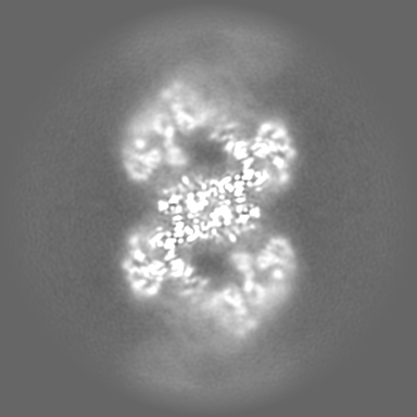
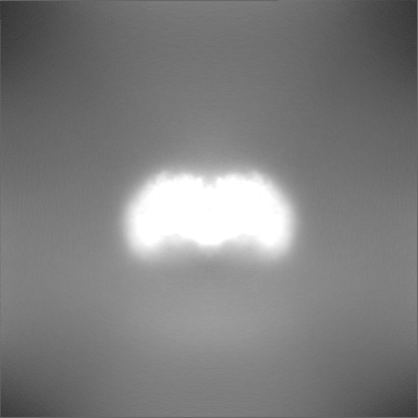
PDB-7u32: MVV cleaved synaptic complex (CSC) intasome at 3.4 A resolution
+
About Yorodumi
-
News
-
Feb 9, 2022. New format data for meta-information of EMDB entries
New format data for meta-information of EMDB entries
Version 3 of the EMDB header file is now the official format.
The previous official version 1.9 will be removed from the archive.
In the structure databanks used in Yorodumi, some data are registered as the other names, "COVID-19 virus" and "2019-nCoV". Here are the details of the virus and the list of structure data.
Jan 31, 2019. EMDB accession codes are about to change! (news from PDBe EMDB page)
EMDB accession codes are about to change! (news from PDBe EMDB page)
The allocation of 4 digits for EMDB accession codes will soon come to an end. Whilst these codes will remain in use, new EMDB accession codes will include an additional digit and will expand incrementally as the available range of codes is exhausted. The current 4-digit format prefixed with “EMD-” (i.e. EMD-XXXX) will advance to a 5-digit format (i.e. EMD-XXXXX), and so on. It is currently estimated that the 4-digit codes will be depleted around Spring 2019, at which point the 5-digit format will come into force.
The EM Navigator/Yorodumi systems omit the EMD- prefix.
Related info.:Q: What is EMD? / ID/Accession-code notation in Yorodumi/EM Navigator
Yorodumi is a browser for structure data from EMDB, PDB, SASBDB, etc.
This page is also the successor to EM Navigator detail page, and also detail information page/front-end page for Omokage search.
The word "yorodu" (or yorozu) is an old Japanese word meaning "ten thousand". "mi" (miru) is to see.
Related info.:EMDB / PDB / SASBDB / Comparison of 3 databanks / Yorodumi Search / Aug 31, 2016. New EM Navigator & Yorodumi / Yorodumi Papers / Jmol/JSmol / Function and homology information / Changes in new EM Navigator and Yorodumi
 Movie
Movie Controller

 Yorodumi
 Open data
 Basic information
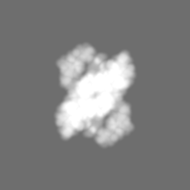
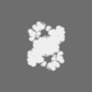
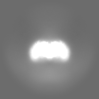
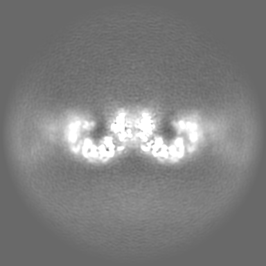
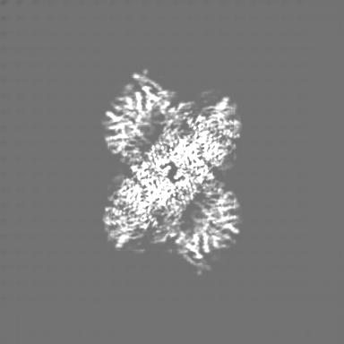
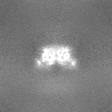
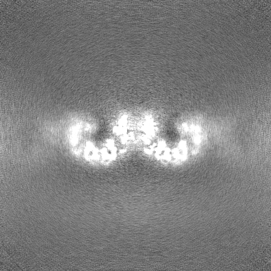
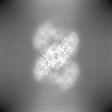
 Map data
 Sample
 Keywords
 hydrolase /
  Function and homology information

 Authors
 United States,
  United Kingdom, 5 items
  Citation

 Structure visualization
 Downloads & links
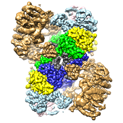 emd_26322.png
 http://ftp.pdbj.org/pub/emdb/structures/EMD-26322


 Z
Z Y
Y X
X


 Sample components
 Processing
 Electron microscopy