 Movie
Movie Controller


+ Open data

- Basic information
| Entry |  | |||||||||
|---|---|---|---|---|---|---|---|---|---|---|
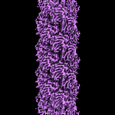
| Title | Cryo-EM of A. pernix flagellum | |||||||||
 Map data Map data | Cryo-EM of A. pernix flagellum | |||||||||
 Sample Sample |
| |||||||||
 Keywords Keywords |  helical symmetry / flagellum / cell appendage / STRUCTURAL PROTEIN helical symmetry / flagellum / cell appendage / STRUCTURAL PROTEIN | |||||||||
| Function / homology | archaeal-type flagellum / Flagellin/pilin, N-terminal / Flagellin, archaea / Archaebacterial flagellin / archaeal or bacterial-type flagellum-dependent cell motility / structural molecule activity / Probable flagellin 1 Function and homology information Function and homology information | |||||||||
| Biological species |   Aeropyrum pernix (archaea) Aeropyrum pernix (archaea) | |||||||||
| Method | helical reconstruction / cryo EM / Resolution: 3.5 Å | |||||||||
 Authors Authors | Wang F / Cvirkaite-Krupovic V | |||||||||
| Funding support |  United States, 2 items United States, 2 items
| |||||||||
 Citation Citation | Journal: Proc Natl Acad Sci U S A / Year: 2023 Title: The evolution of archaeal flagellar filaments. Authors: Mark A B Kreutzberger / Virginija Cvirkaite-Krupovic / Ying Liu / Diana P Baquero / Junfeng Liu / Ravi R Sonani / Chris R Calladine / Fengbin Wang / Mart Krupovic / Edward H Egelman /   Abstract: Flagellar motility has independently arisen three times during evolution: in bacteria, archaea, and eukaryotes. In prokaryotes, the supercoiled flagellar filaments are composed largely of a single ...Flagellar motility has independently arisen three times during evolution: in bacteria, archaea, and eukaryotes. In prokaryotes, the supercoiled flagellar filaments are composed largely of a single protein, bacterial or archaeal flagellin, although these two proteins are not homologous, while in eukaryotes, the flagellum contains hundreds of proteins. Archaeal flagellin and archaeal type IV pilin are homologous, but how archaeal flagellar filaments (AFFs) and archaeal type IV pili (AT4Ps) diverged is not understood, in part, due to the paucity of structures for AFFs and AT4Ps. Despite having similar structures, AFFs supercoil, while AT4Ps do not, and supercoiling is essential for the function of AFFs. We used cryo-electron microscopy to determine the atomic structure of two additional AT4Ps and reanalyzed previous structures. We find that all AFFs have a prominent 10-strand packing, while AT4Ps show a striking structural diversity in their subunit packing. A clear distinction between all AFF and all AT4P structures involves the extension of the N-terminal α-helix with polar residues in the AFFs. Additionally, we characterize a flagellar-like AT4P from with filament and subunit structure similar to that of AFFs which can be viewed as an evolutionary link, showing how the structural diversity of AT4Ps likely allowed for an AT4P to evolve into a supercoiling AFF. | |||||||||
| History |
|
- Structure visualization
| Supplemental images |
|---|
- Downloads & links
-EMDB archive
| Map data | emd_26158.map.gz | 9.8 MB | EMDB map data format | |
|---|---|---|---|---|
| Header (meta data) | emd-26158-v30.xmlemd-26158.xml | 9.6 KB 9.6 KB | Display Display | EMDB header |
| Images |  emd_26158.png emd_26158.png | 143.2 KB | ||
| Archive directory |  http://ftp.pdbj.org/pub/emdb/structures/EMD-26158ftp://ftp.pdbj.org/pub/emdb/structures/EMD-26158 http://ftp.pdbj.org/pub/emdb/structures/EMD-26158ftp://ftp.pdbj.org/pub/emdb/structures/EMD-26158 | HTTPS FTP |
-Related structure data
| Related structure data |  7txiMC  8fj5C  8fjsC  8fk0C  8fk7C  8gi2C M: atomic model generated by this map C: citing same article ( |
|---|---|
| Similar structure data |


-Links
| EMDB pages | EMDB (EBI/PDBe) / EMDataResource |
|---|
-Map
| File | Download / File: emd_26158.map.gz / Format: CCP4 / Size: 125 MB / Type: IMAGE STORED AS FLOATING POINT NUMBER (4 BYTES) | ||||||||||||||||||||||||||||||||||||
|---|---|---|---|---|---|---|---|---|---|---|---|---|---|---|---|---|---|---|---|---|---|---|---|---|---|---|---|---|---|---|---|---|---|---|---|---|---|
| Annotation | Cryo-EM of A. pernix flagellum | ||||||||||||||||||||||||||||||||||||
| Projections & slices | Image control
Images are generated by Spider. | ||||||||||||||||||||||||||||||||||||
| Voxel size | X=Y=Z: 1.08 Å | ||||||||||||||||||||||||||||||||||||
| Density |
| ||||||||||||||||||||||||||||||||||||
| Symmetry | Space group: 1 | ||||||||||||||||||||||||||||||||||||
| Details | EMDB XML:
|
 Z (Sec.)
 Y (Row.)
 X (Col.)


-Supplemental data
- Sample components
-Entire : A. pernix flagellum
| Entire | Name: A. pernix flagellum |
|---|---|
| Components |
|
-Supramolecule #1: A. pernix flagellum
| Supramolecule | Name: A. pernix flagellum / type: complex / ID: 1 / Parent: 0 / Macromolecule list: all |
|---|---|
| Source (natural) | Organism: Aeropyrum pernix (archaea) |
-Macromolecule #1: Probable flagellin 1
| Macromolecule | Name: Probable flagellin 1 / type: protein_or_peptide / ID: 1 / Number of copies: 1 / Enantiomer: LEVO |
|---|---|
| Source (natural) | Organism: Aeropyrum pernix (archaea) |
| Molecular weight | Theoretical: 21.37159 KDa |
| Sequence | String: MRRRRGIVGI EAAIVLIAFV IVAAALAFVA LNMGLFTTQK SKEVMQRGLE EATSALEVDG SVIANVTSGS VDAIAIPIKV SPGREGVDM SVDKTTVRVM LPSKFYENAY CGVFDGSSLS DSKLSTITSS IACTTGWAYL VIFNGDGDNV LELGEKGLLV L ELPTPLNS ...String: MRRRRGIVGI EAAIVLIAFV IVAAALAFVA LNMGLFTTQK SKEVMQRGLE EATSALEVDG SVIANVTSGS VDAIAIPIKV SPGREGVDM SVDKTTVRVM LPSKFYENAY CGVFDGSSLS DSKLSTITSS IACTTGWAYL VIFNGDGDNV LELGEKGLLV L ELPTPLNS YEEFKVEVRP VQGAALTVER IVPASLPTGG AVSLG UniProtKB: Probable flagellin 1 |
-Experimental details
-Structure determination
| Method | cryo EM |
|---|---|
 Processing Processing | helical reconstruction |
| Aggregation state | filament |
-Sample preparation
| Buffer | pH: 6 |
|---|---|
| Vitrification | Cryogen name: ETHANE |
- Electron microscopy
| Microscope | FEI TITAN KRIOS |
|---|---|
| Electron beam | Acceleration voltage: 300 kV / Electron source: FIELD EMISSION GUN |
| Electron optics | Illumination mode: FLOOD BEAM / Imaging mode: BRIGHT FIELDBright-field microscopy / Nominal defocus max: 3.0 µm / Nominal defocus min: 0.5 µm |
| Image recording | Film or detector model: GATAN K3 (6k x 4k) / Average electron dose: 50.0 e/Å2 |
| Experimental equipment |  Model: Titan Krios / Image courtesy: FEI Company |
-Image processing
| Startup model | Type of model: NONE |
|---|---|
| Final angle assignment | Type: NOT APPLICABLE |
| Final reconstruction | Applied symmetry - Helical parameters - Δz: 5.52 Å Applied symmetry - Helical parameters - Δ&Phi: 108 ° Applied symmetry - Helical parameters - Axial symmetry: C1 (asymmetric) Resolution.type: BY AUTHOR / Resolution: 3.5 Å / Resolution method: FSC 0.143 CUT-OFF / Number images used: 59338 |
