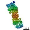
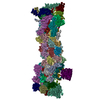
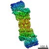
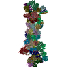
Journal: Proc Natl Acad Sci U S A / Year: 2013 Title: Structure of the 26S proteasome with ATP-γS bound provides insights into the mechanism of nucleotide-dependent substrate translocation. Authors: Paweł Śledź / Pia Unverdorben / Florian Beck / Günter Pfeifer / Andreas Schweitzer / Friedrich Förster / Wolfgang Baumeister / Abstract: The 26S proteasome is a 2.5-MDa, ATP-dependent multisubunit proteolytic complex that processively destroys proteins carrying a degradation signal. The proteasomal ATPase heterohexamer is a key module ...The 26S proteasome is a 2.5-MDa, ATP-dependent multisubunit proteolytic complex that processively destroys proteins carrying a degradation signal. The proteasomal ATPase heterohexamer is a key module of the 19S regulatory particle; it unfolds substrates and translocates them into the 20S core particle where degradation takes place. We used cryoelectron microscopy single-particle analysis to obtain insights into the structural changes of 26S proteasome upon the binding and hydrolysis of ATP. The ATPase ring adopts at least two distinct helical staircase conformations dependent on the nucleotide state. The transition from the conformation observed in the presence of ATP to the predominant conformation in the presence of ATP-γS induces a sliding motion of the ATPase ring over the 20S core particle ring leading to an alignment of the translocation channels of the ATPase and the core particle gate, a conformational state likely to facilitate substrate translocation. Two types of intersubunit modules formed by the large ATPase domain of one ATPase subunit and the small ATPase domain of its neighbor exist. They resemble the contacts observed in the crystal structures of ClpX and proteasome-activating nucleotidase, respectively. The ClpX-like contacts are positioned consecutively and give rise to helical shape in the hexamer, whereas the proteasome-activating nucleotidase-like contact is required to close the ring. Conformational switching between these forms allows adopting different helical conformations in different nucleotide states. We postulate that ATP hydrolysis by the regulatory particle ATPase (Rpt) 5 subunit initiates a cascade of conformational changes, leading to pulling of the substrate, which is primarily executed by Rpt1, Rpt2, and Rpt6.
History
Deposition
Mar 28, 2013
-
Header (metadata) release
Apr 24, 2013
-
Map release
Apr 24, 2013
-
Update
Feb 12, 2014
-
Current status
Feb 12, 2014
Processing site: PDBe / Status: Released
-
Structure visualization
Movie
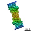
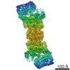
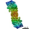
Surface view with section colored by density value
In the structure databanks used in Yorodumi, some data are registered as the other names, "COVID-19 virus" and "2019-nCoV". Here are the details of the virus and the list of structure data.
Jan 31, 2019. EMDB accession codes are about to change! (news from PDBe EMDB page)
EMDB accession codes are about to change! (news from PDBe EMDB page)
The allocation of 4 digits for EMDB accession codes will soon come to an end. Whilst these codes will remain in use, new EMDB accession codes will include an additional digit and will expand incrementally as the available range of codes is exhausted. The current 4-digit format prefixed with “EMD-” (i.e. EMD-XXXX) will advance to a 5-digit format (i.e. EMD-XXXXX), and so on. It is currently estimated that the 4-digit codes will be depleted around Spring 2019, at which point the 5-digit format will come into force.
The EM Navigator/Yorodumi systems omit the EMD- prefix.
Related info.:Q: What is EMD? / ID/Accession-code notation in Yorodumi/EM Navigator
Yorodumi is a browser for structure data from EMDB, PDB, SASBDB, etc.
This page is also the successor to EM Navigator detail page, and also detail information page/front-end page for Omokage search.
The word "yorodu" (or yorozu) is an old Japanese word meaning "ten thousand". "mi" (miru) is to see.
Related info.:EMDB / PDB / SASBDB / Comparison of 3 databanks / Yorodumi Search / Aug 31, 2016. New EM Navigator & Yorodumi / Yorodumi Papers / Jmol/JSmol / Function and homology information / Changes in new EM Navigator and Yorodumi
 Movie
Movie Controller

 Yorodumi
 Open data
 Basic information
 Map data
 Sample
 Keywords
 Function and homology information
 Authors
 Citation
 Structure visualization
 Downloads & links
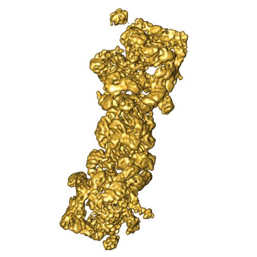 2348-imgMap.jpg
 http://ftp.pdbj.org/pub/emdb/structures/EMD-2348


 Sample components
 Processing
 Electron microscopy
 FIELD EMISSION GUN
