 Movie
Movie Controller
 Structure viewers
 About Yorodumi Papers


+Search query
-Structure paper
| Title | Structure of the 26S proteasome with ATP-γS bound provides insights into the mechanism of nucleotide-dependent substrate translocation. |
|---|---|
| Journal, issue, pages | Proc Natl Acad Sci U S A, Vol. 110, Issue 18, Page 7264-7269, Year 2013 |
| Publish date | Apr 30, 2013 |
 Authors Authors | Paweł Śledź / Pia Unverdorben / Florian Beck / Günter Pfeifer / Andreas Schweitzer / Friedrich Förster / Wolfgang Baumeister /  |
| PubMed Abstract | The 26S proteasome is a 2.5-MDa, ATP-dependent multisubunit proteolytic complex that processively destroys proteins carrying a degradation signal. The proteasomal ATPase heterohexamer is a key module ...The 26S proteasome is a 2.5-MDa, ATP-dependent multisubunit proteolytic complex that processively destroys proteins carrying a degradation signal. The proteasomal ATPase heterohexamer is a key module of the 19S regulatory particle; it unfolds substrates and translocates them into the 20S core particle where degradation takes place. We used cryoelectron microscopy single-particle analysis to obtain insights into the structural changes of 26S proteasome upon the binding and hydrolysis of ATP. The ATPase ring adopts at least two distinct helical staircase conformations dependent on the nucleotide state. The transition from the conformation observed in the presence of ATP to the predominant conformation in the presence of ATP-γS induces a sliding motion of the ATPase ring over the 20S core particle ring leading to an alignment of the translocation channels of the ATPase and the core particle gate, a conformational state likely to facilitate substrate translocation. Two types of intersubunit modules formed by the large ATPase domain of one ATPase subunit and the small ATPase domain of its neighbor exist. They resemble the contacts observed in the crystal structures of ClpX and proteasome-activating nucleotidase, respectively. The ClpX-like contacts are positioned consecutively and give rise to helical shape in the hexamer, whereas the proteasome-activating nucleotidase-like contact is required to close the ring. Conformational switching between these forms allows adopting different helical conformations in different nucleotide states. We postulate that ATP hydrolysis by the regulatory particle ATPase (Rpt) 5 subunit initiates a cascade of conformational changes, leading to pulling of the substrate, which is primarily executed by Rpt1, Rpt2, and Rpt6. |
 External links External links | Proc Natl Acad Sci U S A / PubMed:23589842 / PubMed Central |
| Methods | EM (single particle) |
| Resolution | 9.8 Å |
| Structure data | 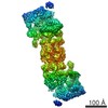 EMDB-2348: |
| Source |
|

 Saccharomyces cerevisiae (brewer's yeast)