 Movie
Movie Controller


+ Open data

- Basic information
| Entry | Database: EMDB / ID: EMD-23380 | ||||||||||||
|---|---|---|---|---|---|---|---|---|---|---|---|---|---|
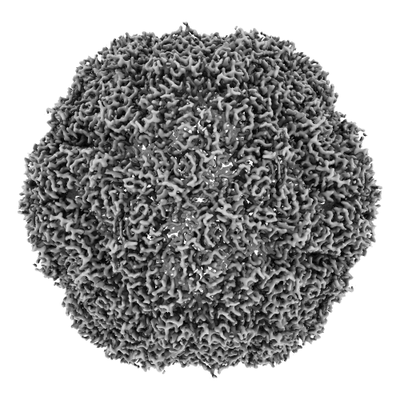
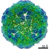
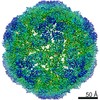
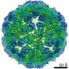
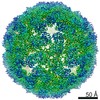
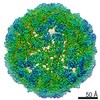
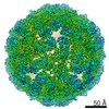
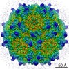
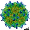
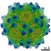
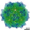
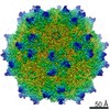
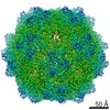
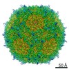
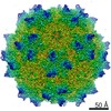
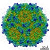
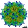
| Title | Thermotoga maritima Encapsulin Nanocompartment Pore Mutant S1K | ||||||||||||
 Map data Map data | |||||||||||||
 Sample Sample |
| ||||||||||||
| Function / homology | Type 1 encapsulin shell protein / Encapsulating protein for peroxidase /  encapsulin nanocompartment / Hydrolases; Acting on peptide bonds (peptidases) / peptidase activity / iron ion transport / intracellular iron ion homeostasis / proteolysis / Type 1 encapsulin shell protein encapsulin nanocompartment / Hydrolases; Acting on peptide bonds (peptidases) / peptidase activity / iron ion transport / intracellular iron ion homeostasis / proteolysis / Type 1 encapsulin shell protein Function and homology information Function and homology information | ||||||||||||
| Biological species |   Thermotoga maritima MSB8 (bacteria) / Thermotoga maritima (strain ATCC 43589 / MSB8 / DSM 3109 / JCM 10099) (bacteria) Thermotoga maritima MSB8 (bacteria) / Thermotoga maritima (strain ATCC 43589 / MSB8 / DSM 3109 / JCM 10099) (bacteria) | ||||||||||||
| Method | single particle reconstruction / cryo EM / Resolution: 2.84 Å | ||||||||||||
 Authors Authors | Andreas MP / Adamson L / Tasneem N / Close W / Giessen T / Lau YH | ||||||||||||
| Funding support |  Australia, Australia,  United States, 3 items United States, 3 items
| ||||||||||||
 Citation Citation | Journal: Sci Adv / Year: 2022 Title: Pore structure controls stability and molecular flux in engineered protein cages. Authors: Lachlan S R Adamson / Nuren Tasneem / Michael P Andreas / William Close / Eric N Jenner / Taylor N Szyszka / Reginald Young / Li Chen Cheah / Alexander Norman / Hugo I MacDermott-Opeskin / ...Authors: Lachlan S R Adamson / Nuren Tasneem / Michael P Andreas / William Close / Eric N Jenner / Taylor N Szyszka / Reginald Young / Li Chen Cheah / Alexander Norman / Hugo I MacDermott-Opeskin / Megan L O'Mara / Frank Sainsbury / Tobias W Giessen / Yu Heng Lau / Abstract: Protein cages are a common architectural motif used by living organisms to compartmentalize and control biochemical reactions. While engineered protein cages have featured in the construction of ...Protein cages are a common architectural motif used by living organisms to compartmentalize and control biochemical reactions. While engineered protein cages have featured in the construction of nanoreactors and synthetic organelles, relatively little is known about the underlying molecular parameters that govern stability and flux through their pores. In this work, we systematically designed 24 variants of the encapsulin cage, featuring pores of different sizes and charges. Twelve pore variants were successfully assembled and purified, including eight designs with exceptional thermal stability. While negatively charged mutations were better tolerated, we were able to form stable assemblies covering a full range of pore sizes and charges, as observed in seven new cryo-EM structures at 2.5- to 3.6-Å resolution. Molecular dynamics simulations and stopped-flow experiments revealed the importance of considering both pore size and charge, together with flexibility and rate-determining steps, when designing protein cages for controlling molecular flux. | ||||||||||||
| History |
|
- Structure visualization
| Movie |
Movie viewer |
|---|---|
| Structure viewer | EM map: SurfViewMolmilJmol/JSmol |
| Supplemental images |
- Downloads & links
-EMDB archive
| Map data | emd_23380.map.gz | 204 MB | EMDB map data format | |
|---|---|---|---|---|
| Header (meta data) | emd-23380-v30.xmlemd-23380.xml | 16.9 KB 16.9 KB | Display Display | EMDB header |
| Images |  emd_23380.png emd_23380.png | 96.3 KB | ||
| Archive directory |  http://ftp.pdbj.org/pub/emdb/structures/EMD-23380ftp://ftp.pdbj.org/pub/emdb/structures/EMD-23380 http://ftp.pdbj.org/pub/emdb/structures/EMD-23380ftp://ftp.pdbj.org/pub/emdb/structures/EMD-23380 | HTTPS FTP |
-Related structure data
| Related structure data | 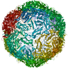 7lijMC 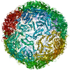 7liiC 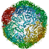 7likC 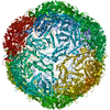 7lilC 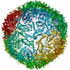 7limC 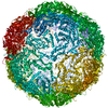 7lisC 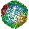 7litC C: citing same article ( M: atomic model generated by this map |
|---|---|
| Similar structure data |
-Links
| EMDB pages | EMDB (EBI/PDBe) / EMDataResource |
|---|---|
| Related items in Molecule of the Month |

-Map
| File | Download / File: emd_23380.map.gz / Format: CCP4 / Size: 216 MB / Type: IMAGE STORED AS FLOATING POINT NUMBER (4 BYTES) | ||||||||||||||||||||||||||||||||||||||||||||||||||||||||||||
|---|---|---|---|---|---|---|---|---|---|---|---|---|---|---|---|---|---|---|---|---|---|---|---|---|---|---|---|---|---|---|---|---|---|---|---|---|---|---|---|---|---|---|---|---|---|---|---|---|---|---|---|---|---|---|---|---|---|---|---|---|---|
| Voxel size | X=Y=Z: 0.98 Å | ||||||||||||||||||||||||||||||||||||||||||||||||||||||||||||
| Density |
| ||||||||||||||||||||||||||||||||||||||||||||||||||||||||||||
| Symmetry | Space group: 1 | ||||||||||||||||||||||||||||||||||||||||||||||||||||||||||||
| Details | EMDB XML:
CCP4 map header:
| ||||||||||||||||||||||||||||||||||||||||||||||||||||||||||||
-Supplemental data
- Sample components
-Entire : Thermotoga maratima Pore Mutant S1K
| Entire | Name: Thermotoga maratima Pore Mutant S1K |
|---|---|
| Components |
|
-Supramolecule #1: Thermotoga maratima Pore Mutant S1K
| Supramolecule | Name: Thermotoga maratima Pore Mutant S1K / type: complex / ID: 1 / Parent: 0 / Macromolecule list: #1 Details: Thermotoga maratima encapsulin pore mutant with H187K mutation |
|---|---|
| Source (natural) | Organism: Thermotoga maritima MSB8 (bacteria) |
| Recombinant expression | Organism: Escherichia coli BL21(DE3) (bacteria) |
| Molecular weight | Theoretical: 1.850714 MDa |
-Macromolecule #1: Maritimacin
| Macromolecule | Name: Maritimacin / type: protein_or_peptide / ID: 1 / Number of copies: 1 / Enantiomer: LEVO / EC number: Hydrolases; Acting on peptide bonds (peptidases) |
|---|---|
| Source (natural) | Organism: Thermotoga maritima (strain ATCC 43589 / MSB8 / DSM 3109 / JCM 10099) (bacteria) Strain: ATCC 43589 / MSB8 / DSM 3109 / JCM 10099 |
| Molecular weight | Theoretical: 30.507818 KDa |
| Recombinant expression | Organism: Escherichia coli BL21(DE3) (bacteria) |
| Sequence | String: MEFLKRSFAP LTEKQWQEID NRAREIFKTQ LYGRKFVDVE GPYGWEYAAH PLGEVEVLSD ENEVVKWGLR KSLPLIELRA TFTLDLWEL DNLERGKPNV DLSSLEETVR KVAEFEDEVI FRGCEKSGVK GLLSFEERKI ECGSTPKDLL EAIVRALSIF S KDGIEGPY ...String: MEFLKRSFAP LTEKQWQEID NRAREIFKTQ LYGRKFVDVE GPYGWEYAAH PLGEVEVLSD ENEVVKWGLR KSLPLIELRA TFTLDLWEL DNLERGKPNV DLSSLEETVR KVAEFEDEVI FRGCEKSGVK GLLSFEERKI ECGSTPKDLL EAIVRALSIF S KDGIEGPY TLVINTDRWI NFLKEEAGKY PLEKRVEECL RGGKIITTPR IEDALVVSER GGDFKLILGQ DLSIGYEDRE KD AVRLFIT ETFTFQVVNP EALILLKF |
-Macromolecule #2: RIBOFLAVIN
| Macromolecule | Name: RIBOFLAVIN / type: ligand / ID: 2 / Number of copies: 1 / Formula: RBF |
|---|---|
| Molecular weight | Theoretical: 376.364 Da |
| Chemical component information |  ChemComp-RBF: |
-Experimental details
-Structure determination
| Method | cryo EM |
|---|---|
 Processing Processing | single particle reconstruction |
| Aggregation state | particle |
-Sample preparation
| Concentration | 2.0 mg/mL | |||||||||
|---|---|---|---|---|---|---|---|---|---|---|
| Buffer | pH: 8 Component:
| |||||||||
| Grid | Model: Quantifoil R1.2/1.3 / Material: COPPER / Mesh: 200 / Support film - Material: CARBON / Support film - topology: HOLEY / Pretreatment - Type: GLOW DISCHARGE / Pretreatment - Atmosphere: AIR / Details: 60 seconds at 5 mA | |||||||||
| Vitrification | Cryogen name: ETHANE / Chamber humidity: 100 % / Chamber temperature: 295 K / Instrument: FEI VITROBOT MARK IV Details: blot 4 seconds, force 20 immediately prior to plunging into ethane. |
- Electron microscopy
| Microscope | TFS GLACIOS |
|---|---|
| Electron beam | Acceleration voltage: 200 kV / Electron source: FIELD EMISSION GUN |
| Electron optics | C2 aperture diameter: 70.0 µm / Illumination mode: FLOOD BEAM / Imaging mode: BRIGHT FIELDBright-field microscopy / Cs: 2.7 mm / Nominal defocus max: -1.8 µm / Nominal defocus min: -1.3 µm / Nominal magnification: 45000 |
| Sample stage | Cooling holder cryogen: NITROGEN |
| Image recording | Film or detector model: GATAN K2 SUMMIT (4k x 4k) / Detector mode: COUNTING / Digitization - Dimensions - Width: 3838 pixel / Digitization - Dimensions - Height: 3710 pixel / Average exposure time: 8.0 sec. / Average electron dose: 62.0 e/Å2 |
-Image processing
| CTF correction | Software - Name: cryoSPARC (ver. 2.15.0) |
|---|---|
| Startup model | Type of model: PDB ENTRY PDB model - PDB ID: Details: Initial map for refinements made from PDB entry 3DKT |
| Initial angle assignment | Type: ANGULAR RECONSTITUTION / Software - Name: cryoSPARC (ver. 2.15.0) |
| Final angle assignment | Type: ANGULAR RECONSTITUTION / Software - Name: cryoSPARC (ver. 2.15.0) |
| Final reconstruction | Applied symmetry - Point group: I (icosahedral) / Resolution.type: BY AUTHOR / Resolution: 2.84 Å / Resolution method: FSC 0.143 CUT-OFF / Software - Name: cryoSPARC (ver. 2.15.0) / Number images used: 58162 |

