Journal: Cell / Year: 2013 Title: The bacterial DnaC helicase loader is a DnaB ring breaker. Authors: Ernesto Arias-Palomo / Valerie L O'Shea / Iris V Hood / James M Berger / Abstract: Dedicated AAA+ ATPases deposit hexameric ring-shaped helicases onto DNA to promote replication in cellular organisms. To understand how loading occurs, we used electron microscopy and small angle X- ...Dedicated AAA+ ATPases deposit hexameric ring-shaped helicases onto DNA to promote replication in cellular organisms. To understand how loading occurs, we used electron microscopy and small angle X-ray scattering (SAXS) to determine the ATP-bound structure of the intact E. coli DnaB⋅DnaC helicase/loader complex. The 480 kDa dodecamer forms a three-tiered assembly, in which DnaC adopts a spiral configuration that remodels N-terminal scaffolding and C-terminal motor regions of DnaB to produce a clear break in the helicase ring. Surprisingly, DnaC's AAA+ fold is dispensable for ring remodeling because the DnaC isolated helicase-binding domain can both load DnaB onto DNA and increase the efficiency by which the helicase acts on substrates in vitro. Our data demonstrate that DnaC opens DnaB by a mechanism akin to that of polymerase clamp loaders and indicate that bacterial replicative helicases, like their eukaryotic counterparts, possess autoregulatory elements that influence how hexameric motor domains are loaded onto and unwind DNA.
History
Deposition
Feb 26, 2013
-
Header (metadata) release
Mar 13, 2013
-
Map release
Apr 17, 2013
-
Update
Apr 24, 2013
-
Current status
Apr 24, 2013
Processing site: PDBe / Status: Released
-
Structure visualization
Movie
Surface view with section colored by density value
The particles were selected using DoG picker as available in APPION. The contrast transfer function of the microscope for each micrograph was estimated using CTFFIND3 and phase-flipped using SPIDER. DnaBC particles were subjected to a multi-model refinement as implemented in SPARX using the 3D averages obtained from the RCT reconstructions as initial references.
CTF correction
Details: Each micrograph
Final reconstruction
Applied symmetry - Point group: C3 (3 fold cyclic) / Algorithm: OTHER / Resolution.type: BY AUTHOR / Resolution: 25.0 Å / Resolution method: FSC 0.5 CUT-OFF / Software - Name: EMAN2, SPARX / Number images used: 9617
+
About Yorodumi
-
News
-
Feb 9, 2022. New format data for meta-information of EMDB entries
New format data for meta-information of EMDB entries
Version 3 of the EMDB header file is now the official format.
The previous official version 1.9 will be removed from the archive.
In the structure databanks used in Yorodumi, some data are registered as the other names, "COVID-19 virus" and "2019-nCoV". Here are the details of the virus and the list of structure data.
Jan 31, 2019. EMDB accession codes are about to change! (news from PDBe EMDB page)
EMDB accession codes are about to change! (news from PDBe EMDB page)
The allocation of 4 digits for EMDB accession codes will soon come to an end. Whilst these codes will remain in use, new EMDB accession codes will include an additional digit and will expand incrementally as the available range of codes is exhausted. The current 4-digit format prefixed with “EMD-” (i.e. EMD-XXXX) will advance to a 5-digit format (i.e. EMD-XXXXX), and so on. It is currently estimated that the 4-digit codes will be depleted around Spring 2019, at which point the 5-digit format will come into force.
The EM Navigator/Yorodumi systems omit the EMD- prefix.
Related info.:Q: What is EMD? / ID/Accession-code notation in Yorodumi/EM Navigator
Yorodumi is a browser for structure data from EMDB, PDB, SASBDB, etc.
This page is also the successor to EM Navigator detail page, and also detail information page/front-end page for Omokage search.
The word "yorodu" (or yorozu) is an old Japanese word meaning "ten thousand". "mi" (miru) is to see.
Related info.:EMDB / PDB / SASBDB / Comparison of 3 databanks / Yorodumi Search / Aug 31, 2016. New EM Navigator & Yorodumi / Yorodumi Papers / Jmol/JSmol / Function and homology information / Changes in new EM Navigator and Yorodumi
 Movie
Movie Controller


 Open data
 Basic information
 Map data
 Sample
 Keywords
 Function and homology information
 Authors
 Citation
 Structure visualization
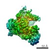
 UCSF Chimera
 Downloads & links
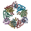
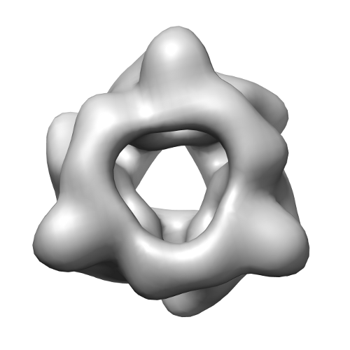 emd_2322.png
 http://ftp.pdbj.org/pub/emdb/structures/EMD-2322


 Sample components
 Processing
 Electron microscopy