National Institutes of Health/National Institute Of Allergy and Infectious Diseases (NIH/NIAID)
AI109025
United States
National Institutes of Health/National Institute of General Medical Sciences (NIH/NIGMS)
P011030036319
United States
National Institutes of Health/National Institute of General Medical Sciences (NIH/NIGMS)
R01GM080139
United States
National Institutes of Health/National Institute of General Medical Sciences (NIH/NIGMS)
U24 GM116789-01A1
United States
Citation
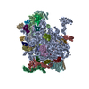
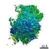
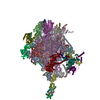
Journal: J Fungi (Basel) / Year: 2021 Title: Cryo-Electron Tomography of Candida glabrata Plasma Membrane Proteins. Authors: Cristina Jiménez-Ortigosa / Jennifer Jiang / Muyuan Chen / Xuyuan Kuang / Kelley R Healey / Paul Castellano / Nikpreet Boparai / Steven J Ludtke / David S Perlin / Wei Dai / Abstract: Fungal plasma membrane proteins have long been recognized as targets for the development of antifungal agents. Despite recent progress in experimental approaches and computational structural ...Fungal plasma membrane proteins have long been recognized as targets for the development of antifungal agents. Despite recent progress in experimental approaches and computational structural predictions, our knowledge of the structural dynamics and spatial distribution of these membrane proteins in the context of their native lipid environment remains limited. By applying cryo-electron tomography (cryoET) and subtomogram analysis, we aim to characterize the structural characteristics and spatial distribution of membrane proteins present in plasma membranes. This study has resulted in the identification of the membrane-embedded structure of the fungal H+-ATPase, Pma1. Tomograms of the plasma membrane revealed that Pma1 complexes are heterogeneously distributed as hexamers that cluster into distinct membrane microdomains. This study characterizes fungal membrane proteins in the native cellular landscape and highlights the unique potential of cryoET to advance our understanding of cellular biology and biological systems.
History
Deposition
Dec 16, 2020
-
Header (metadata) release
Mar 10, 2021
-
Map release
Mar 10, 2021
-
Update
Mar 13, 2024
-
Current status
Mar 13, 2024
Processing site: RCSB / Status: Released
-
Structure visualization
Movie
Surface view with section colored by density value
In the structure databanks used in Yorodumi, some data are registered as the other names, "COVID-19 virus" and "2019-nCoV". Here are the details of the virus and the list of structure data.
Jan 31, 2019. EMDB accession codes are about to change! (news from PDBe EMDB page)
EMDB accession codes are about to change! (news from PDBe EMDB page)
The allocation of 4 digits for EMDB accession codes will soon come to an end. Whilst these codes will remain in use, new EMDB accession codes will include an additional digit and will expand incrementally as the available range of codes is exhausted. The current 4-digit format prefixed with “EMD-” (i.e. EMD-XXXX) will advance to a 5-digit format (i.e. EMD-XXXXX), and so on. It is currently estimated that the 4-digit codes will be depleted around Spring 2019, at which point the 5-digit format will come into force.
The EM Navigator/Yorodumi systems omit the EMD- prefix.
Related info.:Q: What is EMD? / ID/Accession-code notation in Yorodumi/EM Navigator
Yorodumi is a browser for structure data from EMDB, PDB, SASBDB, etc.
This page is also the successor to EM Navigator detail page, and also detail information page/front-end page for Omokage search.
The word "yorodu" (or yorozu) is an old Japanese word meaning "ten thousand". "mi" (miru) is to see.
Related info.:EMDB / PDB / SASBDB / Comparison of 3 databanks / Yorodumi Search / Aug 31, 2016. New EM Navigator & Yorodumi / Yorodumi Papers / Jmol/JSmol / Function and homology information / Changes in new EM Navigator and Yorodumi
 Movie
Movie Controller

 Yorodumi
 Open data
 Basic information
 Map data
 Sample
 Keywords
 hexamer /
 Authors
 United States, 4 items
  Citation
 Structure visualization
 Movie viewer
 Downloads & links
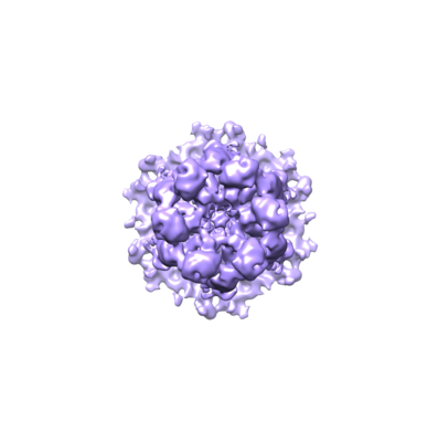 emd_23123.png
 http://ftp.pdbj.org/pub/emdb/structures/EMD-23123
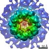


 Sample components
 Processing
 Electron microscopy
