 Movie
Movie Controller

[English] 日本語
 Yorodumi
- EMDB-23122: Subtomogram average of the hexameric fungal plasma membrane proto... -
+ Open data

- Basic information
| Entry | Database: EMDB / ID: EMD-23122 | |||||||||||||||
|---|---|---|---|---|---|---|---|---|---|---|---|---|---|---|---|---|
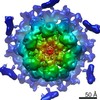
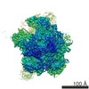
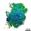
| Title | Subtomogram average of the hexameric fungal plasma membrane proton pump Pma1 from protoplast membrane of Candida glabrata CBS138 strain | |||||||||||||||
 Map data Map data | Plasma membrane proton pump Pma1 from Candida glabrata CBS138 strain protoplast membrane | |||||||||||||||
 Sample Sample |
| |||||||||||||||
 Keywords Keywords | hexamer / complex / MEMBRANE PROTEIN | |||||||||||||||
| Biological species |  [Candida] glabrata (fungus) [Candida] glabrata (fungus) | |||||||||||||||
| Method | subtomogram averaging / cryo EM / Resolution: 18.0 Å | |||||||||||||||
 Authors Authors | Jiang J / Chen M / Jimenez-Ortigosa C / Perlin DS / Dai W | |||||||||||||||
| Funding support |  United States, 4 items United States, 4 items
| |||||||||||||||
 Citation Citation | Journal: J Fungi (Basel) / Year: 2021 Title: Cryo-Electron Tomography of Candida glabrata Plasma Membrane Proteins. Authors: Cristina Jiménez-Ortigosa / Jennifer Jiang / Muyuan Chen / Xuyuan Kuang / Kelley R Healey / Paul Castellano / Nikpreet Boparai / Steven J Ludtke / David S Perlin / Wei Dai /  Abstract: Fungal plasma membrane proteins have long been recognized as targets for the development of antifungal agents. Despite recent progress in experimental approaches and computational structural ...Fungal plasma membrane proteins have long been recognized as targets for the development of antifungal agents. Despite recent progress in experimental approaches and computational structural predictions, our knowledge of the structural dynamics and spatial distribution of these membrane proteins in the context of their native lipid environment remains limited. By applying cryo-electron tomography (cryoET) and subtomogram analysis, we aim to characterize the structural characteristics and spatial distribution of membrane proteins present in plasma membranes. This study has resulted in the identification of the membrane-embedded structure of the fungal H+-ATPase, Pma1. Tomograms of the plasma membrane revealed that Pma1 complexes are heterogeneously distributed as hexamers that cluster into distinct membrane microdomains. This study characterizes fungal membrane proteins in the native cellular landscape and highlights the unique potential of cryoET to advance our understanding of cellular biology and biological systems. | |||||||||||||||
| History |
|
- Structure visualization
| Movie |
 Movie viewer Movie viewer |
|---|---|
| Structure viewer | EM map: SurfViewMolmilJmol/JSmol |
| Supplemental images |

- Downloads & links
-EMDB archive
| Map data | emd_23122.map.gz | 3.9 MB | EMDB map data format | |
|---|---|---|---|---|
| Header (meta data) | emd-23122-v30.xmlemd-23122.xml | 10.7 KB 10.7 KB | Display Display | EMDB header |
| Images |  emd_23122.png emd_23122.png | 97.1 KB | ||
| Filedesc metadata | emd-23122.cif.gz | 4.3 KB | ||
| Archive directory |  http://ftp.pdbj.org/pub/emdb/structures/EMD-23122ftp://ftp.pdbj.org/pub/emdb/structures/EMD-23122 http://ftp.pdbj.org/pub/emdb/structures/EMD-23122ftp://ftp.pdbj.org/pub/emdb/structures/EMD-23122 | HTTPS FTP |
-Validation report
| Summary document | emd_23122_validation.pdf.gz | 381.8 KB | Display | EMDB validaton report |
|---|---|---|---|---|
| Full document | emd_23122_full_validation.pdf.gz | 381.4 KB | Display | |
| Data in XML | emd_23122_validation.xml.gz | 5.7 KB | Display | |
| Data in CIF | emd_23122_validation.cif.gz | 6.5 KB | Display | |
| Arichive directory | https://ftp.pdbj.org/pub/emdb/validation_reports/EMD-23122ftp://ftp.pdbj.org/pub/emdb/validation_reports/EMD-23122 | HTTPS FTP |
-Related structure data
| Related structure data | C: citing same article ( |
|---|---|
| Similar structure data |


-Links
| EMDB pages | EMDB (EBI/PDBe) / EMDataResource |
|---|
-Map
| File | Download / File: emd_23122.map.gz / Format: CCP4 / Size: 8 MB / Type: IMAGE STORED AS FLOATING POINT NUMBER (4 BYTES) | ||||||||||||||||||||||||||||||||||||||||||||||||||||||||||||||||||||
|---|---|---|---|---|---|---|---|---|---|---|---|---|---|---|---|---|---|---|---|---|---|---|---|---|---|---|---|---|---|---|---|---|---|---|---|---|---|---|---|---|---|---|---|---|---|---|---|---|---|---|---|---|---|---|---|---|---|---|---|---|---|---|---|---|---|---|---|---|---|
| Annotation | Plasma membrane proton pump Pma1 from Candida glabrata CBS138 strain protoplast membrane | ||||||||||||||||||||||||||||||||||||||||||||||||||||||||||||||||||||
| Voxel size | X=Y=Z: 2.7975 Å | ||||||||||||||||||||||||||||||||||||||||||||||||||||||||||||||||||||
| Density |
| ||||||||||||||||||||||||||||||||||||||||||||||||||||||||||||||||||||
| Symmetry | Space group: 1 | ||||||||||||||||||||||||||||||||||||||||||||||||||||||||||||||||||||
| Details | EMDB XML:
CCP4 map header:
| ||||||||||||||||||||||||||||||||||||||||||||||||||||||||||||||||||||
-Supplemental data
- Sample components
-Entire : Plasma membrane ATPase Pma1
| Entire | Name: Plasma membrane ATPase Pma1 |
|---|---|
| Components |
|
-Supramolecule #1: Plasma membrane ATPase Pma1
| Supramolecule | Name: Plasma membrane ATPase Pma1 / type: complex / ID: 1 / Parent: 0 Details: On protoplast membranes isolated from Candida glabrata |
|---|---|
| Source (natural) | Organism: [Candida] glabrata (fungus) / Strain: CBS138 |
| Molecular weight | Theoretical: 590 KDa |
-Experimental details
-Structure determination
| Method | cryo EM |
|---|---|
 Processing Processing | subtomogram averaging |
| Aggregation state | particle |
-Sample preparation
| Buffer | pH: 7 |
|---|---|
| Grid | Model: Quantifoil R2/1 / Material: COPPER / Mesh: 200 / Pretreatment - Type: GLOW DISCHARGE / Pretreatment - Time: 30 sec. |
| Vitrification | Cryogen name: ETHANE / Chamber humidity: 95 % / Chamber temperature: 293.15 K / Instrument: LEICA EM GP |
- Electron microscopy
| Microscope | FEI TITAN KRIOS |
|---|---|
| Specialist optics | Phase plate: OTHER / Energy filter - Name: GIF Bioquantum / Energy filter - Slit width: 20 eV |
| Image recording | Film or detector model: GATAN K2 SUMMIT (4k x 4k) / Detector mode: COUNTING / Average electron dose: 1.5 e/Å2 |
| Electron beam | Acceleration voltage: 300 kV / Electron source:  FIELD EMISSION GUN FIELD EMISSION GUN |
| Electron optics | C2 aperture diameter: 100.0 µm / Illumination mode: FLOOD BEAM / Imaging mode: BRIGHT FIELD / Nominal magnification: 42000 |
| Sample stage | Specimen holder model: FEI TITAN KRIOS AUTOGRID HOLDER / Cooling holder cryogen: NITROGEN |
| Experimental equipment |  Model: Titan Krios / Image courtesy: FEI Company |
-Image processing
| Final reconstruction | Applied symmetry - Point group: C6 (6 fold cyclic) / Resolution.type: BY AUTHOR / Resolution: 18.0 Å / Resolution method: FSC 0.143 CUT-OFF / Software - Name: EMAN2 / Number subtomograms used: 1818 |
|---|---|
| Extraction | Number tomograms: 100 / Number images used: 1818 / Software - Name: EMAN2 |
| Final angle assignment | Type: NOT APPLICABLE |
