 Movie
Movie Controller

[English] 日本語
 Yorodumi
- EMDB-23121: Cryo-EM structure of the human 39S mitoribosomal subunit in compl... -
+ Open data

- Basic information
| Entry | Database: EMDB / ID: EMD-23121 | |||||||||
|---|---|---|---|---|---|---|---|---|---|---|
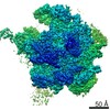
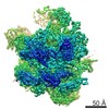
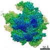
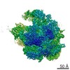
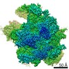
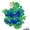
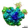
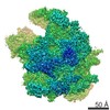
| Title | Cryo-EM structure of the human 39S mitoribosomal subunit in complex with RRFmt and EF-G2mt. | |||||||||
 Map data Map data | Cryo-EM structure of the human 39S mitoribosomal subunit in complex with RRFmt and EF-G2mt. | |||||||||
 Sample Sample |
| |||||||||
| Function / homology |  Function and homology information Function and homology informationribosome disassembly / mitochondrial translational termination / mitochondrial transcription / mitochondrial translational elongation / translation release factor activity, codon nonspecific /  microprocessor complex / Mitochondrial translation elongation / Mitochondrial translation termination / Mitochondrial translation initiation / mitochondrial large ribosomal subunit ...ribosome disassembly / mitochondrial translational termination / mitochondrial transcription / mitochondrial translational elongation / translation release factor activity, codon nonspecific / microprocessor complex / Mitochondrial translation elongation / Mitochondrial translation termination / Mitochondrial translation initiation / mitochondrial large ribosomal subunit / Hydrolases; Acting on ester bonds; Endoribonucleases producing 5'-phosphomonoesters / peptidyl-tRNA hydrolase / mitochondrial small ribosomal subunit / aminoacyl-tRNA hydrolase activity / mitochondrial ribosome / mitochondrial translation / ribosomal large subunit binding / anatomical structure morphogenesis / RNA processing / rescue of stalled ribosome / cellular response to leukemia inhibitory factor / small ribosomal subunit rRNA binding / fibrillar center / large ribosomal subunit rRNA binding / double-stranded RNA binding / large ribosomal subunit / cell junction / endonuclease activity / mitochondrial inner membrane / negative regulation of translation / rRNA binding / nuclear body / ribosome / mitochondrial matrix / structural constituent of ribosome / cell cycle / ribonucleoprotein complex / translation / protein domain specific binding / nucleotide binding / mRNA binding / GTPase activity / synapse / apoptotic process / GTP binding / nucleolus / positive regulation of DNA-templated transcription / mitochondrion / RNA binding / nucleoplasm / nucleus / plasma membrane / cytosol / cytoplasm microprocessor complex / Mitochondrial translation elongation / Mitochondrial translation termination / Mitochondrial translation initiation / mitochondrial large ribosomal subunit ...ribosome disassembly / mitochondrial translational termination / mitochondrial transcription / mitochondrial translational elongation / translation release factor activity, codon nonspecific / microprocessor complex / Mitochondrial translation elongation / Mitochondrial translation termination / Mitochondrial translation initiation / mitochondrial large ribosomal subunit / Hydrolases; Acting on ester bonds; Endoribonucleases producing 5'-phosphomonoesters / peptidyl-tRNA hydrolase / mitochondrial small ribosomal subunit / aminoacyl-tRNA hydrolase activity / mitochondrial ribosome / mitochondrial translation / ribosomal large subunit binding / anatomical structure morphogenesis / RNA processing / rescue of stalled ribosome / cellular response to leukemia inhibitory factor / small ribosomal subunit rRNA binding / fibrillar center / large ribosomal subunit rRNA binding / double-stranded RNA binding / large ribosomal subunit / cell junction / endonuclease activity / mitochondrial inner membrane / negative regulation of translation / rRNA binding / nuclear body / ribosome / mitochondrial matrix / structural constituent of ribosome / cell cycle / ribonucleoprotein complex / translation / protein domain specific binding / nucleotide binding / mRNA binding / GTPase activity / synapse / apoptotic process / GTP binding / nucleolus / positive regulation of DNA-templated transcription / mitochondrion / RNA binding / nucleoplasm / nucleus / plasma membrane / cytosol / cytoplasmSimilarity search - Function | |||||||||
| Biological species |  Homo sapiens (human) / Human (human) Homo sapiens (human) / Human (human) | |||||||||
| Method | single particle reconstruction / cryo EM / Resolution: 3.15 Å | |||||||||
 Authors Authors | Agrawal E / Koripella R | |||||||||
| Funding support |  United States, 1 items United States, 1 items
| |||||||||
 Citation Citation | Journal: Nat Commun / Year: 2021 Title: Distinct mechanisms of the human mitoribosome recycling and antibiotic resistance. Authors: Ravi Kiran Koripella / Ayush Deep / Ekansh K Agrawal / Pooja Keshavan / Nilesh K Banavali / Rajendra K Agrawal / Abstract: Ribosomes are recycled for a new round of translation initiation by dissociation of ribosomal subunits, messenger RNA and transfer RNA from their translational post-termination complex. Here we ...Ribosomes are recycled for a new round of translation initiation by dissociation of ribosomal subunits, messenger RNA and transfer RNA from their translational post-termination complex. Here we present cryo-EM structures of the human 55S mitochondrial ribosome (mitoribosome) and the mitoribosomal large 39S subunit in complex with mitoribosome recycling factor (RRF) and a recycling-specific homolog of elongation factor G (EF-G2). These structures clarify an unusual role of a mitochondria-specific segment of RRF, identify the structural distinctions that confer functional specificity to EF-G2, and show that the deacylated tRNA remains with the dissociated 39S subunit, suggesting a distinct sequence of events in mitoribosome recycling. Furthermore, biochemical and structural analyses reveal that the molecular mechanism of antibiotic fusidic acid resistance for EF-G2 is markedly different from that of mitochondrial elongation factor EF-G1, suggesting that the two human EF-Gs have evolved diversely to negate the effect of a bacterial antibiotic. | |||||||||
| History |
|
- Structure visualization
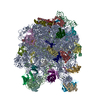
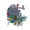
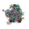
| Movie |
Movie viewer |
|---|---|
| Structure viewer | EM map: SurfViewMolmilJmol/JSmol |
| Supplemental images |

- Downloads & links
-EMDB archive
| Map data | emd_23121.map.gz | 227 MB | EMDB map data format | |
|---|---|---|---|---|
| Header (meta data) | emd-23121-v30.xmlemd-23121.xml | 65.5 KB 65.5 KB | Display Display | EMDB header |
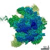
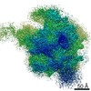
| Images | 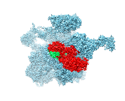 emd_23121.png emd_23121.png | 171.9 KB | ||
| Archive directory |  http://ftp.pdbj.org/pub/emdb/structures/EMD-23121ftp://ftp.pdbj.org/pub/emdb/structures/EMD-23121 http://ftp.pdbj.org/pub/emdb/structures/EMD-23121ftp://ftp.pdbj.org/pub/emdb/structures/EMD-23121 | HTTPS FTP |
-Related structure data
| Related structure data |  7l20MC  7l08C M: atomic model generated by this map C: citing same article ( |
|---|---|
| Similar structure data | |
| EM raw data | EMPIAR-10703 (Title: Distinct mechanisms of the human mitoribosome recycling and antibiotic resistance Data size: 2.3 TB Data #1: Aligned single-frame particles of human mitochondrial 55S-EF-Gmt complex [picked particles - single frame - processed]) |
-Links
| EMDB pages | EMDB (EBI/PDBe) / EMDataResource |
|---|---|
| Related items in Molecule of the Month |


-Map
| File | Download / File: emd_23121.map.gz / Format: CCP4 / Size: 244.1 MB / Type: IMAGE STORED AS FLOATING POINT NUMBER (4 BYTES) | ||||||||||||||||||||||||||||||||||||||||||||||||||||||||||||
|---|---|---|---|---|---|---|---|---|---|---|---|---|---|---|---|---|---|---|---|---|---|---|---|---|---|---|---|---|---|---|---|---|---|---|---|---|---|---|---|---|---|---|---|---|---|---|---|---|---|---|---|---|---|---|---|---|---|---|---|---|---|
| Annotation | Cryo-EM structure of the human 39S mitoribosomal subunit in complex with RRFmt and EF-G2mt. | ||||||||||||||||||||||||||||||||||||||||||||||||||||||||||||
| Voxel size | X=Y=Z: 1.07325 Å | ||||||||||||||||||||||||||||||||||||||||||||||||||||||||||||
| Density |
| ||||||||||||||||||||||||||||||||||||||||||||||||||||||||||||
| Symmetry | Space group: 1 | ||||||||||||||||||||||||||||||||||||||||||||||||||||||||||||
| Details | EMDB XML:
CCP4 map header:
| ||||||||||||||||||||||||||||||||||||||||||||||||||||||||||||
-Supplemental data
- Sample components
+Entire : Cryo-EM structure of Mammalian Mitochondrial ribosome complex wit...
+Supramolecule #1: Cryo-EM structure of Mammalian Mitochondrial ribosome complex wit...
+Macromolecule #1: 16S rRNA mitochondrial
+Macromolecule #2: tRNAval
+Macromolecule #3: 39S ribosomal protein L2, mitochondrial
+Macromolecule #4: 39S ribosomal protein L4, mitochondrial
+Macromolecule #5: 39S ribosomal protein L9, mitochondrial
+Macromolecule #6: 39S ribosomal protein L13, mitochondrial
+Macromolecule #7: 39S ribosomal protein L14, mitochondrial
+Macromolecule #8: 39S ribosomal protein L15, mitochondrial
+Macromolecule #9: 39S ribosomal protein L17, mitochondrial
+Macromolecule #10: 39S ribosomal protein L20, mitochondrial
+Macromolecule #11: 39S ribosomal protein L21, mitochondrial
+Macromolecule #12: 39S ribosomal protein L22, mitochondrial
+Macromolecule #13: 39S ribosomal protein L27, mitochondrial
+Macromolecule #14: 39S ribosomal protein L28, mitochondrial
+Macromolecule #15: 39S ribosomal protein L47, mitochondrial
+Macromolecule #16: 39S ribosomal protein L30, mitochondrial
+Macromolecule #17: 39S ribosomal protein L32, mitochondrial
+Macromolecule #18: 39S ribosomal protein L33, mitochondrial
+Macromolecule #19: 39S ribosomal protein L34, mitochondrial
+Macromolecule #20: 39S ribosomal protein L35, mitochondrial
+Macromolecule #21: 39S ribosomal protein L36, mitochondrial
+Macromolecule #22: 39S ribosomal protein L40, mitochondrial
+Macromolecule #23: 39S ribosomal protein L43, mitochondrial
+Macromolecule #24: 39S ribosomal protein L46, mitochondrial
+Macromolecule #25: 39S ribosomal protein L49, mitochondrial
+Macromolecule #26: 39S ribosomal protein L51, mitochondrial
+Macromolecule #27: cDNA FLJ76418, highly similar to Homo sapiens mitochondrial ribos...
+Macromolecule #28: 39S ribosomal protein L55, mitochondrial
+Macromolecule #29: Ribosomal protein 63, mitochondrial
+Macromolecule #30: Growth arrest and DNA damage-inducible proteins-interacting protein 1
+Macromolecule #31: 39S ribosomal protein S18a, mitochondrial
+Macromolecule #32: 39S ribosomal protein L11, mitochondrial
+Macromolecule #33: 39S ribosomal protein L10, mitochondrial
+Macromolecule #34: 39S ribosomal protein L16, mitochondrial
+Macromolecule #35: Mitochondrial ribosomal protein L18, isoform CRA_b
+Macromolecule #36: 39S ribosomal protein L23, mitochondrial
+Macromolecule #37: 39S ribosomal protein L24, mitochondrial
+Macromolecule #38: 39S ribosomal protein L3, mitochondrial
+Macromolecule #39: 39S ribosomal protein L37, mitochondrial
+Macromolecule #40: 39S ribosomal protein L38, mitochondrial
+Macromolecule #41: 39S ribosomal protein L39, mitochondrial
+Macromolecule #42: 39S ribosomal protein L41, mitochondrial
+Macromolecule #43: 39S ribosomal protein L42, mitochondrial
+Macromolecule #44: 39S ribosomal protein L44, mitochondrial
+Macromolecule #45: 39S ribosomal protein L45, mitochondrial
+Macromolecule #46: 39S ribosomal protein L48, mitochondrial
+Macromolecule #47: 39S ribosomal protein L50, mitochondrial
+Macromolecule #48: 39S ribosomal protein L53, mitochondrial
+Macromolecule #49: 39S ribosomal protein L54, mitochondrial
+Macromolecule #50: Peptidyl-tRNA hydrolase ICT1, mitochondrial
+Macromolecule #51: 39S ribosomal protein S30, mitochondrial
+Macromolecule #52: 39S ribosomal protein L19, mitochondrial
+Macromolecule #53: 39 S P-site finger
+Macromolecule #54: 39S ribosomal protein L12, mitochondrial
+Macromolecule #55: Ribosome-recycling factor, mitochondrial
+Macromolecule #56: Ribosome-releasing factor 2, mitochondrial
+Macromolecule #57: MAGNESIUM ION
+Macromolecule #58: ZINC ION
+Macromolecule #59: PHOSPHOMETHYLPHOSPHONIC ACID GUANYLATE ESTER

-Experimental details
-Structure determination
| Method | cryo EM |
|---|---|
 Processing Processing | single particle reconstruction |
| Aggregation state | particle |
-Sample preparation
| Buffer | pH: 7.5 |
|---|---|
| Vitrification | Cryogen name: ETHANE |
- Electron microscopy
| Microscope | FEI TITAN KRIOS |
|---|---|
| Electron beam | Acceleration voltage: 300 kV / Electron source: OTHER |
| Electron optics | Illumination mode: OTHER / Imaging mode: OTHER |
| Image recording | Film or detector model: DIRECT ELECTRON DE-16 (4k x 4k) / Average electron dose: 71.0 e/Å2 |
| Experimental equipment |  Model: Titan Krios / Image courtesy: FEI Company |
-Image processing
| Startup model | Type of model: PDB ENTRY PDB model - PDB ID: |
|---|---|
| Initial angle assignment | Type: MAXIMUM LIKELIHOOD |
| Final angle assignment | Type: MAXIMUM LIKELIHOOD |
| Final reconstruction | Resolution.type: BY AUTHOR / Resolution: 3.15 Å / Resolution method: FSC 0.143 CUT-OFF / Number images used: 132008 |
-Atomic model buiding 1
| Refinement | Protocol: OTHER |
|---|---|
| Output model | PDB-7l20: |
