 Movie
Movie Controller


+ Open data

- Basic information
| Entry | Database: EMDB / ID: EMD-22982 | |||||||||
|---|---|---|---|---|---|---|---|---|---|---|
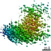
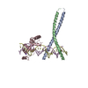
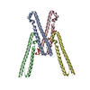
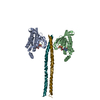
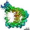
| Title | Nucleocapsid protein from the SARS-CoV-2 coronavirus Virus Virus | |||||||||
 Map data Map data | Nucleocapsid protein from the SARS-CoV-2 CoronavirusVirus | |||||||||
 Sample Sample | SARS-CoV-2 != Severe acute respiratory syndrome coronavirus 2 SARS-CoV-2
| |||||||||
| Biological species |   Severe acute respiratory syndrome coronavirus 2 Severe acute respiratory syndrome coronavirus 2 | |||||||||
| Method | single particle reconstruction / cryo EM / Resolution: 4.3 Å | |||||||||
 Authors Authors | Casasanta MA / Kelly DF | |||||||||
 Citation Citation | Journal: Nanoscale / Year: 2021 Title: Microchip-based structure determination of low-molecular weight proteins using cryo-electron microscopy. Authors: Michael A Casasanta / G M Jonaid / Liam Kaylor / William Y Luqiu / Maria J Solares / Mariah L Schroen / William J Dearnaley / Jarad Wilson / Madeline J Dukes / Deborah F Kelly /  Abstract: Interest in cryo-Electron Microscopy (EM) imaging has skyrocketed in recent years due to its pristine views of macromolecules and materials. As advances in instrumentation and computing algorithms ...Interest in cryo-Electron Microscopy (EM) imaging has skyrocketed in recent years due to its pristine views of macromolecules and materials. As advances in instrumentation and computing algorithms spurred this progress, there is renewed focus to address specimen-related challenges. Here we contribute a microchip-based toolkit to perform complementary structural and biochemical analysis on low-molecular weight proteins. As a model system, we used the SARS-CoV-2 nucleocapsid (N) protein (48 kDa) due to its stability and important role in therapeutic development. Cryo-EM structures of the N protein monomer revealed a flexible N-terminal "top hat" motif and a helical-rich C-terminal domain. To complement our structural findings, we engineered microchip-based immunoprecipitation assays that led to the discovery of the first antibody binding site on the N protein. The data also facilitated molecular modeling of a variety of pandemic and common cold-related coronavirus proteins. Such insights may guide future pandemic-preparedness protocols through immuno-engineering strategies to mitigate viral outbreaks. | |||||||||
| History |
|
- Structure visualization
| Movie |
 Movie viewer Movie viewer |
|---|---|
| Structure viewer | EM map: SurfViewMolmilJmol/JSmol |
| Supplemental images |

- Downloads & links
-EMDB archive
| Map data | emd_22982.map.gz | 304.2 KB | EMDB map data format | |
|---|---|---|---|---|
| Header (meta data) | emd-22982-v30.xmlemd-22982.xml | 12.6 KB 12.6 KB | Display Display | EMDB header |
| Images | 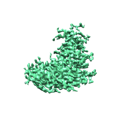 emd_22982.png emd_22982.png | 79.6 KB | ||
| Archive directory |  http://ftp.pdbj.org/pub/emdb/structures/EMD-22982ftp://ftp.pdbj.org/pub/emdb/structures/EMD-22982 http://ftp.pdbj.org/pub/emdb/structures/EMD-22982ftp://ftp.pdbj.org/pub/emdb/structures/EMD-22982 | HTTPS FTP |
-Related structure data
| Related structure data | C: citing same article ( |
|---|---|
| Similar structure data |


-Links
| EMDB pages | EMDB (EBI/PDBe) / EMDataResource |
|---|
-Map
| File | Download / File: emd_22982.map.gz / Format: CCP4 / Size: 12.9 MB / Type: IMAGE STORED AS FLOATING POINT NUMBER (4 BYTES) | ||||||||||||||||||||||||||||||||||||||||||||||||||||||||||||
|---|---|---|---|---|---|---|---|---|---|---|---|---|---|---|---|---|---|---|---|---|---|---|---|---|---|---|---|---|---|---|---|---|---|---|---|---|---|---|---|---|---|---|---|---|---|---|---|---|---|---|---|---|---|---|---|---|---|---|---|---|---|
| Annotation | Nucleocapsid protein from the SARS-CoV-2 Coronavirus | ||||||||||||||||||||||||||||||||||||||||||||||||||||||||||||
| Voxel size | X=Y=Z: 0.93 Å | ||||||||||||||||||||||||||||||||||||||||||||||||||||||||||||
| Density |
| ||||||||||||||||||||||||||||||||||||||||||||||||||||||||||||
| Symmetry | Space group: 1 | ||||||||||||||||||||||||||||||||||||||||||||||||||||||||||||
| Details | EMDB XML:
CCP4 map header:
| ||||||||||||||||||||||||||||||||||||||||||||||||||||||||||||
-Supplemental data
- Sample components
-Entire : SARS-CoV-2
| Entire | Name: SARS-CoV-2 (virus) |
|---|---|
| Components |
|
-Supramolecule #1: Severe acute respiratory syndrome coronavirus 2
| Supramolecule | Name: Severe acute respiratory syndrome coronavirus 2 / type: virus / ID: 1 / Parent: 0 / NCBI-ID: 2697049 Sci species name: Severe acute respiratory syndrome coronavirus 2 Virus type: VIRION / Virus isolate: STRAIN / Virus enveloped: Yes / Virus empty: Yes |
|---|---|
| Host (natural) | Organism:  Homo sapiens (human) Homo sapiens (human) |
| Host system | Organism:  Escherichia coli (E. coli) Escherichia coli (E. coli) |
| Molecular weight | Experimental: 50 KDa |
-Experimental details
-Structure determination
| Method | cryo EM |
|---|---|
 Processing Processing | single particle reconstruction |
| Aggregation state | particle |
-Sample preparation
| Concentration | 0.1 mg/mL | |||||||||||||||
|---|---|---|---|---|---|---|---|---|---|---|---|---|---|---|---|---|
| Buffer | pH: 7.5 Component:
Details: Solutions were made fresh before diluting protein and adding to the EM grids | |||||||||||||||
| Grid | Model: Homemade / Material: SILICON NITRIDE / Support film - topology: CONTINUOUS / Support film - Film thickness: 20.0 nm | |||||||||||||||
| Vitrification | Cryogen name: ETHANE / Chamber humidity: 95 % / Chamber temperature: 293 K / Instrument: FEI VITROBOT MARK III / Details: blot for 7.5 seconds before plunging. | |||||||||||||||
| Details | Sample was monodisperse on a silicon nitride microchip coated with 25% Ni-NTA lipids. |
- Electron microscopy
| Microscope | TFS TALOS F200C |
|---|---|
| Electron beam | Acceleration voltage: 200 kV / Electron source: FIELD EMISSION GUN |
| Electron optics | Illumination mode: FLOOD BEAM / Imaging mode: BRIGHT FIELDBright-field microscopy / Cs: 2.7 mm / Nominal defocus max: 1.5 µm / Nominal defocus min: 0.5 µm / Nominal magnification: 45000 |
| Sample stage | Specimen holder model: GATAN 626 SINGLE TILT LIQUID NITROGEN CRYO TRANSFER HOLDER Cooling holder cryogen: NITROGEN |
| Temperature | Min: 75.0 K / Max: 83.0 K |
| Image recording | Film or detector model: DIRECT ELECTRON DE-12 (4k x 3k) / Detector mode: INTEGRATING / Number grids imaged: 3 / Average exposure time: 0.25 sec. / Average electron dose: 5.0 e/Å2 Details: Images were collected in movie mode with 0.25 second exposures at 30 frames per second. |
| Experimental equipment |  Model: Tecnai F20 / Image courtesy: FEI Company |
-Image processing
| Particle selection | Number selected: 20000 |
|---|---|
| CTF correction | Software - Name: cryoSPARC Details: CTF correction was performed in Cryosparc before 3D reconstruction. |
| Initial angle assignment | Type: MAXIMUM LIKELIHOOD / Software - Name: cryoSPARC |
| Final 3D classification | Number classes: 1 / Avg.num./class: 20000 / Software - Name: cryoSPARC |
| Final angle assignment | Type: MAXIMUM LIKELIHOOD / Software - Name: cryoSPARC |
| Final reconstruction | Applied symmetry - Point group: C1 (asymmetric) / Resolution.type: BY AUTHOR / Resolution: 4.3 Å / Resolution method: FSC 0.143 CUT-OFF / Software - Name: cryoSPARC / Number images used: 20000 |
-Atomic model buiding 1
| Refinement | Space: REAL / Protocol: FLEXIBLE FIT |
|---|
