- EMDB-22143: Cryo-EM structure of NusG-CTD bound to 70S ribosome -
+
Open data
ID or keywords:
Loading...
-
Basic information
Entry
Database: EMDB / ID: EMD-22143
Title
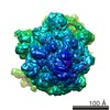
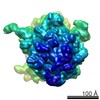
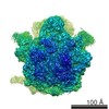
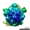
Cryo-EM structure of NusG-CTD bound to 70S ribosome
Map data
UnMasked and UnSharpened EM volume
Sample
Complex: Bacterial Ribosome in complex with NusG-CTD
Protein or peptide: x 21 types
RNA: x 1 types
Keywords
Ribosome / NusG / TRANSLATION
Function / homology
Function and homology information
transcription elongation-coupled chromatin remodeling / mRNA base-pairing translational repressor activity / ornithine decarboxylase inhibitor activity / misfolded RNA binding / transcription antitermination factor activity, RNA binding / Group I intron splicing / RNA folding / four-way junction DNA binding / negative regulation of translational initiation / regulation of mRNA stability ...transcription elongation-coupled chromatin remodeling / mRNA base-pairing translational repressor activity / ornithine decarboxylase inhibitor activity / misfolded RNA binding / transcription antitermination factor activity, RNA binding / Group I intron splicing / RNA folding / four-way junction DNA binding / negative regulation of translational initiation / regulation of mRNA stability / mRNA regulatory element binding translation repressor activity / positive regulation of RNA splicing / DNA endonuclease activity / transcription elongation factor complex / regulation of DNA-templated transcription elongation / transcription antitermination / maintenance of translational fidelity / DNA-templated transcription termination / mRNA 5'-UTR binding / ribosomal small subunit biogenesis / small ribosomal subunit rRNA binding / ribosomal small subunit assembly / cytosolic small ribosomal subunit / ribosome biogenesis / regulation of translation / cytoplasmic translation / small ribosomal subunit / tRNA binding / negative regulation of translation / rRNA binding / molecular adaptor activity / ribosome / structural constituent of ribosome / translation / response to antibiotic / mRNA binding / RNA binding / zinc ion binding / membrane / cytosol / cytoplasm Similarity search - Function
: / Transcription antitermination protein, NusG / Transcription antitermination protein, NusG, bacteria, conserved site / Transcription termination factor nusG signature. / NusG-like / Transcription termination factor nusG / NusG, N-terminal / In Spt5p, this domain may confer affinity for Spt4p. It possesses a RNP-like fold. / NusG, N-terminal domain superfamily / Ribosomal protein S21, conserved site ...: / Transcription antitermination protein, NusG / Transcription antitermination protein, NusG, bacteria, conserved site / Transcription termination factor nusG signature. / NusG-like / Transcription termination factor nusG / NusG, N-terminal / In Spt5p, this domain may confer affinity for Spt4p. It possesses a RNP-like fold. / NusG, N-terminal domain superfamily / Ribosomal protein S21, conserved site / Ribosomal protein S21 signature. / Ribosomal protein S14, bacterial/plastid / Ribosomal protein S21 superfamily / Ribosomal protein S21 / Ribosomal protein S16, conserved site / Ribosomal protein S16 signature. / Ribosomal protein S21 / Ribosomal protein S3, bacterial-type / Ribosomal protein S6, conserved site / Ribosomal protein S6 signature. / Ribosomal protein S19, bacterial-type / Ribosomal protein S7, bacterial/organellar-type / Ribosomal protein S11, bacterial-type / Ribosomal protein S13, bacterial-type / Ribosomal protein S20 / Ribosomal protein S20 superfamily / Ribosomal protein S20 / Ribosomal protein S9, bacterial/plastid / Ribosomal protein S4, bacterial-type / 30S ribosomal protein S17 / Ribosomal protein S5, bacterial-type / Ribosomal protein S6, plastid/chloroplast / Ribosomal protein S2, bacteria/mitochondria/plastid / Ribosomal protein S18, conserved site / Ribosomal protein S18 signature. / Ribosomal protein S16 / Ribosomal protein S16 / Ribosomal protein S16 domain superfamily / Ribosomal protein S15, bacterial-type / Ribosomal protein S2 signature 2. / Ribosomal protein S6 / Ribosomal protein S6 / Ribosomal protein S6 superfamily / Ribosomal protein S12, bacterial-type / Translation elongation factor EF1B/ribosomal protein S6 / Ribosomal protein S18 / Ribosomal protein S18 / Ribosomal protein S18 superfamily / K Homology domain / K homology RNA-binding domain / Ribosomal protein S3, conserved site / Ribosomal protein S14, conserved site / Ribosomal protein S10, conserved site / : / K Homology domain, type 2 / Ribosomal protein S3, C-terminal / Ribosomal protein S3, C-terminal domain superfamily / Ribosomal protein S15/S19, conserved site / KH domain / Ribosomal protein S19/S15 / Ribosomal protein S19/S15, superfamily / Ribosomal protein S10 / Ribosomal protein S3, C-terminal domain / Ribosomal protein S3 signature. / Ribosomal protein S10 signature. / Ribosomal protein S14 signature. / Ribosomal protein S7, conserved site / Ribosomal protein S17, conserved site / K homology domain superfamily, prokaryotic type / Ribosomal protein S19 / Ribosomal protein S2 signature 1. / Ribosomal protein S13, conserved site / Ribosomal protein S2, conserved site / Ribosomal protein S13 / 30s ribosomal protein S13, C-terminal / Ribosomal protein S2 / Ribosomal protein S2, flavodoxin-like domain superfamily / Ribosomal protein S2 / Ribosomal protein S4/S9 N-terminal domain / Ribosomal protein S14 / Ribosomal protein S4/S9, N-terminal / Ribosomal protein S4, conserved site / Type-2 KH domain profile. / Ribosomal protein S4/S9 N-terminal domain / Ribosomal protein S13/S18 / Ribosomal protein S4/S9 / Ribosomal protein S19 signature. / K homology domain-like, alpha/beta / Ribosomal protein S14p/S29e / Ribosomal protein S8 / Ribosomal protein S8 superfamily / Ribosomal protein S5, N-terminal, conserved site / Ribosomal protein S5 signature. / Ribosomal protein S5 / Ribosomal protein S5, N-terminal / Ribosomal S11, conserved site / Ribosomal protein S7 signature. / Ribosomal protein S10p/S20e / Ribosomal protein S13-like, H2TH / S5 double stranded RNA-binding domain profile. Similarity search - Domain/homology
Small ribosomal subunit protein bS6 / Small ribosomal subunit protein uS7 / Small ribosomal subunit protein uS10 / Small ribosomal subunit protein uS11 / Small ribosomal subunit protein uS12 / Small ribosomal subunit protein uS13 / Small ribosomal subunit protein bS16 / Small ribosomal subunit protein bS18 / Small ribosomal subunit protein uS19 / Small ribosomal subunit protein bS20 ...Small ribosomal subunit protein bS6 / Small ribosomal subunit protein uS7 / Small ribosomal subunit protein uS10 / Small ribosomal subunit protein uS11 / Small ribosomal subunit protein uS12 / Small ribosomal subunit protein uS13 / Small ribosomal subunit protein bS16 / Small ribosomal subunit protein bS18 / Small ribosomal subunit protein uS19 / Small ribosomal subunit protein bS20 / Small ribosomal subunit protein uS2 / Small ribosomal subunit protein uS3 / Small ribosomal subunit protein uS4 / Small ribosomal subunit protein uS5 / Small ribosomal subunit protein uS8 / Small ribosomal subunit protein uS9 / Small ribosomal subunit protein uS15 / Transcription termination/antitermination protein NusG / Small ribosomal subunit protein uS14 / Small ribosomal subunit protein uS17 / Small ribosomal subunit protein bS21 Similarity search - Component
National Institutes of Health/National Institute of Mental Health (NIH/NIMH)
R01 GM037219
United States
National Institutes of Health/National Institute of Mental Health (NIH/NIMH)
R01 GM29169
United States
Other private
Amgen
United States
Citation
Journal: iScience / Year: 2020 Title: Escherichia coli NusG Links the Lead Ribosome with the Transcription Elongation Complex. Authors: Robert S Washburn / Philipp K Zuber / Ming Sun / Yaser Hashem / Bingxin Shen / Wen Li / Sho Harvey / Francisco J Acosta Reyes / Max E Gottesman / Stefan H Knauer / Joachim Frank / Abstract: It has been known for more than 50 years that transcription and translation are physically coupled in bacteria, but whether or not this coupling may be mediated by the two-domain protein N- ...It has been known for more than 50 years that transcription and translation are physically coupled in bacteria, but whether or not this coupling may be mediated by the two-domain protein N-utilization substance (Nus) G in Escherichia coli is still heavily debated. Here, we combine integrative structural biology and functional analyses to provide conclusive evidence that NusG can physically link transcription with translation by contacting both RNA polymerase and the ribosome. We present a cryo-electron microscopy structure of a NusG:70S ribosome complex and nuclear magnetic resonance spectroscopy data revealing simultaneous binding of NusG to RNAP and the intact 70S ribosome, providing the first direct structural evidence for NusG-mediated coupling. Furthermore, in vivo reporter assays show that recruitment of NusG occurs late in transcription and strongly depends on translation. Thus, our data suggest that coupling occurs initially via direct RNAP:ribosome contacts and is then mediated by NusG.
History
Deposition
Jun 11, 2020
-
Header (metadata) release
Jul 29, 2020
-
Map release
Jul 29, 2020
-
Update
Mar 6, 2024
-
Current status
Mar 6, 2024
Processing site: RCSB / Status: Released
-
Structure visualization
Movie
Surface view with section colored by density value
Film or detector model: GATAN K2 SUMMIT (4k x 4k) / Detector mode: COUNTING / Digitization - Frames/image: 1-20 / Number grids imaged: 1 / Number real images: 1327 / Average electron dose: 100.0 e/Å2
Experimental equipment
Model: Tecnai Polara / Image courtesy: FEI Company
-
Image processing
Particle selection
Number selected: 188127
Startup model
Type of model: OTHER
Initial angle assignment
Type: MAXIMUM LIKELIHOOD / Software - Name: RELION (ver. 1.2)
Final 3D classification
Software - Name: RELION (ver. 1.2)
Final angle assignment
Type: MAXIMUM LIKELIHOOD / Software - Name: RELION (ver. 1.2)
Final reconstruction
Resolution.type: BY AUTHOR / Resolution: 6.8 Å / Resolution method: FSC 0.143 CUT-OFF / Number images used: 17122
In the structure databanks used in Yorodumi, some data are registered as the other names, "COVID-19 virus" and "2019-nCoV". Here are the details of the virus and the list of structure data.
Jan 31, 2019. EMDB accession codes are about to change! (news from PDBe EMDB page)
EMDB accession codes are about to change! (news from PDBe EMDB page)
The allocation of 4 digits for EMDB accession codes will soon come to an end. Whilst these codes will remain in use, new EMDB accession codes will include an additional digit and will expand incrementally as the available range of codes is exhausted. The current 4-digit format prefixed with “EMD-” (i.e. EMD-XXXX) will advance to a 5-digit format (i.e. EMD-XXXXX), and so on. It is currently estimated that the 4-digit codes will be depleted around Spring 2019, at which point the 5-digit format will come into force.
The EM Navigator/Yorodumi systems omit the EMD- prefix.
Related info.:Q: What is EMD? / ID/Accession-code notation in Yorodumi/EM Navigator
Yorodumi is a browser for structure data from EMDB, PDB, SASBDB, etc.
This page is also the successor to EM Navigator detail page, and also detail information page/front-end page for Omokage search.
The word "yorodu" (or yorozu) is an old Japanese word meaning "ten thousand". "mi" (miru) is to see.
Related info.:EMDB / PDB / SASBDB / Comparison of 3 databanks / Yorodumi Search / Aug 31, 2016. New EM Navigator & Yorodumi / Yorodumi Papers / Jmol/JSmol / Function and homology information / Changes in new EM Navigator and Yorodumi
 Movie
Movie Controller


 Open data
 Basic information
 Map data
 Sample
 Keywords
 Ribosome / NusG /
  Function and homology information
 Authors
 Germany,
  United States, 4 items
  Citation
 Structure visualization
 Downloads & links
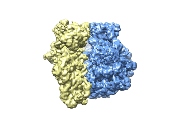 emd_22143.png
 http://ftp.pdbj.org/pub/emdb/structures/EMD-22143


 Sample components
 Processing
 Electron microscopy


