 Movie
Movie Controller

[English] 日本語
 Yorodumi
- EMDB-20828: Cryo-electron tomogram of FIB-milled HEK293 cells treated with Taxol -
+ Open data

- Basic information
| Entry | Database: EMDB / ID: EMD-20828 | |||||||||
|---|---|---|---|---|---|---|---|---|---|---|
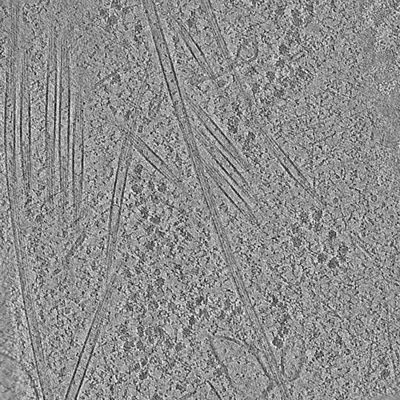
| Title | Cryo-electron tomogram of FIB-milled HEK293 cells treated with Taxol | |||||||||
 Map data Map data | Cryo-electron tomogram of FIB-milled HEK293 cells treated with Taxol | |||||||||
 Sample Sample |
| |||||||||
| Biological species |   Homo sapiens (human) Homo sapiens (human) | |||||||||
| Method | electron tomography / cryo EM | |||||||||
 Authors Authors | Watanabe R / Villa E | |||||||||
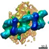
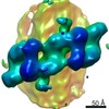
 Citation Citation | Journal: Cell / Year: 2020 Title: The In Situ Structure of Parkinson's Disease-Linked LRRK2. Authors: Reika Watanabe / Robert Buschauer / Jan Böhning / Martina Audagnotto / Keren Lasker / Tsan-Wen Lu / Daniela Boassa / Susan Taylor / Elizabeth Villa /  Abstract: Mutations in leucine-rich repeat kinase 2 (LRRK2) are the most frequent cause of familial Parkinson's disease. LRRK2 is a multi-domain protein containing a kinase and GTPase. Using correlative light ...Mutations in leucine-rich repeat kinase 2 (LRRK2) are the most frequent cause of familial Parkinson's disease. LRRK2 is a multi-domain protein containing a kinase and GTPase. Using correlative light and electron microscopy, in situ cryo-electron tomography, and subtomogram analysis, we reveal a 14-Å structure of LRRK2 bearing a pathogenic mutation that oligomerizes as a right-handed double helix around microtubules, which are left-handed. Using integrative modeling, we determine the architecture of LRRK2, showing that the GTPase and kinase are in close proximity, with the GTPase closer to the microtubule surface, whereas the kinase is exposed to the cytoplasm. We identify two oligomerization interfaces mediated by non-catalytic domains. Mutation of one of these abolishes LRRK2 microtubule-association. Our work demonstrates the power of cryo-electron tomography to generate models of previously unsolved structures in their cellular environment. | |||||||||
| History |
|
- Structure visualization
| Movie |
 Movie viewer Movie viewer |
|---|---|
| Supplemental images |
- Downloads & links
-EMDB archive
| Map data | emd_20828.map.gz | 129.3 MB | EMDB map data format | |
|---|---|---|---|---|
| Header (meta data) | emd-20828-v30.xmlemd-20828.xml | 8.6 KB 8.6 KB | Display Display | EMDB header |
| Images |  emd_20828.png emd_20828.png | 141.5 KB | ||
| Archive directory |  http://ftp.pdbj.org/pub/emdb/structures/EMD-20828ftp://ftp.pdbj.org/pub/emdb/structures/EMD-20828 http://ftp.pdbj.org/pub/emdb/structures/EMD-20828ftp://ftp.pdbj.org/pub/emdb/structures/EMD-20828 | HTTPS FTP |
-Related structure data
| Related structure data | 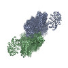 6xr4C C: citing same article ( |
|---|---|
| EM raw data | EMPIAR-10377 (Title: Frame-aligned tilt series of cryo-FIB-milled HEK293 cells treated with taxol Data size: 2.1 Data #1: Frame-aligned tilt series of cryo-FIB-milled HEK293 cells treated with taxol [tilt series]) |
-Links
| EMDB pages | EMDB (EBI/PDBe) / EMDataResource |
|---|
-Map
| File | Download / File: emd_20828.map.gz / Format: CCP4 / Size: 158.9 MB / Type: IMAGE STORED AS SIGNED BYTE | ||||||||||||||||||||||||||||||||||||||||||||||||||||||||||||||||||||
|---|---|---|---|---|---|---|---|---|---|---|---|---|---|---|---|---|---|---|---|---|---|---|---|---|---|---|---|---|---|---|---|---|---|---|---|---|---|---|---|---|---|---|---|---|---|---|---|---|---|---|---|---|---|---|---|---|---|---|---|---|---|---|---|---|---|---|---|---|---|
| Annotation | Cryo-electron tomogram of FIB-milled HEK293 cells treated with Taxol | ||||||||||||||||||||||||||||||||||||||||||||||||||||||||||||||||||||
| Voxel size | X=Y=Z: 10.8 Å | ||||||||||||||||||||||||||||||||||||||||||||||||||||||||||||||||||||
| Density |
| ||||||||||||||||||||||||||||||||||||||||||||||||||||||||||||||||||||
| Symmetry | Space group: 1 | ||||||||||||||||||||||||||||||||||||||||||||||||||||||||||||||||||||
| Details | EMDB XML:
CCP4 map header:
| ||||||||||||||||||||||||||||||||||||||||||||||||||||||||||||||||||||
-Supplemental data
- Sample components
-Entire : Cryo-electron tomogram of FIB-milled HEK293 cells treated with Taxol
| Entire | Name: Cryo-electron tomogram of FIB-milled HEK293 cells treated with Taxol |
|---|---|
| Components |
|
-Supramolecule #1: Cryo-electron tomogram of FIB-milled HEK293 cells treated with Taxol
| Supramolecule | Name: Cryo-electron tomogram of FIB-milled HEK293 cells treated with Taxol type: cell / ID: 1 / Parent: 0 |
|---|---|
| Source (natural) | Organism: Homo sapiens (human) / Cell: HEK293 |
-Experimental details
-Structure determination
| Method | cryo EM |
|---|---|
 Processing Processing | electron tomography |
| Aggregation state | cell |
-Sample preparation
| Buffer | pH: 7 |
|---|---|
| Grid | Details: unspecified |
| Vitrification | Cryogen name: ETHANE-PROPANE |
| Sectioning | Focused ion beam - Instrument: OTHER / Focused ion beam - Ion: OTHER / Focused ion beam - Voltage: 30 kV / Focused ion beam - Current: 0.001 nA / Focused ion beam - Duration: 50 sec. / Focused ion beam - Temperature: 70 K / Focused ion beam - Initial thickness: 4000 nm / Focused ion beam - Final thickness: 80 nm Focused ion beam - Details: The value given for _emd_sectioning_focused_ion_beam.instrument is Aquilos. This is not in a list of allowed values set(['DB235', 'OTHER']) so OTHER is written into the XML file. |
- Electron microscopy
| Microscope | FEI POLARA 300 |
|---|---|
| Electron beam | Acceleration voltage: 300 kV / Electron source: FIELD EMISSION GUN |
| Electron optics | Illumination mode: FLOOD BEAM / Imaging mode: BRIGHT FIELDBright-field microscopy |
| Image recording | Film or detector model: GATAN K2 SUMMIT (4k x 4k) / Detector mode: COUNTING / Average electron dose: 3.0 e/Å2 |
| Experimental equipment |  Model: Tecnai Polara / Image courtesy: FEI Company |
-Image processing
| Final reconstruction | Algorithm: BACK PROJECTION / Software - Name: IMOD / Number images used: 39 |
|---|
