- EMDB-20121: Structure of trans-translation inhibitor bound to E. coli 70S rib... -
+
Open data
ID or keywords:
Loading...
-
Basic information
Entry
Database: EMDB / ID: EMD-20121
Title
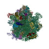
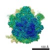
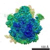
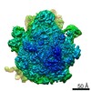
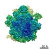
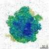
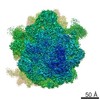
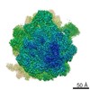
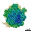
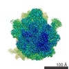
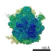
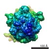
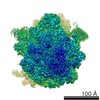
Structure of trans-translation inhibitor bound to E. coli 70S ribosome with P site tRNA
Map data
unfiltered main map
Sample
Complex: Structure of trans-translation inhibitor bound to E. coli 70S ribosome with P site tRNA
Complex: 50S subunitProkaryotic large ribosomal subunit
RNA: x 2 types
Protein or peptide: x 29 types
Complex: 30S subunitProkaryotic small ribosomal subunit
RNA: x 3 types
Protein or peptide: x 20 types
Ligand: x 1 types
Function / homology
Function and homology information
positive regulation of ribosome biogenesis / DnaA-L2 complex / negative regulation of DNA-templated DNA replication initiation / assembly of large subunit precursor of preribosome / cytosolic ribosome assembly / regulation of cell growth / mRNA 5'-UTR binding / ribosomal large subunit assembly / small ribosomal subunit rRNA binding / cytosolic small ribosomal subunit ...positive regulation of ribosome biogenesis / DnaA-L2 complex / negative regulation of DNA-templated DNA replication initiation / assembly of large subunit precursor of preribosome / cytosolic ribosome assembly / regulation of cell growth / mRNA 5'-UTR binding / ribosomal large subunit assembly / small ribosomal subunit rRNA binding / cytosolic small ribosomal subunit / ribosome binding / large ribosomal subunit / cytoplasmic translation / small ribosomal subunit / 5S rRNA binding / cytosolic large ribosomal subunit / transferase activity / tRNA binding / rRNA binding / ribosome / structural constituent of ribosome / ribonucleoprotein complex / translation / mRNA binding / RNA binding / zinc ion binding / metal ion binding / cytosol / cytoplasm Similarity search - Function
Ribosomal protein S21, conserved site / Ribosomal protein S21 signature. / Ribosomal protein L25, short-form / Ribosomal protein S14, bacterial/plastid / Ribosomal protein L31 type A / Ribosomal protein S21 superfamily / Ribosomal protein S21 / Ribosomal protein S16, conserved site / Ribosomal protein S16 signature. / Ribosomal protein L31 signature. ...Ribosomal protein S21, conserved site / Ribosomal protein S21 signature. / Ribosomal protein L25, short-form / Ribosomal protein S14, bacterial/plastid / Ribosomal protein L31 type A / Ribosomal protein S21 superfamily / Ribosomal protein S21 / Ribosomal protein S16, conserved site / Ribosomal protein S16 signature. / Ribosomal protein L31 signature. / Ribosomal protein S21 / Ribosomal protein L31 / Ribosomal protein L31 superfamily / Ribosomal protein L31 / Ribosomal protein L21, conserved site / Ribosomal protein L21 signature. / Ribosomal protein L16 signature 1. / Ribosomal protein L6, conserved site / Ribosomal protein L6 signature 1. / Ribosomal protein L16, conserved site / Ribosomal protein L16 signature 2. / Ribosomal protein L9 signature. / Ribosomal protein L9, bacteria/chloroplast / Ribosomal protein L9, C-terminal / Ribosomal protein L9, C-terminal domain / Ribosomal protein L9, C-terminal domain superfamily / Ribosomal L25p family / Ribosomal protein L25 / Ribosomal protein S14/S29 / Ribosomal protein L28/L24 superfamily / Ribosomal protein L36 signature. / Ribosomal protein L25/Gln-tRNA synthetase, N-terminal / Ribosomal protein L25/Gln-tRNA synthetase, anti-codon-binding domain superfamily / Ribosomal protein L9, N-terminal domain superfamily / Ribosomal protein L9 / Ribosomal protein L9, N-terminal / Ribosomal protein L9, N-terminal domain / Ribosomal protein L28 / Ribosomal protein L35, conserved site / Ribosomal protein L35 signature. / Ribosomal protein L33, conserved site / Ribosomal protein L33 signature. / Ribosomal protein L35, non-mitochondrial / Ribosomal protein L5, bacterial-type / Ribosomal protein L6, bacterial-type / Ribosomal protein L18, bacterial-type / Ribosomal protein L19, conserved site / Ribosomal protein L19 signature. / Ribosomal protein L36 / Ribosomal protein L36 superfamily / Ribosomal protein L36 / Ribosomal protein L9/RNase H1, N-terminal / Ribosomal protein L20 signature. / Ribosomal protein S3, bacterial-type / Ribosomal protein S6, conserved site / Ribosomal protein S6 signature. / Ribosomal protein L27, conserved site / Ribosomal protein L27 signature. / Ribosomal protein S19, bacterial-type / Ribosomal protein S7, bacterial/organellar-type / Ribosomal protein S11, bacterial-type / Ribosomal protein S13, bacterial-type / Ribosomal protein S20 / Ribosomal protein S20 superfamily / Ribosomal protein S20 / Ribosomal protein S9, bacterial/plastid / Ribosomal protein S4, bacterial-type / Ribosomal protein L14P, bacterial-type / Ribosomal protein L34, conserved site / Ribosomal protein L34 signature. / 30S ribosomal protein S17 / Ribosomal protein S5, bacterial-type / Ribosomal protein L22, bacterial/chloroplast-type / Ribosomal protein S6, plastid/chloroplast / Ribosomal protein L35 / Ribosomal protein L35 superfamily / Ribosomal protein L2, bacterial/organellar-type / Ribosomal protein L35 / Ribosomal protein S2, bacteria/mitochondria/plastid / Ribosomal L28 family / Ribosomal protein L33 / Ribosomal protein L33 / Ribosomal protein L28/L24 / Ribosomal protein L33 superfamily / Ribosomal protein L30, bacterial-type / Ribosomal protein L16 / Ribosomal protein L18 / Ribosomal L18 of archaea, bacteria, mitoch. and chloroplast / Ribosomal protein S18, conserved site / Ribosomal protein S18 signature. / L28p-like / Ribosomal protein L20 / Ribosomal protein S16 / Ribosomal protein S16 / Ribosomal protein S16 domain superfamily / Ribosomal protein L20 / Ribosomal protein L20, C-terminal / Ribosomal protein L21 / Ribosomal protein L27 / Ribosomal L27 protein Similarity search - Domain/homology
Large ribosomal subunit protein uL15 / : / Small ribosomal subunit protein bS18 / Large ribosomal subunit protein bL36 / Small ribosomal subunit protein bS21 / 30S ribosomal protein S7 / Large ribosomal subunit protein bL20 / Small ribosomal subunit protein uS11 / Small ribosomal subunit protein uS3 / Large ribosomal subunit protein bL34 ...Large ribosomal subunit protein uL15 / : / Small ribosomal subunit protein bS18 / Large ribosomal subunit protein bL36 / Small ribosomal subunit protein bS21 / 30S ribosomal protein S7 / Large ribosomal subunit protein bL20 / Small ribosomal subunit protein uS11 / Small ribosomal subunit protein uS3 / Large ribosomal subunit protein bL34 / Small ribosomal subunit protein bS16 / 50S ribosomal protein L23 / Large ribosomal subunit protein bL25 / 50S ribosomal protein L35 / 30S ribosomal protein S10 / 50S ribosomal protein L4 / 30S ribosomal protein S19 / 50S ribosomal protein L22 / 50S ribosomal protein L29 / 30S ribosomal protein S17 / 50S ribosomal protein L14 / 50S ribosomal protein L24 / 50S ribosomal protein L5 / 30S ribosomal protein S14 / 30S ribosomal protein S8 / 50S ribosomal protein L6 / 50S ribosomal protein L18 / 30S ribosomal protein S5 / 50S ribosomal protein L30 / 30S ribosomal protein S4 / 50S ribosomal protein L33 / 30S ribosomal protein S20 / 30S ribosomal protein S15 / 50S ribosomal protein L21 / 30S ribosomal protein S9 / 50S ribosomal protein L13 / 50S ribosomal protein L31 / 50S ribosomal protein L32 / 50S ribosomal protein L9 / 30S ribosomal protein S2 / 50S ribosomal protein L19 / 30S ribosomal protein S13 / 30S ribosomal protein S12 / Small ribosomal subunit protein bS6 / Large ribosomal subunit protein bL27 / Large ribosomal subunit protein uL16 / Large ribosomal subunit protein uL2 / Large ribosomal subunit protein uL3 / 50S ribosomal protein L28 Similarity search - Component
Biological species
Escherichia coli (E. coli)
Method
single particle reconstruction / cryo EM / Resolution: 3.1 Å
National Institutes of Health/National Institute of General Medical Sciences (NIH/NIGMS)
GM093278
United States
National Institutes of Health/National Institute of General Medical Sciences (NIH/NIGMS)
GM121650
United States
Citation
Journal: Nat Commun / Year: 2021 Title: trans-Translation inhibitors bind to a novel site on the ribosome and clear Neisseria gonorrhoeae in vivo. Authors: Zachary D Aron / Atousa Mehrani / Eric D Hoffer / Kristie L Connolly / Pooja Srinivas / Matthew C Torhan / John N Alumasa / Mynthia Cabrera / Divya Hosangadi / Jay S Barbor / Steven C ...Authors: Zachary D Aron / Atousa Mehrani / Eric D Hoffer / Kristie L Connolly / Pooja Srinivas / Matthew C Torhan / John N Alumasa / Mynthia Cabrera / Divya Hosangadi / Jay S Barbor / Steven C Cardinale / Steven M Kwasny / Lucas R Morin / Michelle M Butler / Timothy J Opperman / Terry L Bowlin / Ann Jerse / Scott M Stagg / Christine M Dunham / Kenneth C Keiler / Abstract: Bacterial ribosome rescue pathways that remove ribosomes stalled on mRNAs during translation have been proposed as novel antibiotic targets because they are essential in bacteria and are not ...Bacterial ribosome rescue pathways that remove ribosomes stalled on mRNAs during translation have been proposed as novel antibiotic targets because they are essential in bacteria and are not conserved in humans. We previously reported the discovery of a family of acylaminooxadiazoles that selectively inhibit trans-translation, the main ribosome rescue pathway in bacteria. Here, we report optimization of the pharmacokinetic and antibiotic properties of the acylaminooxadiazoles, producing MBX-4132, which clears multiple-drug resistant Neisseria gonorrhoeae infection in mice after a single oral dose. Single particle cryogenic-EM studies of non-stop ribosomes show that acylaminooxadiazoles bind to a unique site near the peptidyl-transfer center and significantly alter the conformation of ribosomal protein bL27, suggesting a novel mechanism for specific inhibition of trans-translation by these molecules. These results show that trans-translation is a viable therapeutic target and reveal a new conformation within the bacterial ribosome that may be critical for ribosome rescue pathways.
History
Deposition
Apr 18, 2019
-
Header (metadata) release
Aug 21, 2019
-
Map release
Feb 10, 2021
-
Update
Sep 22, 2021
-
Current status
Sep 22, 2021
Processing site: RCSB / Status: Released
-
Structure visualization
Movie
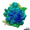
Surface view with section colored by density value
Supramolecule #1: Structure of trans-translation inhibitor bound to E. coli 70S rib...
Supramolecule
Name: Structure of trans-translation inhibitor bound to E. coli 70S ribosome with P site tRNA type: complex / ID: 1 / Parent: 0 / Macromolecule list: #1-#54
Film or detector model: DIRECT ELECTRON DE-64 (8k x 8k) / Detector mode: COUNTING / Digitization - Dimensions - Width: 4096 pixel / Digitization - Dimensions - Height: 4096 pixel / Digitization - Sampling interval: 6.5 µm / Digitization - Frames/image: 1-78 / Number grids imaged: 2 / Number real images: 2197 / Average exposure time: 19.302 sec. / Average electron dose: 58.0 e/Å2 Details: Two separate datesets were combined. The imaging parameter was the same for both datasets. Firth data-set had 1752 micrographs. Second data-set had 445 micrographs.
Experimental equipment
Model: Titan Krios / Image courtesy: FEI Company
-
Image processing
Particle selection
Number selected: 474382 Details: 100,537 particles were selected from first dataset and 373,845 partciles from the second dataset.
In the structure databanks used in Yorodumi, some data are registered as the other names, "COVID-19 virus" and "2019-nCoV". Here are the details of the virus and the list of structure data.
Jan 31, 2019. EMDB accession codes are about to change! (news from PDBe EMDB page)
EMDB accession codes are about to change! (news from PDBe EMDB page)
The allocation of 4 digits for EMDB accession codes will soon come to an end. Whilst these codes will remain in use, new EMDB accession codes will include an additional digit and will expand incrementally as the available range of codes is exhausted. The current 4-digit format prefixed with “EMD-” (i.e. EMD-XXXX) will advance to a 5-digit format (i.e. EMD-XXXXX), and so on. It is currently estimated that the 4-digit codes will be depleted around Spring 2019, at which point the 5-digit format will come into force.
The EM Navigator/Yorodumi systems omit the EMD- prefix.
Related info.:Q: What is EMD? / ID/Accession-code notation in Yorodumi/EM Navigator
Yorodumi is a browser for structure data from EMDB, PDB, SASBDB, etc.
This page is also the successor to EM Navigator detail page, and also detail information page/front-end page for Omokage search.
The word "yorodu" (or yorozu) is an old Japanese word meaning "ten thousand". "mi" (miru) is to see.
Related info.:EMDB / PDB / SASBDB / Comparison of 3 databanks / Yorodumi Search / Aug 31, 2016. New EM Navigator & Yorodumi / Yorodumi Papers / Jmol/JSmol / Function and homology information / Changes in new EM Navigator and Yorodumi
 Movie
Movie Controller

 Yorodumi
 Open data
 Basic information
 Map data
 Sample
 Function and homology information
 regulation of cell growth / mRNA 5'-UTR binding /
 Authors
 United States, 2 items
  Citation
 Structure visualization
 Downloads & links
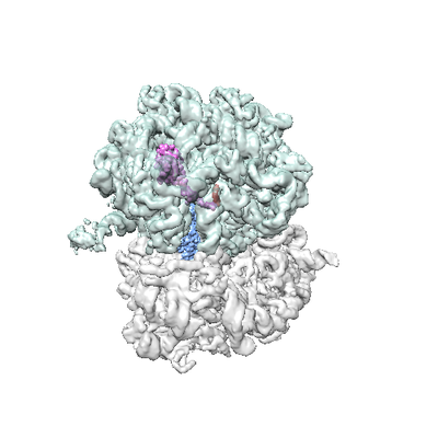 emd_20121.png
 http://ftp.pdbj.org/pub/emdb/structures/EMD-20121
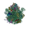


 Z
Z Y
Y X
X


 Sample components
 Processing
 Electron microscopy