 Movie
Movie Controller

[English] 日本語
 Yorodumi
- EMDB-19448: Alpha-synuclein amyloid fibrils with DNAJB1-Helix V mutant, HSC70... -
+ Open data

- Basic information
| Entry | 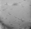 | ||||||||||||
|---|---|---|---|---|---|---|---|---|---|---|---|---|---|
| Title | Alpha-synuclein amyloid fibrils with DNAJB1-Helix V mutant, HSC70, HSP110 and ATP | ||||||||||||
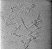
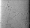
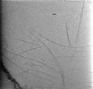
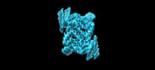
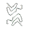
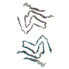
 Map data Map data | Tomogram of alpha-synuclein amyloid fibrils incubated with a variant of DNAJB1 mutated in helix V, Hsc70, Hsp110 and ATP | ||||||||||||
 Sample Sample |
| ||||||||||||
 Keywords Keywords | Chaperones / amyloid fibril / disaggregation / PROTEIN FIBRIL | ||||||||||||
| Biological species |  Homo sapiens (human) Homo sapiens (human) | ||||||||||||
| Method | electron tomography / cryo EM | ||||||||||||
 Authors Authors | Saibil HR / Monistrol J | ||||||||||||
| Funding support |  United Kingdom, 3 items United Kingdom, 3 items
| ||||||||||||
 Citation Citation | Journal: Biorxiv / Year: 2024 Title: Stepwise recruitment of Hsc70 by DNAJB1 produces ordered arrays primed for bursts of amyloid fibre disassembly Authors: Monistrol J / Beton JG / Johnston EC / Saibil HR | ||||||||||||
| History |
|
- Structure visualization
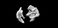
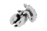
| Supplemental images |
|---|

- Downloads & links
-EMDB archive
| Map data | emd_19448.map.gz | 6 GB |  EMDB map data format EMDB map data format | |
|---|---|---|---|---|
| Header (meta data) | emd-19448-v30.xmlemd-19448.xml | 11.8 KB 11.8 KB | Display Display | EMDB header |
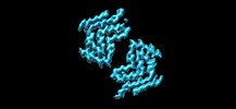
| Images |  emd_19448.png emd_19448.png | 181.2 KB | ||
| Filedesc metadata | emd-19448.cif.gz | 4.2 KB | ||
| Archive directory |  http://ftp.pdbj.org/pub/emdb/structures/EMD-19448ftp://ftp.pdbj.org/pub/emdb/structures/EMD-19448 http://ftp.pdbj.org/pub/emdb/structures/EMD-19448ftp://ftp.pdbj.org/pub/emdb/structures/EMD-19448 | HTTPS FTP |
-Validation report
| Summary document | emd_19448_validation.pdf.gz | 425.8 KB | Display | EMDB validaton report |
|---|---|---|---|---|
| Full document | emd_19448_full_validation.pdf.gz | 425.4 KB | Display | |
| Data in XML | emd_19448_validation.xml.gz | 5.3 KB | Display | |
| Data in CIF | emd_19448_validation.cif.gz | 5.8 KB | Display | |
| Arichive directory | https://ftp.pdbj.org/pub/emdb/validation_reports/EMD-19448ftp://ftp.pdbj.org/pub/emdb/validation_reports/EMD-19448 | HTTPS FTP |
-Related structure data
-Links
| EMDB pages | EMDB (EBI/PDBe) / EMDataResource |
|---|
-Map
| File | Download / File: emd_19448.map.gz / Format: CCP4 / Size: 6.5 GB / Type: IMAGE STORED AS FLOATING POINT NUMBER (4 BYTES) | ||||||||||||||||||||
|---|---|---|---|---|---|---|---|---|---|---|---|---|---|---|---|---|---|---|---|---|---|
| Annotation | Tomogram of alpha-synuclein amyloid fibrils incubated with a variant of DNAJB1 mutated in helix V, Hsc70, Hsp110 and ATP | ||||||||||||||||||||
| Voxel size | X=Y=Z: 3.8 Å | ||||||||||||||||||||
| Density |
| ||||||||||||||||||||
| Symmetry | Space group: 1 | ||||||||||||||||||||
| Details | EMDB XML:
|
-Supplemental data
- Sample components
-Entire : Alpha-synuclein amyloid fibrils incubated with a helix V mutant o...
| Entire | Name: Alpha-synuclein amyloid fibrils incubated with a helix V mutant of DNAJB1, HSC70, HSP110 and ATP |
|---|---|
| Components |
|
-Supramolecule #1: Alpha-synuclein amyloid fibrils incubated with a helix V mutant o...
| Supramolecule | Name: Alpha-synuclein amyloid fibrils incubated with a helix V mutant of DNAJB1, HSC70, HSP110 and ATP type: complex / ID: 1 / Parent: 0 |
|---|---|
| Source (natural) | Organism: Homo sapiens (human) |
-Experimental details
-Structure determination
| Method | cryo EM |
|---|---|
 Processing Processing | electron tomography |
| Aggregation state | filament |
-Sample preparation
| Buffer | pH: 7.5 Component:
| ||||||||||||||||||||||||
|---|---|---|---|---|---|---|---|---|---|---|---|---|---|---|---|---|---|---|---|---|---|---|---|---|---|
| Grid | Model: C-flat-2/2 / Material: COPPER / Mesh: 300 / Support film - Material: CARBON / Support film - topology: HOLEY / Pretreatment - Type: GLOW DISCHARGE | ||||||||||||||||||||||||
| Vitrification | Cryogen name: ETHANE / Chamber temperature: 277.15 K / Instrument: LEICA EM GP | ||||||||||||||||||||||||
| Sectioning | Other: NO SECTIONING | ||||||||||||||||||||||||
| Fiducial marker | Manufacturer: EMS / Diameter: 10 nm |
- Electron microscopy
| Microscope | FEI TITAN KRIOS |
|---|---|
| Specialist optics | Energy filter - Name: TFS Selectris |
| Image recording | Film or detector model: FEI FALCON IV (4k x 4k) / Average electron dose: 116.0 e/Å2 Details: Note: the original pixel size for the tomogram recording was 1.94 A/pixel. This was subsequently rounded to 1.9 A/pixel and the 2x binned tomogram deposited here is listed as 3.8 A/pixel. |
| Electron beam | Acceleration voltage: 300 kV / Electron source:  FIELD EMISSION GUN FIELD EMISSION GUN |
| Electron optics | Illumination mode: FLOOD BEAM / Imaging mode: BRIGHT FIELD / Nominal defocus max: 4.4 µm / Nominal defocus min: 2.0 µm |
| Sample stage | Specimen holder model: FEI TITAN KRIOS AUTOGRID HOLDER / Cooling holder cryogen: NITROGEN |
| Experimental equipment |  Model: Titan Krios / Image courtesy: FEI Company |
-Image processing
| Details | Note: the original tomogram was recorded at 1.94 A/pixel, but this was rounded to 1.9 A/pixel, so the 2x binned tomogram provided here is at 3.8 A/pixel |
|---|---|
| Final reconstruction | Software - Name: RELION (ver. 4) / Number images used: 121 |
