GDP phosphatase activity / non-canonical inflammasome complex assembly / protein localization to vacuole / negative regulation of substrate adhesion-dependent cell spreading / symbiont cell surface / cytolysis in another organism / positive regulation of pyroptosis / vesicle membrane / negative regulation of protein localization to plasma membrane / negative regulation of interleukin-2 production ...GDP phosphatase activity / non-canonical inflammasome complex assembly / protein localization to vacuole / negative regulation of substrate adhesion-dependent cell spreading / symbiont cell surface / cytolysis in another organism / positive regulation of pyroptosis / vesicle membrane / negative regulation of protein localization to plasma membrane / negative regulation of interleukin-2 production / negative regulation of T cell receptor signaling pathway / spectrin binding / cytokine binding / regulation of epidermal cell division / protein kinase C inhibitor activity / positive regulation of epidermal cell differentiation / keratinocyte development / keratinization / defense response to protozoan / Hydrolases; Acting on acid anhydrides; In phosphorus-containing anhydrides / Regulation of localization of FOXO transcription factors / keratinocyte proliferation / phosphoserine residue binding / Activation of BAD and translocation to mitochondria / negative regulation of keratinocyte proliferation / establishment of skin barrier / cellular response to interleukin-1 / SARS-CoV-2 targets host intracellular signalling and regulatory pathways / regulation of protein localization to plasma membrane / Chk1/Chk2(Cds1) mediated inactivation of Cyclin B:Cdk1 complex / protein kinase A signaling / negative regulation of stem cell proliferation / SARS-CoV-1 targets host intracellular signalling and regulatory pathways / regulation of calcium-mediated signaling / RHO GTPases activate PKNs / protein export from nucleus / negative regulation of innate immune response / protein sequestering activity / TP53 Regulates Transcription of Genes Involved in G2 Cell Cycle Arrest / G protein activity / release of cytochrome c from mitochondria / positive regulation of protein export from nucleus / stem cell proliferation / Translocation of SLC2A4 (GLUT4) to the plasma membrane / TP53 Regulates Metabolic Genes / lipopolysaccharide binding / Hydrolases; Acting on acid anhydrides; Acting on GTP to facilitate cellular and subcellular movement / Hsp90 protein binding / negative regulation of protein kinase activity / negative regulation of cysteine-type endopeptidase activity involved in apoptotic process / cytoplasmic vesicle membrane / negative regulation of ERK1 and ERK2 cascade / cellular response to type II interferon / GDP binding / intrinsic apoptotic signaling pathway in response to DNA damage / Interferon gamma signaling / actin cytoskeleton / cellular response to tumor necrosis factor / actin binding / cytoplasmic vesicle / positive regulation of cell growth / defense response to virus / regulation of cell cycle / defense response to bacterium / cadherin binding / Golgi membrane / innate immune response / GTPase activity / GTP binding / protein kinase binding / Golgi apparatus / negative regulation of transcription by RNA polymerase II / enzyme binding / signal transduction / protein homodimerization activity / extracellular space / extracellular exosome / extracellular region / identical protein binding / nucleus / plasma membrane / cytosol / cytoplasm Similarity search - Function
Germany, France, European Union, United Kingdom, 9 items
Organization
Grant number
Country
Boehringer Ingelheim Fonds (BIF)
Germany
Human Frontier Science Program (HFSP)
LT0006/2022-L
France
European Molecular Biology Organization (EMBO)
ALTF 491-2022
European Union
Wellcome Trust
217202/Z/19/Z
United Kingdom
Medical Research Council (MRC, United Kingdom)
MR/V030930/1
United Kingdom
Medical Research Council (MRC, United Kingdom)
MR/T029323/1
United Kingdom
Wellcome Trust
FC001076
United Kingdom
Medical Research Council (MRC, United Kingdom)
FC001076
United Kingdom
Cancer Research UK
FC001076
United Kingdom
Citation
Journal: Science / Year: 2023 Title: PIM1 controls GBP1 activity to limit self-damage and to guard against pathogen infection. Authors: Daniel Fisch / Moritz M Pfleiderer / Eleni Anastasakou / Gillian M Mackie / Fabian Wendt / Xiangyang Liu / Barbara Clough / Samuel Lara-Reyna / Vesela Encheva / Ambrosius P Snijders / ...Authors: Daniel Fisch / Moritz M Pfleiderer / Eleni Anastasakou / Gillian M Mackie / Fabian Wendt / Xiangyang Liu / Barbara Clough / Samuel Lara-Reyna / Vesela Encheva / Ambrosius P Snijders / Hironori Bando / Masahiro Yamamoto / Andrew D Beggs / Jason Mercer / Avinash R Shenoy / Bernd Wollscheid / Kendle M Maslowski / Wojtek P Galej / Eva-Maria Frickel / Abstract: Disruption of cellular activities by pathogen virulence factors can trigger innate immune responses. Interferon-γ (IFN-γ)-inducible antimicrobial factors, such as the guanylate binding proteins ...Disruption of cellular activities by pathogen virulence factors can trigger innate immune responses. Interferon-γ (IFN-γ)-inducible antimicrobial factors, such as the guanylate binding proteins (GBPs), promote cell-intrinsic defense by attacking intracellular pathogens and by inducing programmed cell death. Working in human macrophages, we discovered that GBP1 expression in the absence of IFN-γ killed the cells and induced Golgi fragmentation. IFN-γ exposure improved macrophage survival through the activity of the kinase PIM1. PIM1 phosphorylated GBP1, leading to its sequestration by 14-3-3σ, which thereby prevented GBP1 membrane association. During infection, the virulence protein TgIST interfered with IFN-γ signaling and depleted PIM1, thereby increasing GBP1 activity. Although infected cells can restrain pathogens in a GBP1-dependent manner, this mechanism can protect uninfected bystander cells. Thus, PIM1 can provide a bait for pathogen virulence factors, guarding the integrity of IFN-γ signaling.
Pretreatment - Type: GLOW DISCHARGE / Pretreatment - Time: 20 sec. / Pretreatment - Pressure: 30.0 kPa Details: Glow discharged for 20 seconds at 25 mA and 0.3 bar using a Pelco EasyGlow device.
Vitrification
Cryogen name: ETHANE / Chamber humidity: 100 % / Chamber temperature: 277 K / Instrument: FEI VITROBOT MARK IV
-
Electron microscopy
Microscope
FEI TITAN KRIOS
Electron beam
Acceleration voltage: 300 kV / Electron source: FIELD EMISSION GUN
Film or detector model: GATAN K2 QUANTUM (4k x 4k) / Detector mode: COUNTING / Number grids imaged: 1 / Number real images: 14974 / Average exposure time: 8.0 sec. / Average electron dose: 65.5 e/Å2
In the structure databanks used in Yorodumi, some data are registered as the other names, "COVID-19 virus" and "2019-nCoV". Here are the details of the virus and the list of structure data.
Jan 31, 2019. EMDB accession codes are about to change! (news from PDBe EMDB page)
EMDB accession codes are about to change! (news from PDBe EMDB page)
The allocation of 4 digits for EMDB accession codes will soon come to an end. Whilst these codes will remain in use, new EMDB accession codes will include an additional digit and will expand incrementally as the available range of codes is exhausted. The current 4-digit format prefixed with “EMD-” (i.e. EMD-XXXX) will advance to a 5-digit format (i.e. EMD-XXXXX), and so on. It is currently estimated that the 4-digit codes will be depleted around Spring 2019, at which point the 5-digit format will come into force.
The EM Navigator/Yorodumi systems omit the EMD- prefix.
Related info.:Q: What is EMD? / ID/Accession-code notation in Yorodumi/EM Navigator
Yorodumi is a browser for structure data from EMDB, PDB, SASBDB, etc.
This page is also the successor to EM Navigator detail page, and also detail information page/front-end page for Omokage search.
The word "yorodu" (or yorozu) is an old Japanese word meaning "ten thousand". "mi" (miru) is to see.
Related info.:EMDB / PDB / SASBDB / Comparison of 3 databanks / Yorodumi Search / Aug 31, 2016. New EM Navigator & Yorodumi / Yorodumi Papers / Jmol/JSmol / Function and homology information / Changes in new EM Navigator and Yorodumi
 Movie
Movie Controller


 Open data
 Basic information
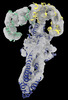
 Map data
 Sample
 Keywords
 Protein complex /
  Function and homology information
 Authors
 Germany,
  France, European Union,
  United Kingdom, 9 items
  Citation


 Structure visualization
 Downloads & links
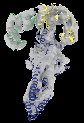 emd_18149.png
 http://ftp.pdbj.org/pub/emdb/structures/EMD-18149


 Z
Z Y
Y X
X


 Sample components
 Processing
 Electron microscopy
