 Movie
Movie Controller


+ Open data

- Basic information
| Entry | 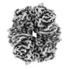 | |||||||||||||||||||||
|---|---|---|---|---|---|---|---|---|---|---|---|---|---|---|---|---|---|---|---|---|---|---|
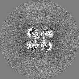
| Title | Structure of GAPDH determined by cryoEM at 100 keV | |||||||||||||||||||||
 Map data Map data | ||||||||||||||||||||||
 Sample Sample |
| |||||||||||||||||||||
 Keywords Keywords |  GAPDH / OXIDOREDUCTASE GAPDH / OXIDOREDUCTASE | |||||||||||||||||||||
| Function / homology |  Function and homology informationglyceraldehyde-3-phosphate dehydrogenase (phosphorylating) / glyceraldehyde-3-phosphate dehydrogenase (NAD+) (phosphorylating) activity / glycolytic process / glucose metabolic process / NAD binding / NADP binding / identical protein binding / metal ion binding Function and homology informationglyceraldehyde-3-phosphate dehydrogenase (phosphorylating) / glyceraldehyde-3-phosphate dehydrogenase (NAD+) (phosphorylating) activity / glycolytic process / glucose metabolic process / NAD binding / NADP binding / identical protein binding / metal ion bindingSimilarity search - Function | |||||||||||||||||||||
| Biological species |   Cryptosporidium parvum (eukaryote) Cryptosporidium parvum (eukaryote) | |||||||||||||||||||||
| Method | single particle reconstruction / cryo EM / Resolution: 2.9 Å | |||||||||||||||||||||
 Authors Authors | McMullan G / Naydenova K / Mihaylov D / Peet MJ / Wilson H / Yamashita K / Dickerson JL / Chen S / Cannone G / Lee Y ...McMullan G / Naydenova K / Mihaylov D / Peet MJ / Wilson H / Yamashita K / Dickerson JL / Chen S / Cannone G / Lee Y / Hutchings KA / Gittins O / Sobhy M / Wells T / El-Gomati MM / Dalby J / Meffert M / Schulze-Briese C / Henderson R / Russo CJ | |||||||||||||||||||||
| Funding support |  United Kingdom, 6 items United Kingdom, 6 items
| |||||||||||||||||||||
 Citation Citation | Journal: Proc Natl Acad Sci U S A / Year: 2023 Title: Structure determination by cryoEM at 100 keV. Authors: Greg McMullan / Katerina Naydenova / Daniel Mihaylov / Keitaro Yamashita / Mathew J Peet / Hugh Wilson / Joshua L Dickerson / Shaoxia Chen / Giuseppe Cannone / Yang Lee / Katherine A ...Authors: Greg McMullan / Katerina Naydenova / Daniel Mihaylov / Keitaro Yamashita / Mathew J Peet / Hugh Wilson / Joshua L Dickerson / Shaoxia Chen / Giuseppe Cannone / Yang Lee / Katherine A Hutchings / Olivia Gittins / Mohamed A Sobhy / Torquil Wells / Mohamed M El-Gomati / Jason Dalby / Matthias Meffert / Clemens Schulze-Briese / Richard Henderson / Christopher J Russo /   Abstract: Electron cryomicroscopy can, in principle, determine the structures of most biological molecules but is currently limited by access, specimen preparation difficulties, and cost. We describe a purpose- ...Electron cryomicroscopy can, in principle, determine the structures of most biological molecules but is currently limited by access, specimen preparation difficulties, and cost. We describe a purpose-built instrument operating at 100 keV-including advances in electron optics, detection, and processing-that makes structure determination fast and simple at a fraction of current costs. The instrument attains its theoretical performance limits, allowing atomic resolution imaging of gold test specimens and biological molecular structure determination in hours. We demonstrate its capabilities by determining the structures of eleven different specimens, ranging in size from 140 kDa to 2 MDa, using a fraction of the data normally required. CryoEM with a microscope designed specifically for high-efficiency, on-the-spot imaging of biological molecules will expand structural biology to a wide range of previously intractable problems. | |||||||||||||||||||||
| History |
|
- Structure visualization
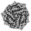
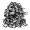
| Supplemental images |
|---|

- Downloads & links
-EMDB archive
| Map data | emd_17964.map.gz | 6 MB | EMDB map data format | |
|---|---|---|---|---|
| Header (meta data) | emd-17964-v30.xmlemd-17964.xml | 17.9 KB 17.9 KB | Display Display | EMDB header |
| FSC (resolution estimation) | emd_17964_fsc.xml | 9.1 KB | Display | FSC data file |
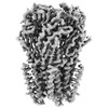
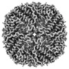
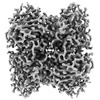
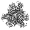
| Images |  emd_17964.png emd_17964.png | 91.9 KB | ||
| Masks | emd_17964_msk_1.map | 64 MB | Mask map | |
| Filedesc metadata | emd-17964.cif.gz | 6.2 KB | ||
| Others | emd_17964_half_map_1.map.gzemd_17964_half_map_2.map.gz | 48.5 MB 48.5 MB | ||
| Archive directory |  http://ftp.pdbj.org/pub/emdb/structures/EMD-17964ftp://ftp.pdbj.org/pub/emdb/structures/EMD-17964 http://ftp.pdbj.org/pub/emdb/structures/EMD-17964ftp://ftp.pdbj.org/pub/emdb/structures/EMD-17964 | HTTPS FTP |
-Related structure data
| Related structure data | 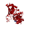 8pvfMC 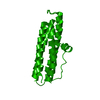 8pv9C  8pvaC 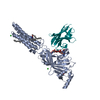 8pvbC 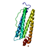 8pvcC 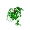 8pvdC 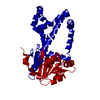 8pveC 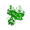 8pvgC 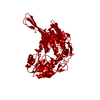 8pvhC  8pviC 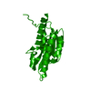 8pvjC M: atomic model generated by this map C: citing same article ( |
|---|---|
| Similar structure data |

-Links
| EMDB pages | EMDB (EBI/PDBe) / EMDataResource |
|---|---|
| Related items in Molecule of the Month |

-Map
| File | Download / File: emd_17964.map.gz / Format: CCP4 / Size: 64 MB / Type: IMAGE STORED AS FLOATING POINT NUMBER (4 BYTES) | ||||||||||||||||||||
|---|---|---|---|---|---|---|---|---|---|---|---|---|---|---|---|---|---|---|---|---|---|
| Voxel size | X=Y=Z: 0.8415 Å | ||||||||||||||||||||
| Density |
| ||||||||||||||||||||
| Symmetry | Space group: 1 | ||||||||||||||||||||
| Details | EMDB XML:
|
-Supplemental data
-Mask #1
| File | emd_17964_msk_1.map | ||||||||||||
|---|---|---|---|---|---|---|---|---|---|---|---|---|---|
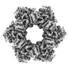
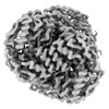
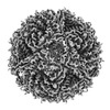
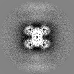
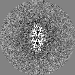
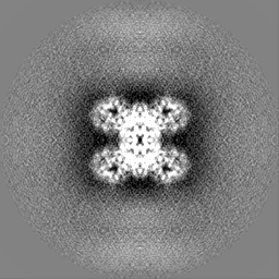
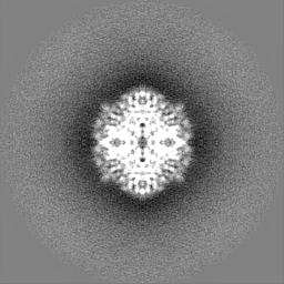
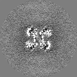
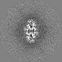
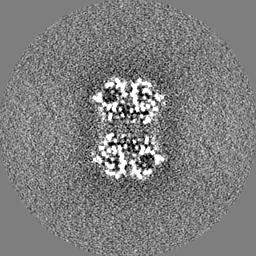
| Projections & Slices |
| ||||||||||||
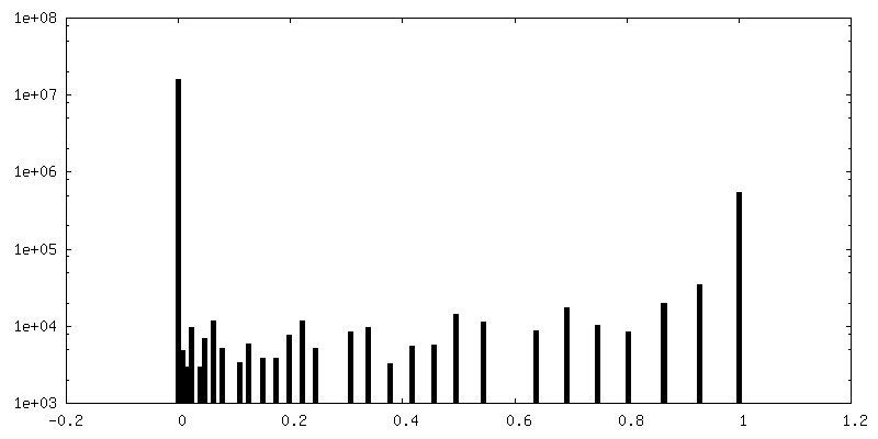
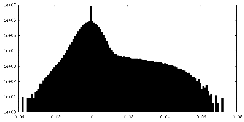
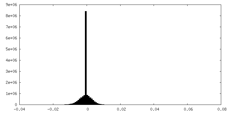
| Density Histograms |
 Z
Z Y
Y X
X


-Half map: #1
| File | emd_17964_half_map_1.map | ||||||||||||
|---|---|---|---|---|---|---|---|---|---|---|---|---|---|
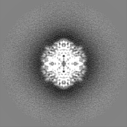
| Projections & Slices |
| ||||||||||||
| Density Histograms |


-Half map: #2
| File | emd_17964_half_map_2.map | ||||||||||||
|---|---|---|---|---|---|---|---|---|---|---|---|---|---|
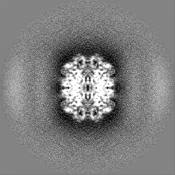
| Projections & Slices |
| ||||||||||||
| Density Histograms |


- Sample components
-Entire : Glyceraldehyde 3-phosphate dehydrogenase
| Entire | Name: Glyceraldehyde 3-phosphate dehydrogenase |
|---|---|
| Components |
|
-Supramolecule #1: Glyceraldehyde 3-phosphate dehydrogenase
| Supramolecule | Name: Glyceraldehyde 3-phosphate dehydrogenase / type: complex / ID: 1 / Parent: 0 / Macromolecule list: #1 |
|---|---|
| Source (natural) | Organism: Cryptosporidium parvum (eukaryote) |
-Macromolecule #1: Glyceraldehyde-3-phosphate dehydrogenase
| Macromolecule | Name: Glyceraldehyde-3-phosphate dehydrogenase / type: protein_or_peptide / ID: 1 / Number of copies: 1 / Enantiomer: LEVO EC number: glyceraldehyde-3-phosphate dehydrogenase (phosphorylating) |
|---|---|
| Source (natural) | Organism: Cryptosporidium parvum (eukaryote) |
| Molecular weight | Theoretical: 38.059484 KDa |
| Recombinant expression | Organism:  Escherichia coli (E. coli) Escherichia coli (E. coli) |
| Sequence | String: MHHHHHHSSG RENLYFQGTL GINGFGRIGR LVLRACMERN DITVVAINDP FMDVEYMAYL LKYDSVHGNF NGTVEVSGKD LCINGKVVK VFQAKDPAEI PWGASGAQIV CESTGVFTTE EKASLHLKGG AKKVIISAPP KDNVPMYVMG VNNTEYDPSK F NVISNASC ...String: MHHHHHHSSG RENLYFQGTL GINGFGRIGR LVLRACMERN DITVVAINDP FMDVEYMAYL LKYDSVHGNF NGTVEVSGKD LCINGKVVK VFQAKDPAEI PWGASGAQIV CESTGVFTTE EKASLHLKGG AKKVIISAPP KDNVPMYVMG VNNTEYDPSK F NVISNASC TTNCLAPLAK IINDKFGIVE GLMTTVHSLT ANQLTVDGPS KGGKDWRAGR CAGNNIIPAS TGAAKAVGKV IP ALNGKLT GMAIRVPTPD VSVVDLTCKL AKPASIEEIY QAVKEASNGP MKGIMGYTSD DVVSTDFIGC KYSSIFDKNA CIA LNDSFV KLISWYDNES GYSNRLVDLA VYVASRGL UniProtKB: Glyceraldehyde-3-phosphate dehydrogenase |
-Macromolecule #2: NICOTINAMIDE-ADENINE-DINUCLEOTIDE
| Macromolecule | Name: NICOTINAMIDE-ADENINE-DINUCLEOTIDE / type: ligand / ID: 2 / Number of copies: 1 / Formula: NAD |
|---|---|
| Molecular weight | Theoretical: 663.425 Da |
| Chemical component information |  ChemComp-NAD: |
-Experimental details
-Structure determination
| Method | cryo EM |
|---|---|
 Processing Processing | single particle reconstruction |
| Aggregation state | particle |
-Sample preparation
| Buffer | pH: 7.4 |
|---|---|
| Grid | Model: UltrAuFoil R0./1 / Material: GOLD |
| Vitrification | Cryogen name: ETHANE |
- Electron microscopy
| Microscope | JEOL 1400/HR + YPS FEG |
|---|---|
| Electron beam | Acceleration voltage: 100 kV / Electron source: FIELD EMISSION GUN |
| Electron optics | Illumination mode: FLOOD BEAM / Imaging mode: BRIGHT FIELDBright-field microscopy / Nominal defocus max: 2.0 µm / Nominal defocus min: 0.5 µm |
| Sample stage | Specimen holder model: GATAN 626 SINGLE TILT LIQUID NITROGEN CRYO TRANSFER HOLDER Cooling holder cryogen: NITROGEN |
| Image recording | Film or detector model: DECTRIS SINGLA (1k x 1k) / Average electron dose: 40.0 e/Å2 |
-Image processing
| Startup model | Type of model: EMDB MAP EMDB ID: |
|---|---|
| Initial angle assignment | Type: MAXIMUM LIKELIHOOD |
| Final angle assignment | Type: MAXIMUM LIKELIHOOD |
| Final reconstruction | Applied symmetry - Point group: D2 (2x2 fold dihedral) / Resolution.type: BY AUTHOR / Resolution: 2.9 Å / Resolution method: FSC 0.143 CUT-OFF / Number images used: 19411 |
| FSC plot (resolution estimation) | 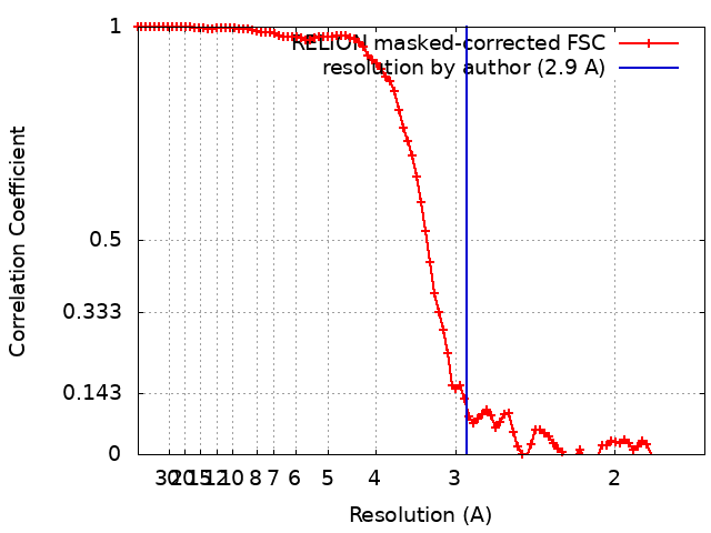 |


