[English] 日本語
Yorodumi
- EMDB-15560: membrane region of the Trypanosoma brucei mitochondrial ATP synth... -

+
Open data


ID or keywords:

Loading...

-
Basic information

Entry
Database: EMDB / ID: EMD-15560
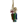
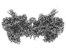
Titlemembrane region of the Trypanosoma brucei mitochondrial ATP synthase dimer
Map data
Sample
  • Complex: mitochondrial ATP synthase dimer from Trypanosoma brucei
    • Protein or peptide: x 15 types
  • Ligand: x 6 types
Function / homology
Function and homology information


kinetoplast / ATP biosynthetic process / nuclear lumen / ciliary plasm / mitochondrial proton-transporting ATP synthase complex / proton-transporting ATP synthase complex, coupling factor F(o) / proton transmembrane transport / mitochondrial membrane / mitochondrial inner membrane / mitochondrion ...kinetoplast / ATP biosynthetic process / nuclear lumen / ciliary plasm / mitochondrial proton-transporting ATP synthase complex / proton-transporting ATP synthase complex, coupling factor F(o) / proton transmembrane transport / mitochondrial membrane / mitochondrial inner membrane / mitochondrion / membrane / cytoplasm
Similarity search - Function
T. brucei spp.-specific protein / Uncharacterized protein / Transmembrane protein / ATP synthase subunit a / Uncharacterized protein / Letm1 RBD domain-containing protein / Uncharacterized protein / Uncharacterized protein / Uncharacterized protein / Uncharacterized protein ...T. brucei spp.-specific protein / Uncharacterized protein / Transmembrane protein / ATP synthase subunit a / Uncharacterized protein / Letm1 RBD domain-containing protein / Uncharacterized protein / Uncharacterized protein / Uncharacterized protein / Uncharacterized protein / Uncharacterized protein / Uncharacterized protein / Uncharacterized protein / Uncharacterized protein
Similarity search - Component
Biological speciesTrypanosoma brucei brucei (eukaryote)
Methodsingle particle reconstruction / cryo EM / Resolution: 2.7 Å
AuthorsMuehleip A / Gahura O / Zikova A / Amunts A
Funding supportEuropean Union, 1 items
OrganizationGrant numberCountry
European Research Council (ERC)European Union
CitationJournal: Nat Commun / Year: 2022
Title: An ancestral interaction module promotes oligomerization in divergent mitochondrial ATP synthases.
Authors: Ondřej Gahura / Alexander Mühleip / Carolina Hierro-Yap / Brian Panicucci / Minal Jain / David Hollaus / Martina Slapničková / Alena Zíková / Alexey Amunts /
Abstract: Mitochondrial ATP synthase forms stable dimers arranged into oligomeric assemblies that generate the inner-membrane curvature essential for efficient energy conversion. Here, we report cryo-EM ...Mitochondrial ATP synthase forms stable dimers arranged into oligomeric assemblies that generate the inner-membrane curvature essential for efficient energy conversion. Here, we report cryo-EM structures of the intact ATP synthase dimer from Trypanosoma brucei in ten different rotational states. The model consists of 25 subunits, including nine lineage-specific, as well as 36 lipids. The rotary mechanism is influenced by the divergent peripheral stalk, conferring a greater conformational flexibility. Proton transfer in the lumenal half-channel occurs via a chain of five ordered water molecules. The dimerization interface is formed by subunit-g that is critical for interactions but not for the catalytic activity. Although overall dimer architecture varies among eukaryotes, we find that subunit-g together with subunit-e form an ancestral oligomerization motif, which is shared between the trypanosomal and mammalian lineages. Therefore, our data defines the subunit-g/e module as a structural component determining ATP synthase oligomeric assemblies.
History
DepositionAug 9, 2022-
Header (metadata) releaseOct 26, 2022-
Map releaseOct 26, 2022-
UpdateOct 26, 2022-
Current statusOct 26, 2022Processing site: PDBe / Status: Released

-
Structure visualization

Supplemental images

Downloads & links

-
Map

FileDownload / File: emd_15560.map.gz / Format: CCP4 / Size: 669.9 MB / Type: IMAGE STORED AS FLOATING POINT NUMBER (4 BYTES)
Voxel sizeX=Y=Z: 0.83 Å
Density
Contour LevelBy AUTHOR: 0.04
Minimum - Maximum-0.10553464 - 0.20181364
Average (Standard dev.)0.00074083573 (±0.0033686697)
SymmetrySpace group: 1
Details

EMDB XML:

Map geometry
Axis orderXYZ
Origin000
Dimensions560560560
Spacing560560560
CellA=B=C: 464.8 Å
α=β=γ: 90.0 °

-
Supplemental data

-
Mask #1

Fileemd_15560_msk_1.map
Projections & Slices
AxesZYX

Projections

Slices (1/2)
Density Histograms

-
Half map: #1

Fileemd_15560_half_map_1.map
Projections & Slices
AxesZYX

Projections

Slices (1/2)
Density Histograms

-
Half map: #2

Fileemd_15560_half_map_2.map
Projections & Slices
AxesZYX

Projections

Slices (1/2)
Density Histograms

-
Sample components

+
Entire : mitochondrial ATP synthase dimer from Trypanosoma brucei

EntireName: mitochondrial ATP synthase dimer from Trypanosoma brucei
Components
  • Complex: mitochondrial ATP synthase dimer from Trypanosoma brucei
    • Protein or peptide: ATP synthase subunit a
    • Protein or peptide: subunit-8
    • Protein or peptide: subunit-d
    • Protein or peptide: ATPTB1
    • Protein or peptide: subunit-f
    • Protein or peptide: subunit-i/j
    • Protein or peptide: ATPTB6
    • Protein or peptide: subunit-k
    • Protein or peptide: subunit-e
    • Protein or peptide: subunit-g
    • Protein or peptide: ATPTB11
    • Protein or peptide: ATPTB12
    • Protein or peptide: subunit-b
    • Protein or peptide: ATPEG3
    • Protein or peptide: ATPEG4
  • Ligand: CARDIOLIPIN
  • Ligand: DODECYL-BETA-D-MALTOSIDE
  • Ligand: 2-{[(4-O-alpha-D-glucopyranosyl-alpha-D-glucopyranosyl)oxy]methyl}-4-{[(3beta,9beta,14beta,17beta,25R)-spirost-5-en-3-yl]oxy}butyl 4-O-alpha-D-glucopyranosyl-alpha-D-glucopyranoside
  • Ligand: 1,2-dioleoyl-sn-glycero-3-phosphoethanolamine
  • Ligand: 1,2-DIACYL-SN-GLYCERO-3-PHOSPHOCHOLINE
  • Ligand: water

+
Supramolecule #1: mitochondrial ATP synthase dimer from Trypanosoma brucei

SupramoleculeName: mitochondrial ATP synthase dimer from Trypanosoma brucei
type: complex / Chimera: Yes / ID: 1 / Parent: 0 / Macromolecule list: #1-#15
Source (natural)Organism: Trypanosoma brucei brucei (eukaryote) / Strain: Lister427 / Organelle: Mitochondrion

+
Macromolecule #1: ATP synthase subunit a

MacromoleculeName: ATP synthase subunit a / type: protein_or_peptide / ID: 1 / Number of copies: 2 / Enantiomer: LEVO
Source (natural)Organism: Trypanosoma brucei brucei (eukaryote)
Molecular weightTheoretical: 28.708406 KDa
SequenceString: MFLFFFCDLF WLRLLLCMYY CVWSRLCFIV YFNCLMLIFD FLLFCLFDLY LFVGLCLFLL LWFMLFNLYS LILYYCITYL NLYLLFCIV FLLYIAFLFL FCFLCDFFLF NNLLVGDSFM DVFFIRFLLC FLECFSLLCR CLSTFLRLFC NLLSSHFLLL M FFDFFYFI ...String:
MFLFFFCDLF WLRLLLCMYY CVWSRLCFIV YFNCLMLIFD FLLFCLFDLY LFVGLCLFLL LWFMLFNLYS LILYYCITYL NLYLLFCIV FLLYIAFLFL FCFLCDFFLF NNLLVGDSFM DVFFIRFLLC FLECFSLLCR CLSTFLRLFC NLLSSHFLLL M FFDFFYFI FVFFFYGVFC YWFILFIFVF CFCLLFYVFL YLLDLFAAIL QLFIFCNMIL QLIMDFLLFL LFV

+
Macromolecule #2: subunit-8

MacromoleculeName: subunit-8 / type: protein_or_peptide / ID: 2 / Number of copies: 2 / Enantiomer: LEVO
Source (natural)Organism: Trypanosoma brucei brucei (eukaryote)
Molecular weightTheoretical: 13.750958 KDa
SequenceString:
MLRRLGANVS NMARPMNKYA VTVSPRRHLE PMSTWYLASW AMVWYYAFFF WMPMVWTDIM VPSFVYNKLP VIHFLQEKRA EQKLRRVLD ETYTEWTEEL DQAHVTDAIT RSLNI

+
Macromolecule #3: subunit-d

MacromoleculeName: subunit-d / type: protein_or_peptide / ID: 3 / Number of copies: 2 / Enantiomer: LEVO
Source (natural)Organism: Trypanosoma brucei brucei (eukaryote)
Molecular weightTheoretical: 43.379324 KDa
SequenceString: MRRVSSPNIT IQSVRWISGV SPLLYFPPTT TSTTNREDQI NKNTNIAIQM IKRYKGEVPP HYTRKSSATI EQVEKEIDAL LGGAEKLRK TSTDDQPMDK LTLMERCLRH ALWSYHKEEG RYDFDQIGRW VVYTPEDEVK LAQLKREVEA KEKLAALRKR R EEEGLPGG ...String:
MRRVSSPNIT IQSVRWISGV SPLLYFPPTT TSTTNREDQI NKNTNIAIQM IKRYKGEVPP HYTRKSSATI EQVEKEIDAL LGGAEKLRK TSTDDQPMDK LTLMERCLRH ALWSYHKEEG RYDFDQIGRW VVYTPEDEVK LAQLKREVEA KEKLAALRKR R EEEGLPGG PVPRINWPQE YSSFIDREPV VAKRIRYDTL ASTTLERDEK QIESTLQQYR RASQDKRLDD LVDLLERFKP VL AREAIMQ RLTIKHLEGQ LGVWRYMDWC PEVRDRAELE VDITGWQWWS PLEERRLLPV RLRSVNEVRE IMSKTQAKKS AEA AERNPI VTQTSTGDNA RDRLLKEVLA LQARINQRDE VEPSQTEQKK KAHH

+
Macromolecule #4: ATPTB1

MacromoleculeName: ATPTB1 / type: protein_or_peptide / ID: 4 / Number of copies: 2 / Enantiomer: LEVO
Source (natural)Organism: Trypanosoma brucei brucei (eukaryote) / Strain: 927/4 GUTat10.1
Molecular weightTheoretical: 46.883637 KDa
SequenceString: (AME)QGSWSVLKK NCSNFFPGLL AFAQQTQEAY GIWLRIYNRQ QKYGPTDFVE QSETFSPDYH KRFHSQDKNM WVDKEL CTE VSQKEVARLM TYKLDMWRMA HCAGALLATG GYAIPFGLFW LANDTWVPSS FNLTGEELRA WREAQDLYRY RSAPSYL TD TKWHFDFHAY ...String:
(AME)QGSWSVLKK NCSNFFPGLL AFAQQTQEAY GIWLRIYNRQ QKYGPTDFVE QSETFSPDYH KRFHSQDKNM WVDKEL CTE VSQKEVARLM TYKLDMWRMA HCAGALLATG GYAIPFGLFW LANDTWVPSS FNLTGEELRA WREAQDLYRY RSAPSYL TD TKWHFDFHAY PWNETQERAW DDLFEKNDVR RDPKVVRPAA EMYDGFIKFE LIRRKSLRHL CRSMNIPTFP MLARLCNG T RVRDYWNLAW CEDYMVITQR LHESMTDEEL YDYAWRRYLA PYDKNLNREQ LMERVEDYFE FLGPDFVAHG KAPNLVILT NYVLGYYNDP AYLEGDISEL DKNDYDHLAS WGKDAFLRRL EFENGPLRDQ VEAHTQRLLA ERAAIAKGDN AAAVEGRHTA

+
Macromolecule #5: subunit-f

MacromoleculeName: subunit-f / type: protein_or_peptide / ID: 5 / Number of copies: 2 / Enantiomer: LEVO
Source (natural)Organism: Trypanosoma brucei brucei (eukaryote) / Strain: 927/4 GUTat10.1
Molecular weightTheoretical: 17.182668 KDa
SequenceString:
MVLFSTYRSS RLVSKEFLHG PVMRFRALGE YYFQRAWNGT LNWALPGEYR LYAVMIPFIY FYHRWHNDHT LDRDHVEKAM IMRWGGTLE DVRKLSAKDQ LRVRCFTDIE KLYSAYGPKD TYLQPPGDTL PGKDFYRKAG GAQAHH

+
Macromolecule #6: subunit-i/j

MacromoleculeName: subunit-i/j / type: protein_or_peptide / ID: 6 / Number of copies: 2 / Enantiomer: LEVO
Source (natural)Organism: Trypanosoma brucei brucei (eukaryote) / Strain: 927/4 GUTat10.1
Molecular weightTheoretical: 12.661607 KDa
SequenceString:
MVYTRWKCDR LPVFQLKLFT QEYPMHAAVG IFTIIFLWKH MSHCSEETER KYGWWAGYPY WRDPIARRNE TKYKQMIINN DVDITHPKW TGCSVEQLEE LSRVV

+
Macromolecule #7: ATPTB6

MacromoleculeName: ATPTB6 / type: protein_or_peptide / ID: 7 / Number of copies: 2 / Enantiomer: LEVO
Source (natural)Organism: Trypanosoma brucei brucei (eukaryote) / Strain: MHOM/CI/86/DAL972
Molecular weightTheoretical: 20.307389 KDa
SequenceString:
MTKYELKMQY FDEWMIRWRK FQTESDWEIE KGRQWWRRFN MAVSGALFCG LVLYTSGTAT LKRQYGLPHF FDIGVDGQAK ETMLKTLTS RWRYTPQGYG RVLITGVPTY ILFVTLEHYR ERRRMQQYLQ QNTVFGEQMR RLLSTGKIEE YLPVNIKATL P ASQQAIYN Y

+
Macromolecule #8: subunit-k

MacromoleculeName: subunit-k / type: protein_or_peptide / ID: 8 / Number of copies: 2 / Enantiomer: LEVO
Source (natural)Organism: Trypanosoma brucei brucei (eukaryote) / Strain: 927/4 GUTat10.1
Molecular weightTheoretical: 14.531121 KDa
SequenceString:
MLRRSSAALI RRTPVRHSGG ELFVRPKLEE IPPADQCRGF FGPLNDSLKF LRLLDIKWMM NRAVAMRREY LIATPTLFTF IWMFTWKGA VIYFWGDRAP PRRMDWNTEE TGRLPLGFKP TPAPL

+
Macromolecule #9: subunit-e

MacromoleculeName: subunit-e / type: protein_or_peptide / ID: 9 / Number of copies: 2 / Enantiomer: LEVO
Source (natural)Organism: Trypanosoma brucei brucei (eukaryote) / Strain: 927/4 GUTat10.1
Molecular weightTheoretical: 10.448932 KDa
SequenceString:
MSAKAAPKTL HQVRNVAYFF AAWLGVQKGY IEKSANDRLW VEHQRKVRQQ NVERQQALDS IKLMQQGVRA TTPGQLEGVP AELQQLAEA FTK

+
Macromolecule #10: subunit-g

MacromoleculeName: subunit-g / type: protein_or_peptide / ID: 10 / Number of copies: 2 / Enantiomer: LEVO
Source (natural)Organism: Trypanosoma brucei brucei (eukaryote) / Strain: MHOM/CI/86/DAL972
Molecular weightTheoretical: 16.092668 KDa
SequenceString:
MSSTKCAVAC KIMTPLCNAA SKVQARSAKK LAALTDAGIQ KTISEHNANG TDAAVSSTKR YLAEQRQLFH YRVVRFFDEC HYIISGEYF AQYTKVNLIW DLRFLTKLVV LFLIGTVLGR QSIFPPIDPD SPLVEALVTK VNPNY

+
Macromolecule #11: ATPTB11

MacromoleculeName: ATPTB11 / type: protein_or_peptide / ID: 11 / Number of copies: 2 / Enantiomer: LEVO
Source (natural)Organism: Trypanosoma brucei brucei (eukaryote) / Strain: 927/4 GUTat10.1
Molecular weightTheoretical: 17.929576 KDa
SequenceString:
MLRKTPLFAM ATTRKALVGN GPTFSTGGEC MNTCDIQNAF PMNDRGVRSS SPFQEPNTAI YDSYLAWTYF QPMDVHIEKL PAPEAKYYQ RHTKKPWDVS STELTEIQSR KKYFQTLGYL VAFIYLYFLM PKEKSFSGLS GPDGHWIMLP KGRPELF

+
Macromolecule #12: ATPTB12

MacromoleculeName: ATPTB12 / type: protein_or_peptide / ID: 12 / Number of copies: 2 / Enantiomer: LEVO
Source (natural)Organism: Trypanosoma brucei brucei (eukaryote) / Strain: 927/4 GUTat10.1
Molecular weightTheoretical: 11.676294 KDa
SequenceString:
MSSGFHFHDV SNDAIKGMPP SEALHKHLEN AQLAHRICLA KALKAGEPPV EKCALTWGEV LIRYQAWSEY RPPFQDSVAQ AKYKKYWSK KRQEEDDKNP FK

+
Macromolecule #13: subunit-b

MacromoleculeName: subunit-b / type: protein_or_peptide / ID: 13 / Number of copies: 2 / Enantiomer: LEVO
Source (natural)Organism: Trypanosoma brucei brucei (eukaryote) / Strain: MHOM/CI/86/DAL972
Molecular weightTheoretical: 12.325279 KDa
SequenceString:
MLRRLVPRVM MAPMGGATAL CTSRGYNMLV FRDPKRRPQL SEEERAKVVV NQAEWPEEFK DFDPDDPYKN SPEIIKGMSS WNLFLWGVE CAFIYQFYEL VFPKSI

+
Macromolecule #14: ATPEG3

MacromoleculeName: ATPEG3 / type: protein_or_peptide / ID: 14 / Number of copies: 2 / Enantiomer: LEVO
Source (natural)Organism: Trypanosoma brucei brucei (eukaryote) / Strain: 927/4 GUTat10.1
Molecular weightTheoretical: 12.293796 KDa
SequenceString:
MTENIEAVMS DFWSNPADHF RPNLKALTLY AERQHYVDRW LHVKERWLAP WYLPWWSPLF QLGTWYSQRS RNLFLVENHL SYRPYKFRR NDEDRNNPY

+
Macromolecule #15: ATPEG4

MacromoleculeName: ATPEG4 / type: protein_or_peptide / ID: 15 / Number of copies: 2 / Enantiomer: LEVO
Source (natural)Organism: Trypanosoma brucei brucei (eukaryote) / Strain: Lister427
Molecular weightTheoretical: 7.649776 KDa
SequenceString:
MLLGGFVPRR FSQFNRDPCW MFFIFSVGFW LGEYPAMMIK YNARDLVYDP HRYVWSHHDD HH

+
Macromolecule #16: CARDIOLIPIN

MacromoleculeName: CARDIOLIPIN / type: ligand / ID: 16 / Number of copies: 24 / Formula: CDL
Molecular weightTheoretical: 1.464043 KDa
Chemical component information

ChemComp-CDL:
CARDIOLIPIN / phospholipid*YM / Cardiolipin

+
Macromolecule #17: DODECYL-BETA-D-MALTOSIDE

MacromoleculeName: DODECYL-BETA-D-MALTOSIDE / type: ligand / ID: 17 / Number of copies: 4 / Formula: LMT
Molecular weightTheoretical: 510.615 Da
Chemical component information

ChemComp-LMT:
DODECYL-BETA-D-MALTOSIDE / detergent*YM

+
Macromolecule #18: 2-{[(4-O-alpha-D-glucopyranosyl-alpha-D-glucopyranosyl)oxy]methyl...

MacromoleculeName: 2-{[(4-O-alpha-D-glucopyranosyl-alpha-D-glucopyranosyl)oxy]methyl}-4-{[(3beta,9beta,14beta,17beta,25R)-spirost-5-en-3-yl]oxy}butyl 4-O-alpha-D-glucopyranosyl-alpha-D-glucopyranoside
type: ligand / ID: 18 / Number of copies: 4 / Formula: Q7G
Molecular weightTheoretical: 1.165315 KDa
Chemical component information

ChemComp-Q7G:
2-{[(4-O-alpha-D-glucopyranosyl-alpha-D-glucopyranosyl)oxy]methyl}-4-{[(3beta,9beta,14beta,17beta,25R)-spirost-5-en-3-yl]oxy}butyl 4-O-alpha-D-glucopyranosyl-alpha-D-glucopyranoside

+
Macromolecule #19: 1,2-dioleoyl-sn-glycero-3-phosphoethanolamine

MacromoleculeName: 1,2-dioleoyl-sn-glycero-3-phosphoethanolamine / type: ligand / ID: 19 / Number of copies: 4 / Formula: PEE
Molecular weightTheoretical: 744.034 Da
Chemical component information

ChemComp-PEE:
1,2-dioleoyl-sn-glycero-3-phosphoethanolamine / DOPE, phospholipid*YM / Discrete optimized protein energy

+
Macromolecule #20: 1,2-DIACYL-SN-GLYCERO-3-PHOSPHOCHOLINE

MacromoleculeName: 1,2-DIACYL-SN-GLYCERO-3-PHOSPHOCHOLINE / type: ligand / ID: 20 / Number of copies: 8 / Formula: PC1
Molecular weightTheoretical: 790.145 Da
Chemical component information

ChemComp-PC1:
1,2-DIACYL-SN-GLYCERO-3-PHOSPHOCHOLINE / phospholipid*YM / Phosphatidylcholine

+
Macromolecule #21: water

MacromoleculeName: water / type: ligand / ID: 21 / Number of copies: 24 / Formula: HOH
Molecular weightTheoretical: 18.015 Da
Chemical component information

ChemComp-HOH:
WATER / Water

-
Experimental details

-
Structure determination

Methodcryo EM
Processingsingle particle reconstruction
Aggregation stateparticle

-
Sample preparation

BufferpH: 8
GridModel: Quantifoil R1.2/1.3 / Material: GOLD / Mesh: 300
VitrificationCryogen name: ETHANE / Chamber humidity: 100 %

-
Electron microscopy

MicroscopeFEI TITAN KRIOS
Electron beamAcceleration voltage: 300 kV / Electron source: FIELD EMISSION GUN
Electron opticsC2 aperture diameter: 70.0 µm / Illumination mode: FLOOD BEAM / Imaging mode: BRIGHT FIELDBright-field microscopy / Cs: 2.7 mm / Nominal defocus max: 3.2 µm / Nominal defocus min: 1.6 µm
Sample stageSpecimen holder model: FEI TITAN KRIOS AUTOGRID HOLDER / Cooling holder cryogen: NITROGEN
TemperatureMin: 70.0 K / Max: 70.0 K
Image recordingFilm or detector model: GATAN K2 QUANTUM (4k x 4k) / Detector mode: COUNTING / Average electron dose: 33.0 e/Å2
Experimental equipment
Model: Titan Krios / Image courtesy: FEI Company

-
Image processing

Initial angle assignmentType: MAXIMUM LIKELIHOOD
Final angle assignmentType: MAXIMUM LIKELIHOOD
Final reconstructionApplied symmetry - Point group: C2 (2 fold cyclic) / Resolution.type: BY AUTHOR / Resolution: 2.7 Å / Resolution method: FSC 0.143 CUT-OFF / Number images used: 100605
FSC plot (resolution estimation)

+
About Yorodumi

-
News

-
Feb 9, 2022. New format data for meta-information of EMDB entries

New format data for meta-information of EMDB entries

  • Version 3 of the EMDB header file is now the official format.
  • The previous official version 1.9 will be removed from the archive.

Related info.:EMDB header

External links:wwPDB to switch to version 3 of the EMDB data model

-
Aug 12, 2020. Covid-19 info

Covid-19 info

URL: https://pdbj.org/emnavi/covid19.php

New page: Covid-19 featured information page in EM Navigator.

Related info.:Covid-19 info / Mar 5, 2020. Novel coronavirus structure data

+
Mar 5, 2020. Novel coronavirus structure data

Novel coronavirus structure data

Related info.:Yorodumi Speices / Aug 12, 2020. Covid-19 info

External links:COVID-19 featured content - PDBj / Molecule of the Month (242):Coronavirus Proteases

+
Jan 31, 2019. EMDB accession codes are about to change! (news from PDBe EMDB page)

EMDB accession codes are about to change! (news from PDBe EMDB page)

  • The allocation of 4 digits for EMDB accession codes will soon come to an end. Whilst these codes will remain in use, new EMDB accession codes will include an additional digit and will expand incrementally as the available range of codes is exhausted. The current 4-digit format prefixed with “EMD-” (i.e. EMD-XXXX) will advance to a 5-digit format (i.e. EMD-XXXXX), and so on. It is currently estimated that the 4-digit codes will be depleted around Spring 2019, at which point the 5-digit format will come into force.
  • The EM Navigator/Yorodumi systems omit the EMD- prefix.

Related info.:Q: What is EMD? / ID/Accession-code notation in Yorodumi/EM Navigator

External links:EMDB Accession Codes are Changing Soon! / Contact to PDBj

+
Jul 12, 2017. Major update of PDB

Major update of PDB

  • wwPDB released updated PDB data conforming to the new PDBx/mmCIF dictionary.
  • This is a major update changing the version number from 4 to 5, and with Remediation, in which all the entries are updated.
  • In this update, many items about electron microscopy experimental information are reorganized (e.g. em_software).
  • Now, EM Navigator and Yorodumi are based on the updated data.

External links:wwPDB Remediation / Enriched Model Files Conforming to OneDep Data Standards Now Available in the PDB FTP Archive

-
Yorodumi

Thousand views of thousand structures

  • Yorodumi is a browser for structure data from EMDB, PDB, SASBDB, etc.
  • This page is also the successor to EM Navigator detail page, and also detail information page/front-end page for Omokage search.
  • The word "yorodu" (or yorozu) is an old Japanese word meaning "ten thousand". "mi" (miru) is to see.

Related info.:EMDB / PDB / SASBDB / Comparison of 3 databanks / Yorodumi Search / Aug 31, 2016. New EM Navigator & Yorodumi / Yorodumi Papers / Jmol/JSmol / Function and homology information / Changes in new EM Navigator and Yorodumi

Read more