 Movie
Movie Controller

[English] 日本語
 Yorodumi
- EMDB-1525: Structure of Thermusphage P23-77 using electron cryomicroscopy an... -
+ Open data

- Basic information
| Entry | Database: EMDB / ID: EMD-1525 | |||||||||
|---|---|---|---|---|---|---|---|---|---|---|
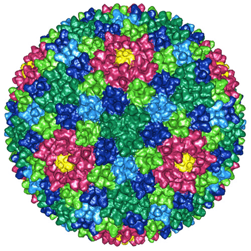
| Title | Structure of Thermusphage P23-77 using electron cryomicroscopy and three-dimensional image reconstruction | |||||||||
 Map data Map data | This is a 14 A resolution cryo-EM reconstruction of Thermusphage P23-77 | |||||||||
 Sample Sample |
| |||||||||
 Keywords Keywords | Icosahedral thermophilic bacterial virus | |||||||||
| Biological species |   Thermus phage P23-77 (virus) Thermus phage P23-77 (virus) | |||||||||
| Method |  single particle reconstruction / cryo EM / negative staining / Resolution: 14.0 Å single particle reconstruction / cryo EM / negative staining / Resolution: 14.0 Å | |||||||||
 Authors Authors | Jaatinen ST / Happonen LJ / Laurinmaki P / Butcher SJ / Bamford DH | |||||||||
 Citation Citation | Journal: Virology / Year: 2008 Title: Biochemical and structural characterisation of membrane-containing icosahedral dsDNA bacteriophages infecting thermophilic Thermus thermophilus. Authors: S T Jaatinen / L J Happonen / P Laurinmäki / S J Butcher / D H Bamford /  Abstract: Icosahedral dsDNA viruses isolated from hot springs and proposed to belong to the Tectiviridae family infect the gram-negative thermophilic Thermus thermophilus bacterium. Seven such viruses were ...Icosahedral dsDNA viruses isolated from hot springs and proposed to belong to the Tectiviridae family infect the gram-negative thermophilic Thermus thermophilus bacterium. Seven such viruses were obtained from the Promega Corporation collection. The structural protein patterns of three of these viruses, growing to a high titer, appeared very similar but not identical. The most stable virus, P23-77, was chosen for more detailed studies. Analysis of highly purified P23-77 by thin layer chromatography for neutral lipids showed lipid association with the virion. Cryo-EM based three-dimensional image reconstruction of P23-77 to 1.4 nm resolution revealed an icosahedrally-ordered protein coat, with spikes on the vertices, and an internal membrane. The capsid architecture of P23-77 is most similar to that of the archaeal virus SH1. These findings further complicate the grouping of icosahedrally-symmetric viruses containing an inner membrane. We propose a single superfamily or order with members in several viral families. | |||||||||
| History |
|
- Structure visualization
| Movie |
 Movie viewer Movie viewer |
|---|---|
| Structure viewer | EM map: SurfViewMolmilJmol/JSmol |
| Supplemental images |

- Downloads & links
-EMDB archive
| Map data | emd_1525.map.gz | 60.2 MB | EMDB map data format | |
|---|---|---|---|---|
| Header (meta data) | emd-1525-v30.xmlemd-1525.xml | 9.6 KB 9.6 KB | Display Display | EMDB header |
| Images | 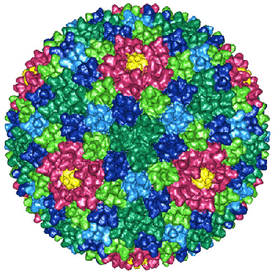 1525.gif 1525.gif emd_1525.gif emd_1525.gif | 108.1 KB 162.2 KB | ||
| Archive directory |  http://ftp.pdbj.org/pub/emdb/structures/EMD-1525ftp://ftp.pdbj.org/pub/emdb/structures/EMD-1525 http://ftp.pdbj.org/pub/emdb/structures/EMD-1525ftp://ftp.pdbj.org/pub/emdb/structures/EMD-1525 | HTTPS FTP |
-Related structure data
| Similar structure data |
|---|


-Links
| EMDB pages | EMDB (EBI/PDBe) / EMDataResource |
|---|
-Map
| File | Download / File: emd_1525.map.gz / Format: CCP4 / Size: 120.1 MB / Type: IMAGE STORED AS SIGNED INTEGER (2 BYTES) | ||||||||||||||||||||||||||||||||||||||||||||||||||||||||||||||||||||
|---|---|---|---|---|---|---|---|---|---|---|---|---|---|---|---|---|---|---|---|---|---|---|---|---|---|---|---|---|---|---|---|---|---|---|---|---|---|---|---|---|---|---|---|---|---|---|---|---|---|---|---|---|---|---|---|---|---|---|---|---|---|---|---|---|---|---|---|---|---|
| Annotation | This is a 14 A resolution cryo-EM reconstruction of Thermusphage P23-77 | ||||||||||||||||||||||||||||||||||||||||||||||||||||||||||||||||||||
| Projections & slices | Image control
Images are generated by Spider. | ||||||||||||||||||||||||||||||||||||||||||||||||||||||||||||||||||||
| Voxel size | X=Y=Z: 2.8 Å | ||||||||||||||||||||||||||||||||||||||||||||||||||||||||||||||||||||
| Density |
| ||||||||||||||||||||||||||||||||||||||||||||||||||||||||||||||||||||
| Symmetry | Space group: 1 | ||||||||||||||||||||||||||||||||||||||||||||||||||||||||||||||||||||
| Details | EMDB XML:
CCP4 map header:
| ||||||||||||||||||||||||||||||||||||||||||||||||||||||||||||||||||||
 Z (Sec.)
 Y (Row.)
 X (Col.)


-Supplemental data
- Sample components
-Entire : Thermusphage P23-77
| Entire | Name: Thermusphage P23-77 |
|---|---|
| Components |
|
-Supramolecule #1000: Thermusphage P23-77
| Supramolecule | Name: Thermusphage P23-77 / type: sample / ID: 1000 Details: The sample was purified on a sucrose gradient, and stored at 28 C prior to plunging. Oligomeric state: 1 / Number unique components: 1 |
|---|
-Supramolecule #1: Thermus phage P23-77
| Supramolecule | Name: Thermus phage P23-77 / type: virus / ID: 1 / Name.synonym: Thermusphage P23-77 / NCBI-ID: 668994 / Sci species name: Thermus phage P23-77 / Virus type: VIRION / Virus isolate: STRAIN / Virus enveloped: Yes / Virus empty: No / Syn species name: Thermusphage P23-77 |
|---|---|
| Host (natural) | Organism:  Thermus thermophilus (bacteria) / synonym: BACTERIA(EUBACTERIA) Thermus thermophilus (bacteria) / synonym: BACTERIA(EUBACTERIA) |
| Virus shell | Shell ID: 1 / Diameter: 780 Å / T number (triangulation number): 28 |
-Experimental details
-Structure determination
| Method | negative staining, cryo EM |
|---|---|
 Processing Processing | single particle reconstruction |
| Aggregation state | particle |
-Sample preparation
| Buffer | pH: 7.5 / Details: 20 mM Tris-HCl pH 7.5, 1mM MgCl2, 0.1 mM CaCl2 |
|---|---|
| Staining | Type: NEGATIVE Details: Vitrified. Grids were blotted for roughly one second before being plunged into liquid ethane. |
| Grid | Details: 400 mesh copper grid, Quantifoil R2/2 holey |
| Vitrification | Cryogen name: ETHANE / Instrument: HOMEMADE PLUNGER / Details: Vitrification instrument: Guillotine Method: A small vial of ethane is placed inside a larger liquid nitrogen reservoir. The grid holding 3 microliters of the sample is held in place at the bottom of a plunger by the means of fine ...Method: A small vial of ethane is placed inside a larger liquid nitrogen reservoir. The grid holding 3 microliters of the sample is held in place at the bottom of a plunger by the means of fine tweezers. When the liquid ethane is ready, a piece of filter paper is then pressed against the sample to blot off excess buffer, sufficient to leave a thin layer on the grid. The filter paper is removed, and the plunger is allowed to drop into the liquid ethane. Once the grid enters the liquid ethane, the sample is rapidly frozen, and the grid is transferred under liquid nitrogen to a storage box immersed in liquid nitrogen for later use in the microscope. |
- Electron microscopy
| Microscope | FEI TECNAI 20 |
|---|---|
| Electron beam | Acceleration voltage: 200 kV / Electron source: FIELD EMISSION GUN |
| Electron optics | Calibrated magnification: 49300 / Illumination mode: FLOOD BEAM / Imaging mode: BRIGHT FIELDBright-field microscopy / Cs: 2.0 mm / Nominal defocus max: 3.0 µm / Nominal defocus min: 1.0 µm / Nominal magnification: 50000 |
| Sample stage | Specimen holder: Side entry liquid nitrogen-cooled cryo specimen holder Specimen holder model: GATAN LIQUID NITROGEN |
| Details | Low dose conditions. |
| Date | Mar 19, 2007 |
| Image recording | Category: FILM / Film or detector model: KODAK SO-163 FILM / Digitization - Scanner: ZEISS SCAI / Digitization - Sampling interval: 14 µm / Number real images: 25 Details: Images were scanned at 7 microns, and binned by 2 to 14 microns step size. |
-Image processing
| CTF correction | Details: Each micrograph |
|---|---|
| Final reconstruction | Applied symmetry - Point group: I (icosahedral) / Algorithm: OTHER / Resolution.type: BY AUTHOR / Resolution: 14.0 Å / Resolution method: FSC 0.5 CUT-OFF / Software - Name: PFT, POR, EM3DR2, P3DR / Number images used: 880 |
| Details | The particles were selected using an automatic selection program (ETHAN). |
