 ムービー
ムービー コントローラー


+ データを開く

- 基本情報
基本情報
| 登録情報 | データベース: EMDB / ID: EMD-1525 | |||||||||
|---|---|---|---|---|---|---|---|---|---|---|
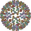
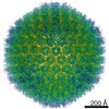
| タイトル | Structure of Thermusphage P23-77 using electron cryomicroscopy and three-dimensional image reconstruction | |||||||||
 マップデータ マップデータ | This is a 14 A resolution cryo-EM reconstruction of Thermusphage P23-77 | |||||||||
 試料 試料 |
| |||||||||
 キーワード キーワード | Icosahedral thermophilic bacterial virus | |||||||||
| 生物種 |   Thermus phage P23-77 (ファージ) Thermus phage P23-77 (ファージ) | |||||||||
| 手法 |  単粒子再構成法 / クライオ電子顕微鏡法 / ネガティブ染色法 / 解像度: 14.0 Å 単粒子再構成法 / クライオ電子顕微鏡法 / ネガティブ染色法 / 解像度: 14.0 Å | |||||||||
 データ登録者 データ登録者 | Jaatinen ST / Happonen LJ / Laurinmaki P / Butcher SJ / Bamford DH | |||||||||
 引用 引用 | ジャーナル: Virology / 年: 2008 タイトル: Biochemical and structural characterisation of membrane-containing icosahedral dsDNA bacteriophages infecting thermophilic Thermus thermophilus. 著者: S T Jaatinen / L J Happonen / P Laurinmäki / S J Butcher / D H Bamford /  要旨: Icosahedral dsDNA viruses isolated from hot springs and proposed to belong to the Tectiviridae family infect the gram-negative thermophilic Thermus thermophilus bacterium. Seven such viruses were ...Icosahedral dsDNA viruses isolated from hot springs and proposed to belong to the Tectiviridae family infect the gram-negative thermophilic Thermus thermophilus bacterium. Seven such viruses were obtained from the Promega Corporation collection. The structural protein patterns of three of these viruses, growing to a high titer, appeared very similar but not identical. The most stable virus, P23-77, was chosen for more detailed studies. Analysis of highly purified P23-77 by thin layer chromatography for neutral lipids showed lipid association with the virion. Cryo-EM based three-dimensional image reconstruction of P23-77 to 1.4 nm resolution revealed an icosahedrally-ordered protein coat, with spikes on the vertices, and an internal membrane. The capsid architecture of P23-77 is most similar to that of the archaeal virus SH1. These findings further complicate the grouping of icosahedrally-symmetric viruses containing an inner membrane. We propose a single superfamily or order with members in several viral families. | |||||||||
| 履歴 |
|
- 構造の表示
構造の表示
| ムービー |
 ムービービューア ムービービューア |
|---|---|
| 構造ビューア | EMマップ: SurfViewMolmilJmol/JSmol |
| 添付画像 |

- ダウンロードとリンク
-EMDBアーカイブ
| マップデータ | emd_1525.map.gz | 60.2 MB | EMDBマップデータ形式 | |
|---|---|---|---|---|
| ヘッダ (付随情報) | emd-1525-v30.xmlemd-1525.xml | 9.6 KB 9.6 KB | 表示 表示 | EMDBヘッダ |
| 画像 |  1525.gif 1525.gif emd_1525.gif emd_1525.gif | 108.1 KB 162.2 KB | ||
| アーカイブディレクトリ |  http://ftp.pdbj.org/pub/emdb/structures/EMD-1525ftp://ftp.pdbj.org/pub/emdb/structures/EMD-1525 http://ftp.pdbj.org/pub/emdb/structures/EMD-1525ftp://ftp.pdbj.org/pub/emdb/structures/EMD-1525 | HTTPS FTP |
-関連構造データ


-リンク
| EMDBのページ | EMDB (EBI/PDBe) / EMDataResource |
|---|
-マップ
| ファイル | ダウンロード / ファイル: emd_1525.map.gz / 形式: CCP4 / 大きさ: 120.1 MB / タイプ: IMAGE STORED AS SIGNED INTEGER (2 BYTES) | ||||||||||||||||||||||||||||||||||||||||||||||||||||||||||||||||||||
|---|---|---|---|---|---|---|---|---|---|---|---|---|---|---|---|---|---|---|---|---|---|---|---|---|---|---|---|---|---|---|---|---|---|---|---|---|---|---|---|---|---|---|---|---|---|---|---|---|---|---|---|---|---|---|---|---|---|---|---|---|---|---|---|---|---|---|---|---|---|
| 注釈 | This is a 14 A resolution cryo-EM reconstruction of Thermusphage P23-77 | ||||||||||||||||||||||||||||||||||||||||||||||||||||||||||||||||||||
| ボクセルのサイズ | X=Y=Z: 2.8 Å | ||||||||||||||||||||||||||||||||||||||||||||||||||||||||||||||||||||
| 密度 |
| ||||||||||||||||||||||||||||||||||||||||||||||||||||||||||||||||||||
| 対称性 | 空間群: 1 | ||||||||||||||||||||||||||||||||||||||||||||||||||||||||||||||||||||
| 詳細 | EMDB XML:
CCP4マップ ヘッダ情報:
| ||||||||||||||||||||||||||||||||||||||||||||||||||||||||||||||||||||
-添付データ
- 試料の構成要素
-全体 : Thermusphage P23-77
| 全体 | 名称: Thermusphage P23-77 |
|---|---|
| 要素 |
|
-超分子 #1000: Thermusphage P23-77
| 超分子 | 名称: Thermusphage P23-77 / タイプ: sample / ID: 1000 詳細: The sample was purified on a sucrose gradient, and stored at 28 C prior to plunging. 集合状態: 1 / Number unique components: 1 |
|---|
-超分子 #1: Thermus phage P23-77
| 超分子 | 名称: Thermus phage P23-77 / タイプ: virus / ID: 1 / Name.synonym: Thermusphage P23-77 / NCBI-ID: 668994 / 生物種: Thermus phage P23-77 / ウイルスタイプ: VIRION / ウイルス・単離状態: STRAIN / ウイルス・エンベロープ: Yes / ウイルス・中空状態: No / Syn species name: Thermusphage P23-77 |
|---|---|
| 宿主 | 生物種:  Thermus thermophilus (サーマス・サーモフィルス) Thermus thermophilus (サーマス・サーモフィルス)別称: BACTERIA(EUBACTERIA) |
| ウイルス殻 | Shell ID: 1 / 直径: 780 Å / T番号(三角分割数): 28 |
-実験情報
-構造解析
| 手法 | ネガティブ染色法, クライオ電子顕微鏡法 |
|---|---|
 解析 解析 | 単粒子再構成法 |
| 試料の集合状態 | particle |
-試料調製
| 緩衝液 | pH: 7.5 / 詳細: 20 mM Tris-HCl pH 7.5, 1mM MgCl2, 0.1 mM CaCl2 |
|---|---|
| 染色 | タイプ: NEGATIVE 詳細: Vitrified. Grids were blotted for roughly one second before being plunged into liquid ethane. |
| グリッド | 詳細: 400 mesh copper grid, Quantifoil R2/2 holey |
| 凍結 | 凍結剤: ETHANE / 装置: HOMEMADE PLUNGER / 詳細: Vitrification instrument: Guillotine 手法: A small vial of ethane is placed inside a larger liquid nitrogen reservoir. The grid holding 3 microliters of the sample is held in place at the bottom of a plunger by the means of fine ...手法: A small vial of ethane is placed inside a larger liquid nitrogen reservoir. The grid holding 3 microliters of the sample is held in place at the bottom of a plunger by the means of fine tweezers. When the liquid ethane is ready, a piece of filter paper is then pressed against the sample to blot off excess buffer, sufficient to leave a thin layer on the grid. The filter paper is removed, and the plunger is allowed to drop into the liquid ethane. Once the grid enters the liquid ethane, the sample is rapidly frozen, and the grid is transferred under liquid nitrogen to a storage box immersed in liquid nitrogen for later use in the microscope. |
- 電子顕微鏡法
| 顕微鏡 | FEI TECNAI 20 |
|---|---|
| 電子線 | 加速電圧: 200 kV / 電子線源: FIELD EMISSION GUN |
| 電子光学系 | 倍率(補正後): 49300 / 照射モード: FLOOD BEAM / 撮影モード: BRIGHT FIELDBright-field microscopy / Cs: 2.0 mm / 最大 デフォーカス(公称値): 3.0 µm / 最小 デフォーカス(公称値): 1.0 µm / 倍率(公称値): 50000 |
| 試料ステージ | 試料ホルダー: Side entry liquid nitrogen-cooled cryo specimen holder 試料ホルダーモデル: GATAN LIQUID NITROGEN |
| 詳細 | Low dose conditions. |
| 日付 | 2007年3月19日 |
| 撮影 | カテゴリ: FILM / フィルム・検出器のモデル: KODAK SO-163 FILM / デジタル化 - スキャナー: ZEISS SCAI / デジタル化 - サンプリング間隔: 14 µm / 実像数: 25 詳細: Images were scanned at 7 microns, and binned by 2 to 14 microns step size. |
-画像解析
| CTF補正 | 詳細: Each micrograph |
|---|---|
| 最終 再構成 | 想定した対称性 - 点群: I (正20面体型対称) / アルゴリズム: OTHER / 解像度のタイプ: BY AUTHOR / 解像度: 14.0 Å / 解像度の算出法: FSC 0.5 CUT-OFF / ソフトウェア - 名称: PFT, POR, EM3DR2, P3DR / 使用した粒子像数: 880 |
| 詳細 | The particles were selected using an automatic selection program (ETHAN). |
