 Movie
Movie Controller


+ Open data

- Basic information
| Entry |  | |||||||||
|---|---|---|---|---|---|---|---|---|---|---|
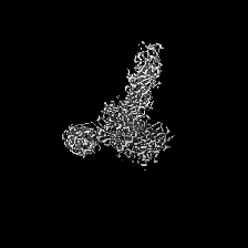
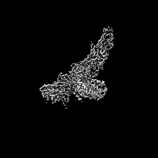
| Title | S-layer protein SlaA from Sulfolobus acidocaldarius at pH 4.0 | |||||||||
 Map data Map data | ||||||||||
 Sample Sample |
| |||||||||
 Keywords Keywords | S-layer / dimer / N-glycosylation / STRUCTURAL PROTEIN | |||||||||
| Function / homology | S-layer / extracellular region / S-layer protein A Function and homology information Function and homology information | |||||||||
| Biological species |   Sulfolobus acidocaldarius DSM 639 (acidophilic) / Sulfolobus acidocaldarius (acidophilic) Sulfolobus acidocaldarius DSM 639 (acidophilic) / Sulfolobus acidocaldarius (acidophilic) | |||||||||
| Method | single particle reconstruction / cryo EM / Resolution: 3.1 Å | |||||||||
 Authors Authors | Gambelli L / Isupov MN / Daum B | |||||||||
| Funding support | European Union, 1 items
| |||||||||
 Citation Citation | Journal: Elife / Year: 2024 Title: Structure of the two-component S-layer of the archaeon . Authors: Lavinia Gambelli / Mathew McLaren / Rebecca Conners / Kelly Sanders / Matthew C Gaines / Lewis Clark / Vicki A M Gold / Daniel Kattnig / Mateusz Sikora / Cyril Hanus / Michail N Isupov / Bertram Daum /     Abstract: Surface layers (S-layers) are resilient two-dimensional protein lattices that encapsulate many bacteria and most archaea. In archaea, S-layers usually form the only structural component of the cell ...Surface layers (S-layers) are resilient two-dimensional protein lattices that encapsulate many bacteria and most archaea. In archaea, S-layers usually form the only structural component of the cell wall and thus act as the final frontier between the cell and its environment. Therefore, S-layers are crucial for supporting microbial life. Notwithstanding their importance, little is known about archaeal S-layers at the atomic level. Here, we combined single-particle cryo electron microscopy, cryo electron tomography, and Alphafold2 predictions to generate an atomic model of the two-component S-layer of . The outer component of this S-layer (SlaA) is a flexible, highly glycosylated, and stable protein. Together with the inner and membrane-bound component (SlaB), they assemble into a porous and interwoven lattice. We hypothesise that jackknife-like conformational changes in SlaA play important roles in S-layer assembly. | |||||||||
| History |
|
- Structure visualization
| Supplemental images |
|---|

- Downloads & links
-EMDB archive
| Map data | emd_14635.map.gz | 38.1 MB | EMDB map data format | |
|---|---|---|---|---|
| Header (meta data) | emd-14635-v30.xmlemd-14635.xml | 16.1 KB 16.1 KB | Display Display | EMDB header |
| FSC (resolution estimation) | emd_14635_fsc.xml | 8 KB | Display | FSC data file |
| Images | 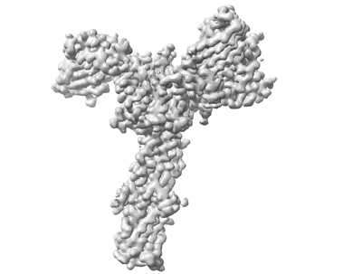 emd_14635.png emd_14635.png | 52 KB | ||
| Filedesc metadata | emd-14635.cif.gz | 6.7 KB | ||
| Others | emd_14635_half_map_1.map.gzemd_14635_half_map_2.map.gz | 33.1 MB 33.1 MB | ||
| Archive directory |  http://ftp.pdbj.org/pub/emdb/structures/EMD-14635ftp://ftp.pdbj.org/pub/emdb/structures/EMD-14635 http://ftp.pdbj.org/pub/emdb/structures/EMD-14635ftp://ftp.pdbj.org/pub/emdb/structures/EMD-14635 | HTTPS FTP |
-Related structure data
| Related structure data | 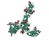 7zcxMC  8an2C  8an3C  8qoxC  8qp0C M: atomic model generated by this map C: citing same article ( |
|---|---|
| Similar structure data |


-Links
| EMDB pages | EMDB (EBI/PDBe) / EMDataResource |
|---|
-Map
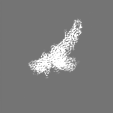
| File | Download / File: emd_14635.map.gz / Format: CCP4 / Size: 42.9 MB / Type: IMAGE STORED AS FLOATING POINT NUMBER (4 BYTES) | ||||||||||||||||||||||||||||||||||||
|---|---|---|---|---|---|---|---|---|---|---|---|---|---|---|---|---|---|---|---|---|---|---|---|---|---|---|---|---|---|---|---|---|---|---|---|---|---|
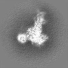
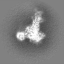
| Projections & slices | Image control
Images are generated by Spider. | ||||||||||||||||||||||||||||||||||||
| Voxel size | X=Y=Z: 1.05 Å | ||||||||||||||||||||||||||||||||||||
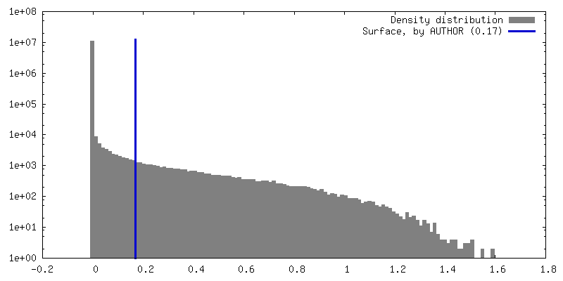
| Density |
| ||||||||||||||||||||||||||||||||||||
| Symmetry | Space group: 1 | ||||||||||||||||||||||||||||||||||||
| Details | EMDB XML:
|
 Z (Sec.)
 Y (Row.)
 X (Col.)


-Supplemental data
-Half map: #2
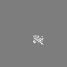
| File | emd_14635_half_map_1.map | ||||||||||||
|---|---|---|---|---|---|---|---|---|---|---|---|---|---|
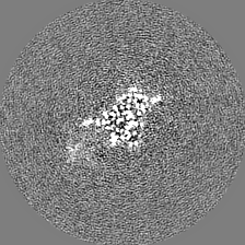
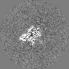
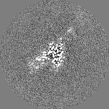
| Projections & Slices |
| ||||||||||||
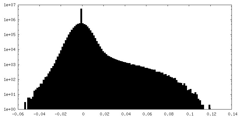
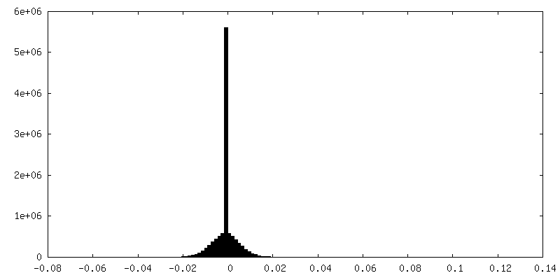
| Density Histograms |


-Half map: #1
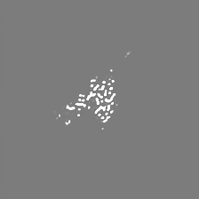
| File | emd_14635_half_map_2.map | ||||||||||||
|---|---|---|---|---|---|---|---|---|---|---|---|---|---|
| Projections & Slices |
| ||||||||||||
| Density Histograms |


- Sample components
-Entire : Monomeric S-layer protein SlaA with N-glycosylation
| Entire | Name: Monomeric S-layer protein SlaA with N-glycosylation |
|---|---|
| Components |
|
-Supramolecule #1: Monomeric S-layer protein SlaA with N-glycosylation
| Supramolecule | Name: Monomeric S-layer protein SlaA with N-glycosylation / type: organelle_or_cellular_component / ID: 1 / Parent: 0 / Macromolecule list: #1 |
|---|---|
| Source (natural) | Organism: Sulfolobus acidocaldarius DSM 639 (acidophilic) / Location in cell: Surface layer |
-Macromolecule #1: S-layer protein A
| Macromolecule | Name: S-layer protein A / type: protein_or_peptide / ID: 1 / Number of copies: 1 / Enantiomer: LEVO |
|---|---|
| Source (natural) | Organism: Sulfolobus acidocaldarius (acidophilic) Strain: ATCC 33909 / DSM 639 / JCM 8929 / NBRC 15157 / NCIMB 11770 |
| Molecular weight | Theoretical: 151.078406 KDa |
| Recombinant expression | Organism: Sulfolobus acidocaldarius (acidophilic) |
| Sequence | String: MNKLVGLLVS SLFLASILIG IAPAITTTAL TPPVSAGGIQ AYLLTGSGAP ASGLVLFVVN VSNIQVSSSN VTNVISTVVS NIQINAKTE NAQTGATTGS VTVRFPTSGY NAYYDSVDKV VFVVVSFLYP YTTTSVNIPL SYLSKYLPGL LTAQPYDETG A QVTSVSST ...String: MNKLVGLLVS SLFLASILIG IAPAITTTAL TPPVSAGGIQ AYLLTGSGAP ASGLVLFVVN VSNIQVSSSN VTNVISTVVS NIQINAKTE NAQTGATTGS VTVRFPTSGY NAYYDSVDKV VFVVVSFLYP YTTTSVNIPL SYLSKYLPGL LTAQPYDETG A QVTSVSST PFGSLIDTST GQQILGTNPV LTSYNSYTTQ ANTNMQEGVV SGTLTSFTLG GQSFSGSTVP VILYAPFIFS NS PYQAGLY NPMQVNGNLG SLSSEAYYHP VIWGRALINT TLIDTYASGS VPFTFQLNYS VPGPLTINMA QLAWIASINN LPT SFTYLS YKFSNGYESF LGIISNSTQL TAGALTINPS GNFTINGKKF YVYLLVVGST NSTTPVEYVT KLVVEYPSST NFLP QGVTV TTSSNKYTLP VYEIGGPAGT TITLTGNWYS TPYTVQITVG STPTLTNYVS QILLKAVAYE GINVSTTQSP YYSTA ILST PPSEISITGS STITAQGKLT ATSASATVNL LTNATLTYEN IPLTQYSFNG IIVTPGYAAI NGTTAMAYVI GALYNK TSD YVLSFAGSQE PMQVMNNNLT EVTTLAPFGL TLLAPSVPAT ETGTSPLQLE FFTVPSTSYI ALVDFGLWGN LTSVTVS AY DTVNNKLSVN LGYFYGIVIP PSISTAPYNY QNFICPNNYV TVTIYDPDAV LDPYPSGSFT TSSLPLKYGN MNITGAVI F PGSSVYNPSG VFGYSNFNKG AAVTTFTYTA QSGPFSPVAL TGNTNYLSQY ADNNPTDNYY FIQTVNGMPV LMGGLSIVA SPVSASLPSS TSSPGFMYLL PSAAQVPSPL PGMATPNYNL NIYITYKIDG ATVGNNMING LYVASQNTLI YVVPNGSFVG SNIKLTYTT TDYAVLHYFY STGQYKVFKT VSVPNVTANL YFPSSTTPLY QLSVPLYLSE PYYGSPLPTY IGLGTNGTSL W NSPNYVLF GVSAVQQYLG FIKSISVTLS NGTTVVIPLT TSNMQTLFPQ LVGQELQACN GTFQFGISIT GLEKLLNLNV QQ LNNSILS VTYHDYVTGE TLTATTKLVA LSTLSLVAKG AGVVEFLLTA YPYTGNITFA PPWFIAENVV KQPFMTYSDL QFA KTNPSA ILSLSTVNIT VVGLGGKASV YYNSTSGQTV ITNIYGQTVA TLSGNVLPTL TELAAGNGTF TGSLQFTIVP NNTV VQIPS SLTKTSFAVY TNGSLAIVLN GKAYSLGPAG LFLLPFVTYT GSAIGANATA IITVSDGVGT STTQVPITAE NFTPI RLAP FQVPAQVPLP NAPKLKYEYN GSIVITPQQQ VLKIYVTSIL PYPQEFQIQA FVYEASQFNV HTGSPTAAPV YFSYSA VRA YPALGIGTSV PNLLVYVQLQ GISNLPAGKY VIVLSAVPFA GGPVLSEYPA QLIFTNVTLT Q UniProtKB: S-layer protein A |
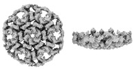
-Macromolecule #8: 2-acetamido-2-deoxy-beta-D-glucopyranose
| Macromolecule | Name: 2-acetamido-2-deoxy-beta-D-glucopyranose / type: ligand / ID: 8 / Number of copies: 1 / Formula: NAG |
|---|---|
| Molecular weight | Theoretical: 221.208 Da |
| Chemical component information |  ChemComp-NAG: |
-Macromolecule #9: 6-deoxy-6-sulfo-beta-D-glucopyranose
| Macromolecule | Name: 6-deoxy-6-sulfo-beta-D-glucopyranose / type: ligand / ID: 9 / Number of copies: 1 / Formula: YZT |
|---|---|
| Molecular weight | Theoretical: 244.22 Da |
| Chemical component information |  ChemComp-YZT: |
-Experimental details
-Structure determination
| Method | cryo EM |
|---|---|
 Processing Processing | single particle reconstruction |
| Aggregation state | particle |
-Sample preparation
| Buffer | pH: 3.8 |
|---|---|
| Vitrification | Cryogen name: ETHANE / Chamber humidity: 100 % / Chamber temperature: 277 K / Instrument: FEI VITROBOT MARK IV Details: Grid type: lacey with graphene oxide Blotting paper: Whatman 597. |
- Electron microscopy
| Microscope | FEI TALOS ARCTICA |
|---|---|
| Electron beam | Acceleration voltage: 200 kV / Electron source: FIELD EMISSION GUN |
| Electron optics | Illumination mode: FLOOD BEAM / Imaging mode: BRIGHT FIELDBright-field microscopy / Nominal defocus max: 2.4 µm / Nominal defocus min: 1.2 µm / Nominal magnification: 130000 |
| Sample stage | Cooling holder cryogen: NITROGEN |
| Image recording | Film or detector model: GATAN K2 SUMMIT (4k x 4k) / Detector mode: COUNTING / Average electron dose: 59.12 e/Å2 |
| Experimental equipment |  Model: Talos Arctica / Image courtesy: FEI Company |
-Image processing
| Startup model | Type of model: NONE |
|---|---|
| Initial angle assignment | Type: MAXIMUM LIKELIHOOD |
| Final angle assignment | Type: MAXIMUM LIKELIHOOD |
| Final reconstruction | Resolution.type: BY AUTHOR / Resolution: 3.1 Å / Resolution method: FSC 0.143 CUT-OFF / Number images used: 66040 |
| FSC plot (resolution estimation) |  |
-Atomic model buiding 1
| Refinement | Protocol: AB INITIO MODEL |
|---|---|
| Output model | PDB-7zcx: |
