 Movie
Movie Controller

[English] 日本語
 Yorodumi
- EMDB-14176: TMEM106B filaments with Fold I-d from Multiple system atrophy (ca... -
+ Open data

- Basic information
| Entry | Database: EMDB / ID: EMD-14176 | |||||||||
|---|---|---|---|---|---|---|---|---|---|---|
| Title | TMEM106B filaments with Fold I-d from Multiple system atrophy (case 18) | |||||||||
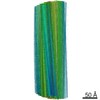
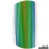
 Map data Map data | Sharpened map of TMEM106B filaments with Fold I-d from Multiple system atrophy (case 18) | |||||||||
 Sample Sample |
| |||||||||
| Function / homology |  Function and homology information Function and homology information lysosomal protein catabolic process / regulation of lysosome organization / lysosomal lumen acidification / lysosome localization / positive regulation of dendrite development / dendrite morphogenesis / lysosomal transport / lysosome organization / neuron cellular homeostasis / late endosome membrane ...lysosomal protein catabolic process / regulation of lysosome organization / lysosomal lumen acidification / lysosome localization / positive regulation of dendrite development / dendrite morphogenesis / lysosomal transport / lysosome organization / neuron cellular homeostasis / late endosome membrane / ATPase binding / lysosome / endosome / lysosomal membrane / plasma membrane lysosomal protein catabolic process / regulation of lysosome organization / lysosomal lumen acidification / lysosome localization / positive regulation of dendrite development / dendrite morphogenesis / lysosomal transport / lysosome organization / neuron cellular homeostasis / late endosome membrane ...lysosomal protein catabolic process / regulation of lysosome organization / lysosomal lumen acidification / lysosome localization / positive regulation of dendrite development / dendrite morphogenesis / lysosomal transport / lysosome organization / neuron cellular homeostasis / late endosome membrane / ATPase binding / lysosome / endosome / lysosomal membrane / plasma membraneSimilarity search - Function | |||||||||
| Biological species |  Homo sapiens (human) / human (human) Homo sapiens (human) / human (human) | |||||||||
| Method | helical reconstruction / cryo EM / Resolution: 3.64 Å | |||||||||
 Authors Authors | Lovestam S / Schweighauser M / Scheres SHW | |||||||||
| Funding support |  United Kingdom, 1 items United Kingdom, 1 items
| |||||||||
 Citation Citation | Journal: Nature / Year: 2022 Title: Age-dependent formation of TMEM106B amyloid filaments in human brains. Authors: Manuel Schweighauser / Diana Arseni / Mehtap Bacioglu / Melissa Huang / Sofia Lövestam / Yang Shi / Yang Yang / Wenjuan Zhang / Abhay Kotecha / Holly J Garringer / Ruben Vidal / Grace I ...Authors: Manuel Schweighauser / Diana Arseni / Mehtap Bacioglu / Melissa Huang / Sofia Lövestam / Yang Shi / Yang Yang / Wenjuan Zhang / Abhay Kotecha / Holly J Garringer / Ruben Vidal / Grace I Hallinan / Kathy L Newell / Airi Tarutani / Shigeo Murayama / Masayuki Miyazaki / Yuko Saito / Mari Yoshida / Kazuko Hasegawa / Tammaryn Lashley / Tamas Revesz / Gabor G Kovacs / John van Swieten / Masaki Takao / Masato Hasegawa / Bernardino Ghetti / Maria Grazia Spillantini / Benjamin Ryskeldi-Falcon / Alexey G Murzin / Michel Goedert / Sjors H W Scheres /      Abstract: Many age-dependent neurodegenerative diseases, such as Alzheimer's and Parkinson's, are characterized by abundant inclusions of amyloid filaments. Filamentous inclusions of the proteins tau, amyloid- ...Many age-dependent neurodegenerative diseases, such as Alzheimer's and Parkinson's, are characterized by abundant inclusions of amyloid filaments. Filamentous inclusions of the proteins tau, amyloid-β, α-synuclein and transactive response DNA-binding protein (TARDBP; also known as TDP-43) are the most common. Here we used structure determination by cryogenic electron microscopy to show that residues 120-254 of the lysosomal type II transmembrane protein 106B (TMEM106B) also form amyloid filaments in human brains. We determined the structures of TMEM106B filaments from a number of brain regions of 22 individuals with abundant amyloid deposits, including those resulting from sporadic and inherited tauopathies, amyloid-β amyloidoses, synucleinopathies and TDP-43 proteinopathies, as well as from the frontal cortex of 3 individuals with normal neurology and no or only a few amyloid deposits. We observed three TMEM106B folds, with no clear relationships between folds and diseases. TMEM106B filaments correlated with the presence of a 29-kDa sarkosyl-insoluble fragment and globular cytoplasmic inclusions, as detected by an antibody specific to the carboxy-terminal region of TMEM106B. The identification of TMEM106B filaments in the brains of older, but not younger, individuals with normal neurology indicates that they form in an age-dependent manner. | |||||||||
| History |
|
- Structure visualization
| Movie |
Movie viewer |
|---|---|
| Structure viewer | EM map: SurfViewMolmilJmol/JSmol |
| Supplemental images |

- Downloads & links
-EMDB archive
| Map data | emd_14176.map.gz | 71.9 MB | EMDB map data format | |
|---|---|---|---|---|
| Header (meta data) | emd-14176-v30.xmlemd-14176.xml | 15.3 KB 15.3 KB | Display Display | EMDB header |
| FSC (resolution estimation) | emd_14176_fsc.xml | 17.5 KB | Display | FSC data file |
| Images | 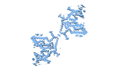 emd_14176.png emd_14176.png | 35.5 KB | ||
| Others | emd_14176_half_map_1.map.gzemd_14176_half_map_2.map.gz | 365.9 MB 365.8 MB | ||
| Archive directory |  http://ftp.pdbj.org/pub/emdb/structures/EMD-14176ftp://ftp.pdbj.org/pub/emdb/structures/EMD-14176 http://ftp.pdbj.org/pub/emdb/structures/EMD-14176ftp://ftp.pdbj.org/pub/emdb/structures/EMD-14176 | HTTPS FTP |
-Related structure data
| Related structure data |  7qvfMC  7qvcC  7qwgC 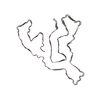 7qwlC 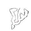 7qwmC M: atomic model generated by this map C: citing same article ( |
|---|---|
| Similar structure data | |
| EM raw data | EMPIAR-10968 (Title: Cryo-EM reconstruction of TMEM106B fold I-d filaments from case 18 with multiple system atrophy Data size: 1.0 TB Data #1: Unaligned multi-frame movies [micrographs - multiframe]) |

-Links
| EMDB pages | EMDB (EBI/PDBe) / EMDataResource |
|---|---|
| Related items in Molecule of the Month |

-Map
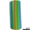
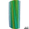
| File | Download / File: emd_14176.map.gz / Format: CCP4 / Size: 459.9 MB / Type: IMAGE STORED AS FLOATING POINT NUMBER (4 BYTES) | ||||||||||||||||||||||||||||||||||||||||||||||||||||||||||||||||||||
|---|---|---|---|---|---|---|---|---|---|---|---|---|---|---|---|---|---|---|---|---|---|---|---|---|---|---|---|---|---|---|---|---|---|---|---|---|---|---|---|---|---|---|---|---|---|---|---|---|---|---|---|---|---|---|---|---|---|---|---|---|---|---|---|---|---|---|---|---|---|
| Annotation | Sharpened map of TMEM106B filaments with Fold I-d from Multiple system atrophy (case 18) | ||||||||||||||||||||||||||||||||||||||||||||||||||||||||||||||||||||
| Projections & slices | Image control
Images are generated by Spider. | ||||||||||||||||||||||||||||||||||||||||||||||||||||||||||||||||||||
| Voxel size | X=Y=Z: 1.15 Å | ||||||||||||||||||||||||||||||||||||||||||||||||||||||||||||||||||||
| Density |
| ||||||||||||||||||||||||||||||||||||||||||||||||||||||||||||||||||||
| Symmetry | Space group: 1 | ||||||||||||||||||||||||||||||||||||||||||||||||||||||||||||||||||||
| Details | EMDB XML:
CCP4 map header:
| ||||||||||||||||||||||||||||||||||||||||||||||||||||||||||||||||||||
 Z (Sec.)
 Y (Row.)
 X (Col.)


-Supplemental data
-Half map: Half map 1 of TMEM106B filaments with Fold...
| File | emd_14176_half_map_1.map | ||||||||||||
|---|---|---|---|---|---|---|---|---|---|---|---|---|---|
| Annotation | Half map 1 of TMEM106B filaments with Fold I-d from Multiple system atrophy (case 18) | ||||||||||||
| Projections & Slices |
| ||||||||||||
| Density Histograms |


-Half map: Half map 2 of TMEM106B filaments with Fold...
| File | emd_14176_half_map_2.map | ||||||||||||
|---|---|---|---|---|---|---|---|---|---|---|---|---|---|
| Annotation | Half map 2 of TMEM106B filaments with Fold I-d from Multiple system atrophy (case 18) | ||||||||||||
| Projections & Slices |
| ||||||||||||
| Density Histograms |


- Sample components
-Entire : TMEM106B
| Entire | Name: TMEM106B |
|---|---|
| Components |
|
-Supramolecule #1: TMEM106B
| Supramolecule | Name: TMEM106B / type: tissue / ID: 1 / Parent: 0 / Macromolecule list: all |
|---|---|
| Source (natural) | Organism: Homo sapiens (human) |
-Macromolecule #1: Transmembrane protein 106B
| Macromolecule | Name: Transmembrane protein 106B / type: protein_or_peptide / ID: 1 / Number of copies: 6 / Enantiomer: LEVO |
|---|---|
| Source (natural) | Organism: human (human) |
| Molecular weight | Theoretical: 31.156318 KDa |
| Sequence | String: MGKSLSHLPL HSSKEDAYDG VTSENMRNGL VNSEVHNEDG RNGDVSQFPY VEFTGRDSVT CPTCQGTGRI PRGQENQLVA LIPYSDQRL RPRRTKLYVM ASVFVCLLLS GLAVFFLFPR SIDVKYIGVK SAYVSYDVQK RTIYLNITNT LNITNNNYYS V EVENITAQ ...String: MGKSLSHLPL HSSKEDAYDG VTSENMRNGL VNSEVHNEDG RNGDVSQFPY VEFTGRDSVT CPTCQGTGRI PRGQENQLVA LIPYSDQRL RPRRTKLYVM ASVFVCLLLS GLAVFFLFPR SIDVKYIGVK SAYVSYDVQK RTIYLNITNT LNITNNNYYS V EVENITAQ VQFSKTVIGK ARLNNITIIG PLDMKQIDYT VPTVIAEEMS YMYDFCTLIS IKVHNIVLMM QVTVTTTYFG HS EQISQER YQYVDCGRNT TYQLGQSEYL NVLQPQQ |
-Experimental details
-Structure determination
| Method | cryo EM |
|---|---|
 Processing Processing | helical reconstruction |
| Aggregation state | filament |
-Sample preparation
| Buffer | pH: 7.4 |
|---|---|
| Vitrification | Cryogen name: ETHANE |
- Electron microscopy
| Microscope | FEI TITAN KRIOS |
|---|---|
| Electron beam | Acceleration voltage: 300 kV / Electron source: FIELD EMISSION GUN |
| Electron optics | Illumination mode: FLOOD BEAM / Imaging mode: BRIGHT FIELDBright-field microscopy / Nominal defocus max: 3.0 µm / Nominal defocus min: 1.2 µm |
| Image recording | Film or detector model: FEI FALCON IV (4k x 4k) / Average electron dose: 40.0 e/Å2 |
| Experimental equipment |  Model: Titan Krios / Image courtesy: FEI Company |
-Image processing
| Final angle assignment | Type: NOT APPLICABLE |
|---|---|
| Final reconstruction | Applied symmetry - Helical parameters - Δz: 4.78 Å Applied symmetry - Helical parameters - Δ&Phi: -0.41 ° Applied symmetry - Helical parameters - Axial symmetry: C2 (2 fold cyclic )Resolution.type: BY AUTHOR / Resolution: 3.64 Å / Resolution method: FSC 0.143 CUT-OFF / Software - Name: RELION (ver. 4.0) / Software - details: Excellent stuff. / Number images used: 12057 |
| FSC plot (resolution estimation) |  |
