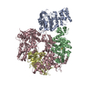
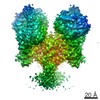
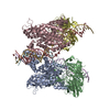
- EMDB-13021: CryoEM structure of DNA polymerase alpha - primase bound to SARS ... -
+
Open data
ID or keywords:
Loading...
-
Basic information
Entry
Database: EMDB / ID: EMD-13021
Title
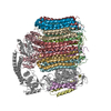
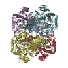
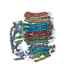
CryoEM structure of DNA polymerase alpha - primase bound to SARS CoV nsp1 protein
Map data
3D refinement map
Sample
Complex: CryoEM structure of DNA polymerase alpha - primase bound to SARS CoV nsp1 protein
Function / homology
Function and homology information
DNA primase AEP / positive regulation of DNA primase activity / ribonucleotide binding / DNA replication initiation / DNA/RNA hybrid binding / Telomere C-strand synthesis initiation / Inhibition of replication initiation of damaged DNA by RB1/E2F1 / Polymerase switching / alpha DNA polymerase:primase complex / regulation of type I interferon production ...DNA primase AEP / positive regulation of DNA primase activity / ribonucleotide binding / DNA replication initiation / DNA/RNA hybrid binding / Telomere C-strand synthesis initiation / Inhibition of replication initiation of damaged DNA by RB1/E2F1 / Polymerase switching / alpha DNA polymerase:primase complex / regulation of type I interferon production / Processive synthesis on the lagging strand / Assembly of the SARS-CoV-1 Replication-Transcription Complex (RTC) / Maturation of replicase proteins / Removal of the Flap Intermediate / Transcription of SARS-CoV-1 sgRNAs / DNA primase activity / Polymerase switching on the C-strand of the telomere / lagging strand elongation / mitotic DNA replication initiation / DNA replication, synthesis of primer / Translation of Replicase and Assembly of the Replication Transcription Complex / Replication of the SARS-CoV-1 genome / K48-linked deubiquitinase activity / DNA strand elongation involved in DNA replication / host cell endoplasmic reticulum / K63-linked deubiquitinase activity / leading strand elongation / DNA synthesis involved in DNA repair / G1/S-Specific Transcription / DNA replication origin binding / SARS-CoV-1 modulates host translation machinery / DNA replication initiation / Activation of the pre-replicative complex / viral genome replication / Defective pyroptosis / methyltransferase activity / nuclear matrix / double-strand break repair via nonhomologous end joining / protein import into nucleus / SARS-CoV-1 activates/modulates innate immune responses / single-stranded DNA binding / nuclear envelope / 4 iron, 4 sulfur cluster binding / methylation / double membrane vesicle viral factory outer membrane / SARS coronavirus main proteinase / host cell endosome / symbiont-mediated degradation of host mRNA / mRNA guanylyltransferase / symbiont-mediated suppression of host ISG15-protein conjugation / G-quadruplex RNA binding / omega peptidase activity / symbiont-mediated suppression of host cytoplasmic pattern recognition receptor signaling pathway via inhibition of IRF3 activity / host cell Golgi apparatus / symbiont-mediated perturbation of host ubiquitin-like protein modification / endonuclease activity / ubiquitinyl hydrolase 1 / cysteine-type deubiquitinase activity / Hydrolases; Acting on peptide bonds (peptidases); Cysteine endopeptidases / DNA replication / DNA-directed DNA polymerase / single-stranded RNA binding / host cell perinuclear region of cytoplasm / DNA-directed DNA polymerase activity / viral protein processing / lyase activity / induction by virus of host autophagy / symbiont-mediated suppression of host gene expression / cysteine-type endopeptidase activity / RNA-dependent RNA polymerase activity / nucleotide binding / DNA repair / symbiont-mediated suppression of host type I interferon-mediated signaling pathway / chromatin binding / chromatin / nucleolus / protein kinase binding / magnesium ion binding / proteolysis / DNA binding / zinc ion binding / nucleoplasm / membrane / identical protein binding / metal ion binding / nucleus / cytosol Similarity search - Function
DNA polymerase alpha, subunit B, N-terminal domain superfamily / DNA polymerase alpha subunit B N-terminal / DNA polymerase alpha, subunit B, N-terminal / DNA polymerase alpha, subunit B / DNA primase, small subunit, eukaryotic/archaeal / DNA primase, large subunit, eukaryotic / DNA primase, small subunit / DNA primase small subunit / DNA primase large subunit, eukaryotic/archaeal / DNA polymerase alpha catalytic subunit, N-terminal domain ...DNA polymerase alpha, subunit B, N-terminal domain superfamily / DNA polymerase alpha subunit B N-terminal / DNA polymerase alpha, subunit B, N-terminal / DNA polymerase alpha, subunit B / DNA primase, small subunit, eukaryotic/archaeal / DNA primase, large subunit, eukaryotic / DNA primase, small subunit / DNA primase small subunit / DNA primase large subunit, eukaryotic/archaeal / DNA polymerase alpha catalytic subunit, N-terminal domain / DNA polymerase alpha, zinc finger domain superfamily / Eukaryotic and archaeal DNA primase, large subunit / DNA Polymerase alpha zinc finger / DNA polymerase alpha subunit p180 N terminal / Zinc finger, DNA-directed DNA polymerase, family B, alpha / DNA polymerase alpha catalytic subunit, catalytic domain / DNA polymerase alpha/delta/epsilon, subunit B / DNA polymerase alpha/epsilon subunit B / Non-structural protein 3, SUD-N macrodomain, SARS-CoV / DNA polymerase family B, thumb domain / DNA polymerase family B signature. / DNA-directed DNA polymerase, family B, conserved site / DNA polymerase family B / DNA polymerase family B, exonuclease domain / DNA-directed DNA polymerase, family B, exonuclease domain / DNA-directed DNA polymerase, family B, multifunctional domain / DNA polymerase, palm domain superfamily / DNA polymerase type-B family / DNA-directed DNA polymerase, family B / Non-structural protein NSP3, SUD-N (Mac2) domain, betacoronavirus / Sarbecovirus Nsp3c-N domain profile. / Non-structural protein NSP3, N-terminal, betacoronavirus / Polyprotein cleavage domain PL2pro superfamily, betacoronavirus / Non-structural protein NSP3, SUD-N (Mac2) domain superfamily, betacoronavirus / Betacoronavirus SUD-C domain / Betacoronavirus replicase NSP3, N-terminal / NSP1 globular domain superfamily, betacoronavirus / Non-structural protein 2, SARS-CoV-like / Coronavirus 3Ecto domain profile. / : / Betacoronavirus Nsp3e group 2-specific marker (G2M) domain profile. / NSP1, C-terminal domain, betacoronavirus / Betacoronavirus Nsp3c-M domain profile. / NSP1, globular domain, betacoronavirus / Non-structural protein NSP3, SUD-M domain, betacoronavirus / Non-structural protein NSP3, SUD-M domain superfamily, betacoronavirus / Betacoronavirus replicase NSP1 / Betacoronavirus single-stranded poly(A) binding domain / Betacoronavirus (BetaCoV) Nsp1 C-terminal domain profile. / Betacoronavirus Nsp3c-C domain profile. / Betacoronavirus Nsp3e nucleic acid-binding (NAB) domain profile. / DPUP/SUD, C-terminal, betacoronavirus / Non-structural protein NSP3, nucleic acid-binding domain superfamily, betacoronavirus / Non-structural protein 6, betacoronavirus / Betacoronavirus nucleic acid-binding (NAB) / Non-structural protein NSP3, nucleic acid-binding domain, betacoronavirus / Non-structural protein NSP3A domain-like superfamily / Papain-like protease, N-terminal domain superfamily, coronavirus / Papain-like viral protease, palm and finger domains, coronavirus / : / Coronavirus (CoV) Nsp2 middle domain profile. / Coronavirus (CoV) Nsp2 N-terminal domain profile. / Coronavirus (CoV) Nsp2 C-terminal domain profile. / NSP1, globular domain, alpha/betacoronavirus / : / Coronavirus (CoV) Nsp3 Y domain profile. / Coronavirus (CoV) Nsp1 globular domain profile. / Coronavirus replicase NSP2, N-terminal / Nonstructural protein 2, N-terminal domain, coronavirus / Coronavirus replicase NSP2, C-terminal / Non-structural protein 2, C-terminal domain, coronavirus / Coronavirus Nsp3a Ubl domain profile. / Coronavirus Nsp3d Ubl domain profile. / Coronavirus RNA-dependent RNA polymerase (RdRp) Nsp7 cofactor domain profile. / Coronavirus RNA-dependent RNA polymerase (RdRp) Nsp8 cofactor domain profile. / Coronavirus Nsp9 single-stranded RNA (ssRNA)-binding domain profile. / Coronavirus (CoV) ExoN/MTase coactivator domain profile. / NSP3, first ubiquitin-like (Ubl) domain, coronavirus / NSP3, second ubiquitin-like (Ubl) domain, coronavirus / Coronavirus Nsp4 C-terminal (Nsp4C) domain profile. / Papain-like protease, thumb domain superfamily, coronavirus / Coronavirus replicase NSP7 / Peptidase family C16 domain profile. / Non-structural protein NSP7, coronavirus / Peptidase C30, coronavirus / Peptidase C16, coronavirus / Non-structural protein NSP9, coronavirus / Non-structural protein NSP8, coronavirus / RNA synthesis protein NSP10, coronavirus / Non-structural protein NSP4, C-terminal, coronavirus / RNA synthesis protein NSP10 superfamily, coronavirus / Non-structural protein NSP9 superfamily, coronavirus / Non-structural protein NSP7 superfamily, coronavirus / Non-structural protein NSP8 superfamily, coronavirus / Non-structural protein NSP4, C-terminal superfamily, coronavirus / Peptidase C30, domain 3, coronavirus / Non-structural protein 6, coronavirus / Coronavirus replicase NSP3, C-terminal / Non-structural protein NSP4, N-terminal, coronavirus / Coronavirus endopeptidase C30 Similarity search - Domain/homology
DNA polymerase alpha catalytic subunit / Replicase polyprotein 1a / DNA primase small subunit / DNA primase large subunit / DNA polymerase alpha subunit B Similarity search - Component
Biological species
Homo sapiens (human)
Method
single particle reconstruction / cryo EM / Resolution: 4.4 Å
Journal: Protein Sci / Year: 2022 Title: Structural basis for the interaction of SARS-CoV-2 virulence factor nsp1 with DNA polymerase α-primase. Authors: Mairi L Kilkenny / Charlotte E Veale / Amir Guppy / Steven W Hardwick / Dimitri Y Chirgadze / Neil J Rzechorzek / Joseph D Maman / Luca Pellegrini / Abstract: The molecular mechanisms that drive the infection by the severe acute respiratory syndrome coronavirus 2 (SARS-CoV-2)-the causative agent of coronavirus disease 2019 (COVID-19)-are under intense ...The molecular mechanisms that drive the infection by the severe acute respiratory syndrome coronavirus 2 (SARS-CoV-2)-the causative agent of coronavirus disease 2019 (COVID-19)-are under intense current scrutiny to understand how the virus operates and to uncover ways in which the disease can be prevented or alleviated. Recent proteomic screens of the interactions between viral and host proteins have identified the human proteins targeted by SARS-CoV-2. The DNA polymerase α (Pol α)-primase complex or primosome-responsible for initiating DNA synthesis during genomic duplication-was identified as a target of nonstructural protein 1 (nsp1), a major virulence factor in the SARS-CoV-2 infection. Here, we validate the published reports of the interaction of nsp1 with the primosome by demonstrating direct binding with purified recombinant components and providing a biochemical characterization of their interaction. Furthermore, we provide a structural basis for the interaction by elucidating the cryo-electron microscopy structure of nsp1 bound to the primosome. Our findings provide biochemical evidence for the reported targeting of Pol α by the virulence factor nsp1 and suggest that SARS-CoV-2 interferes with Pol α's putative role in the immune response during the viral infection.
History
Deposition
Jun 1, 2021
-
Header (metadata) release
Nov 10, 2021
-
Map release
Nov 10, 2021
-
Update
Feb 23, 2022
-
Current status
Feb 23, 2022
Processing site: PDBe / Status: Released
-
Structure visualization
Movie
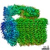
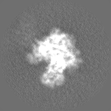
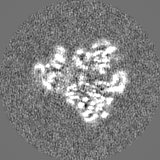
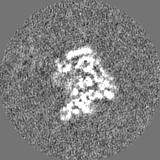
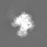
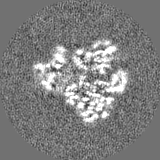
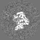
Surface view with section colored by density value
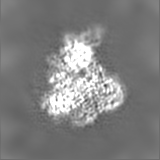
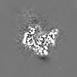
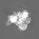
Entire : CryoEM structure of DNA polymerase alpha - primase bound to SARS ...
Entire
Name: CryoEM structure of DNA polymerase alpha - primase bound to SARS CoV nsp1 protein
Components
Complex: CryoEM structure of DNA polymerase alpha - primase bound to SARS CoV nsp1 protein
-
Supramolecule #1: CryoEM structure of DNA polymerase alpha - primase bound to SARS ...
Supramolecule
Name: CryoEM structure of DNA polymerase alpha - primase bound to SARS CoV nsp1 protein type: complex / ID: 1 / Parent: 0 / Macromolecule list: #1-#5
Source (natural)
Organism: Homo sapiens (human)
Recombinant expression
Organism: Spodoptera frugiperda (fall armyworm)
Molecular weight
Theoretical: 310 KDa
-
Experimental details
-
Structure determination
Method
cryo EM
Processing
single particle reconstruction
Aggregation state
particle
-
Sample preparation
Concentration
0.3 mg/mL
Buffer
pH: 7.2 Component:
Concentration
Name
25.0 mM
Hepes
150.0 mM
KCl
1.0 mM
DTT
Grid
Model: UltrAuFoil R1.2/1.3 / Material: GOLD / Mesh: 300 / Support film - Material: GOLD / Support film - topology: HOLEY / Support film - Film thickness: 50.0 nm / Pretreatment - Type: GLOW DISCHARGE
Vitrification
Cryogen name: ETHANE / Instrument: FEI VITROBOT MARK IV
Details
The nsp1 protein was added in 10-fold stoichiometric excess
-
Electron microscopy
Microscope
FEI TITAN KRIOS
Electron beam
Acceleration voltage: 300 kV / Electron source: FIELD EMISSION GUN
Film or detector model: GATAN K3 BIOQUANTUM (6k x 4k) / Number real images: 2919 / Average exposure time: 1.31 sec. / Average electron dose: 46.91 e/Å2
Experimental equipment
Model: Titan Krios / Image courtesy: FEI Company
-
Image processing
Particle selection
Number selected: 709068
CTF correction
Software - Name: CTFFIND (ver. 4.1)
Startup model
Type of model: OTHER Details: The initial 3D model was generated using a Stochastic Gradient Descent algorithm as implemented in Relion 3.1.
Initial angle assignment
Type: OTHER / Software - Name: RELION (ver. 3.1) / Details: As implemented in Relion 3.1.
Final 3D classification
Software - Name: RELION (ver. 3.1)
Final angle assignment
Type: OTHER / Software - Name: RELION (ver. 3.1) / Details: As implemented in Relion 3.1.
Final reconstruction
Applied symmetry - Point group: C1 (asymmetric) / Resolution.type: BY AUTHOR / Resolution: 4.4 Å / Resolution method: FSC 0.143 CUT-OFF / Software - Name: RELION (ver. 3.1) / Number images used: 233476
In the structure databanks used in Yorodumi, some data are registered as the other names, "COVID-19 virus" and "2019-nCoV". Here are the details of the virus and the list of structure data.
Jan 31, 2019. EMDB accession codes are about to change! (news from PDBe EMDB page)
EMDB accession codes are about to change! (news from PDBe EMDB page)
The allocation of 4 digits for EMDB accession codes will soon come to an end. Whilst these codes will remain in use, new EMDB accession codes will include an additional digit and will expand incrementally as the available range of codes is exhausted. The current 4-digit format prefixed with “EMD-” (i.e. EMD-XXXX) will advance to a 5-digit format (i.e. EMD-XXXXX), and so on. It is currently estimated that the 4-digit codes will be depleted around Spring 2019, at which point the 5-digit format will come into force.
The EM Navigator/Yorodumi systems omit the EMD- prefix.
Related info.:Q: What is EMD? / ID/Accession-code notation in Yorodumi/EM Navigator
Yorodumi is a browser for structure data from EMDB, PDB, SASBDB, etc.
This page is also the successor to EM Navigator detail page, and also detail information page/front-end page for Omokage search.
The word "yorodu" (or yorozu) is an old Japanese word meaning "ten thousand". "mi" (miru) is to see.
Related info.:EMDB / PDB / SASBDB / Comparison of 3 databanks / Yorodumi Search / Aug 31, 2016. New EM Navigator & Yorodumi / Yorodumi Papers / Jmol/JSmol / Function and homology information / Changes in new EM Navigator and Yorodumi
 Movie
Movie Controller

 Yorodumi
 Open data
 Basic information
 Map data
 Sample
 Function and homology information
 ribonucleotide binding / DNA replication initiation / DNA/RNA hybrid binding / Telomere C-strand synthesis initiation / Inhibition of replication initiation of damaged DNA by RB1/E2F1 / Polymerase switching / alpha DNA polymerase:primase complex / regulation of type I interferon production ...DNA primase AEP / positive regulation of DNA primase activity /
 Authors
 United Kingdom, 1 items
  Citation
 Structure visualization
 Downloads & links
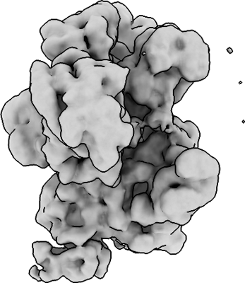 emd_13021.png
 http://ftp.pdbj.org/pub/emdb/structures/EMD-13021
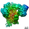


 Z
Z Y
Y X
X


 Sample components

 Processing
 Electron microscopy