 Movie
Movie Controller


+ Open data

- Basic information
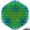
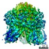
| Entry | Database: EMDB / ID: EMD-11112 | |||||||||||||||
|---|---|---|---|---|---|---|---|---|---|---|---|---|---|---|---|---|
| Title | The atomic structure of the HAdVF-41 penton base in solution | |||||||||||||||
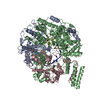
 Map data Map data | 3D reconstruction of a recombinantely expressed human Adenovirus 41 penton base. | |||||||||||||||
 Sample Sample |
| |||||||||||||||
| Function / homology | Adenovirus penton base protein / Adenovirus penton base protein / T=25 icosahedral viral capsid / endocytosis involved in viral entry into host cell / host cell nucleus / virion attachment to host cell / structural molecule activity /  Penton protein Penton protein Function and homology information Function and homology information | |||||||||||||||
| Biological species |  Human adenovirus F serotype 41 / Human adenovirus 41 Human adenovirus F serotype 41 / Human adenovirus 41 | |||||||||||||||
| Method | single particle reconstruction / cryo EM / Resolution: 3.8 Å | |||||||||||||||
 Authors Authors | Carlson L-A / Rafie K | |||||||||||||||
| Funding support |  Sweden, 4 items Sweden, 4 items
| |||||||||||||||
 Citation Citation | Journal: Sci Adv / Year: 2021 Title: The structure of enteric human adenovirus 41-A leading cause of diarrhea in children. Authors: K Rafie / A Lenman / J Fuchs / A Rajan / N Arnberg / L-A Carlson /  Abstract: Human adenovirus (HAdV) types F40 and F41 are a prominent cause of diarrhea and diarrhea-associated mortality in young children worldwide. These enteric HAdVs differ notably in tissue tropism and ...Human adenovirus (HAdV) types F40 and F41 are a prominent cause of diarrhea and diarrhea-associated mortality in young children worldwide. These enteric HAdVs differ notably in tissue tropism and pathogenicity from respiratory and ocular adenoviruses, but the structural basis for this divergence has been unknown. Here, we present the first structure of an enteric HAdV-HAdV-F41-determined by cryo-electron microscopy to a resolution of 3.8 Å. The structure reveals extensive alterations to the virion exterior as compared to nonenteric HAdVs, including a unique arrangement of capsid protein IX. The structure also provides new insights into conserved aspects of HAdV architecture such as a proposed location of core protein V, which links the viral DNA to the capsid, and assembly-induced conformational changes in the penton base protein. Our findings provide the structural basis for adaptation of enteric HAdVs to a fundamentally different tissue tropism. | |||||||||||||||
| History |
|
- Structure visualization
| Movie |
Movie viewer |
|---|---|
| Structure viewer | EM map: SurfViewMolmilJmol/JSmol |
| Supplemental images |

- Downloads & links
-EMDB archive
| Map data | emd_11112.map.gz | 17.7 MB | EMDB map data format | |
|---|---|---|---|---|
| Header (meta data) | emd-11112-v30.xmlemd-11112.xml | 19.2 KB 19.2 KB | Display Display | EMDB header |
| Images | 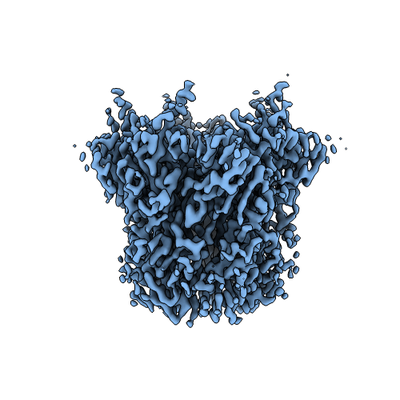 emd_11112.png emd_11112.png | 108.4 KB | ||
| Others | emd_11112_half_map_1.map.gzemd_11112_half_map_2.map.gz | 14.8 MB 14.8 MB | ||
| Archive directory |  http://ftp.pdbj.org/pub/emdb/structures/EMD-11112ftp://ftp.pdbj.org/pub/emdb/structures/EMD-11112 http://ftp.pdbj.org/pub/emdb/structures/EMD-11112ftp://ftp.pdbj.org/pub/emdb/structures/EMD-11112 | HTTPS FTP |
-Related structure data
| Related structure data |  6z7qMC  6z7nC M: atomic model generated by this map C: citing same article ( |
|---|---|
| Similar structure data |


-Links
| EMDB pages | EMDB (EBI/PDBe) / EMDataResource |
|---|---|
| Related items in Molecule of the Month |


-Map
| File | Download / File: emd_11112.map.gz / Format: CCP4 / Size: 19.4 MB / Type: IMAGE STORED AS FLOATING POINT NUMBER (4 BYTES) | ||||||||||||||||||||||||||||||||||||||||||||||||||||||||||||||||||||
|---|---|---|---|---|---|---|---|---|---|---|---|---|---|---|---|---|---|---|---|---|---|---|---|---|---|---|---|---|---|---|---|---|---|---|---|---|---|---|---|---|---|---|---|---|---|---|---|---|---|---|---|---|---|---|---|---|---|---|---|---|---|---|---|---|---|---|---|---|---|
| Annotation | 3D reconstruction of a recombinantely expressed human Adenovirus 41 penton base. | ||||||||||||||||||||||||||||||||||||||||||||||||||||||||||||||||||||
| Voxel size | X=Y=Z: 1.041 Å | ||||||||||||||||||||||||||||||||||||||||||||||||||||||||||||||||||||
| Density |
| ||||||||||||||||||||||||||||||||||||||||||||||||||||||||||||||||||||
| Symmetry | Space group: 1 | ||||||||||||||||||||||||||||||||||||||||||||||||||||||||||||||||||||
| Details | EMDB XML:
CCP4 map header:
| ||||||||||||||||||||||||||||||||||||||||||||||||||||||||||||||||||||
-Supplemental data
-Half map: Half Map 1
| File | emd_11112_half_map_1.map | ||||||||||||
|---|---|---|---|---|---|---|---|---|---|---|---|---|---|
| Annotation | Half Map 1 | ||||||||||||
| Projections & Slices |
| ||||||||||||
| Density Histograms |
 Z
Z Y
Y X
X


-Half map: Half Map 2
| File | emd_11112_half_map_2.map | ||||||||||||
|---|---|---|---|---|---|---|---|---|---|---|---|---|---|
| Annotation | Half Map 2 | ||||||||||||
| Projections & Slices |
| ||||||||||||
| Density Histograms |


- Sample components
-Entire : Human adenovirus 41
| Entire | Name: Human adenovirus 41 |
|---|---|
| Components |
|
-Supramolecule #1: Human adenovirus 41
| Supramolecule | Name: Human adenovirus 41 / type: virus / ID: 1 / Parent: 0 / Macromolecule list: all Details: The HAdVF-41 penton base was recombinantely expressed in Sf9 cells. NCBI-ID: 10524 / Sci species name: Human adenovirus 41 / Virus type: VIRION / Virus isolate: SEROTYPE / Virus enveloped: No / Virus empty: No |
|---|---|
| Host (natural) | Organism:  Homo sapiens (human) Homo sapiens (human) |
| Host system | Organism:   Spodoptera frugiperda (fall armyworm) / Recombinant cell: Sf9 Spodoptera frugiperda (fall armyworm) / Recombinant cell: Sf9 |
| Molecular weight | Theoretical: 285 KDa |
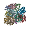
-Macromolecule #1: Penton protein
| Macromolecule | Name: Penton protein / type: protein_or_peptide / ID: 1 / Number of copies: 5 / Enantiomer: LEVO |
|---|---|
| Source (natural) | Organism: Human adenovirus F serotype 41 |
| Molecular weight | Theoretical: 57.14216 KDa |
| Recombinant expression | Organism: Spodoptera frugiperda (fall armyworm) |
| Sequence | String: MRRAVGVPPV MAYAEGPPPS YESVMGSADS PATLEALYVP PRYLGPTEGR NSIRYSELAP LYDTTRVYLV DNKSADIASL NYQNDHSNF QTTVVQNNDF TPAEAGTQTI NFDERSRWGA DLKTILRTNM PNINEFMSTN KFKARLMVEK KNKETGLPRY E WFEFTLPE ...String: MRRAVGVPPV MAYAEGPPPS YESVMGSADS PATLEALYVP PRYLGPTEGR NSIRYSELAP LYDTTRVYLV DNKSADIASL NYQNDHSNF QTTVVQNNDF TPAEAGTQTI NFDERSRWGA DLKTILRTNM PNINEFMSTN KFKARLMVEK KNKETGLPRY E WFEFTLPE GNYSETMTID LMNNAIVDNY LEVGRQNGVL ESDIGVKFDT RNFRLGWDPV TKLVMPGVYT NEAFHPDIVL LP GCGVDFT QSRLSNLLGI RKRLPFQEGF QIMYEDLEGG NIPALLDVAK YEASIQKAKE EGKEIGDDTF ATRPQDLVIE PVA KDSKNR SYNLLPNDQN NTAYRSWFLA YNYGDPKKGV QSWTLLTTAD VTCGSQQVYW SLPDMMQDPV TFRPSTQVSN YPVV GVELL PVHAKSFYNE QAVYSQLIRQ STALTHVFNR FPENQILVRP PAPTITTVSE NVPALTDHGT LPLRSSISGV QRVTI TDAR RRTCPYVHKA LGIVAPKVLS SRTF |
-Experimental details
-Structure determination
| Method | cryo EM |
|---|---|
 Processing Processing | single particle reconstruction |
| Aggregation state | particle |
-Sample preparation
| Concentration | 1 mg/mL |
|---|---|
| Buffer | pH: 7.4 |
| Grid | Model: Quantifoil R1.2/1.3 / Material: COPPER / Mesh: 200 / Pretreatment - Type: GLOW DISCHARGE / Pretreatment - Atmosphere: OTHER |
| Vitrification | Cryogen name: ETHANE / Chamber humidity: 80 % / Chamber temperature: 295.15 K / Instrument: FEI VITROBOT MARK IV |
| Details | The sample was monodisperse with a preferred orientation in the vitrious ice. |
- Electron microscopy
| Microscope | FEI TITAN KRIOS |
|---|---|
| Electron beam | Acceleration voltage: 300 kV / Electron source: FIELD EMISSION GUN |
| Electron optics | C2 aperture diameter: 100.0 µm / Illumination mode: FLOOD BEAM / Imaging mode: BRIGHT FIELDBright-field microscopy / Nominal defocus max: 3.0 µm / Nominal defocus min: 0.5 µm / Nominal magnification: 130000 |
| Specialist optics | Energy filter - Name: GIF Bioquantum / Energy filter - Slit width: 20 eV |
| Sample stage | Specimen holder model: FEI TITAN KRIOS AUTOGRID HOLDER / Cooling holder cryogen: NITROGEN |
| Details | Data were collected at a 30 degree tilt of the specimen stage due to a preferred orientation of the sample in the vitrified ice. |
| Image recording | Film or detector model: GATAN K2 QUANTUM (4k x 4k) / Detector mode: COUNTING / Digitization - Frames/image: 1-40 / Number grids imaged: 1 / Number real images: 948 / Average electron dose: 0.93 e/Å2 |
| Experimental equipment |  Model: Titan Krios / Image courtesy: FEI Company |
-Image processing
| Particle selection | Number selected: 198491 |
|---|---|
| CTF correction | Software - Name: Gctf (ver. 1.18) Software - details: per-micrograph and initial per-particle CTF correction Details: per-micrograph CTF correction as well the initial per-particle CTF correction before 2D classification were performed in GCTF. Later per-particle CTF-refinement were performed in RELION 3.1-beta. |
| Startup model | Type of model: OTHER Details: An initial dataset of the HAdV-F41 penton base collected at no tilt of the specimen stage was used to generate a low-resolution input model. |
| Initial angle assignment | Type: MAXIMUM LIKELIHOOD / Software - Name: RELION (ver. 3.1-beta) |
| Final 3D classification | Software - Name: RELION (ver. 3.1-beta) |
| Final angle assignment | Type: MAXIMUM LIKELIHOOD / Software - Name: RELION (ver. 3.1-beta) |
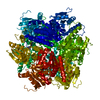
| Final reconstruction | Applied symmetry - Point group: C5 (5 fold cyclic) / Resolution.type: BY AUTHOR / Resolution: 3.8 Å / Resolution method: FSC 0.143 CUT-OFF / Software - Name: RELION (ver. 3.1-beta) / Number images used: 107110 |
-Atomic model buiding 1
| Refinement | Space: REAL / Protocol: RIGID BODY FIT |
|---|---|
| Output model | PDB-6z7q: |
