 Movie
Movie Controller

[English] 日本語
 Yorodumi
- EMDB-11110: Local asymmetric reconstruction of the HVR2-containing loop of HA... -
+ Open data

- Basic information
| Entry | Database: EMDB / ID: EMD-11110 | |||||||||||||||
|---|---|---|---|---|---|---|---|---|---|---|---|---|---|---|---|---|
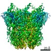
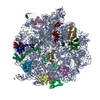
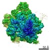
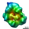
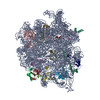
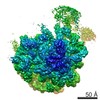
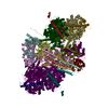
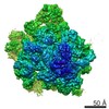
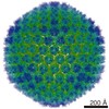
| Title | Local asymmetric reconstruction of the HVR2-containing loop of HAdV-F41. | |||||||||||||||
 Map data Map data | Full unmasked volume | |||||||||||||||
 Sample Sample |
| |||||||||||||||
| Biological species |   Human adenovirus 41 Human adenovirus 41 | |||||||||||||||
| Method | single particle reconstruction / cryo EM / Resolution: 3.35 Å | |||||||||||||||
 Authors Authors | Carlson L-A / Rafie K | |||||||||||||||
| Funding support |  Sweden, 4 items Sweden, 4 items
| |||||||||||||||
 Citation Citation | Journal: Sci Adv / Year: 2021 Title: The structure of enteric human adenovirus 41-A leading cause of diarrhea in children. Authors: K Rafie / A Lenman / J Fuchs / A Rajan / N Arnberg / L-A Carlson /  Abstract: Human adenovirus (HAdV) types F40 and F41 are a prominent cause of diarrhea and diarrhea-associated mortality in young children worldwide. These enteric HAdVs differ notably in tissue tropism and ...Human adenovirus (HAdV) types F40 and F41 are a prominent cause of diarrhea and diarrhea-associated mortality in young children worldwide. These enteric HAdVs differ notably in tissue tropism and pathogenicity from respiratory and ocular adenoviruses, but the structural basis for this divergence has been unknown. Here, we present the first structure of an enteric HAdV-HAdV-F41-determined by cryo-electron microscopy to a resolution of 3.8 Å. The structure reveals extensive alterations to the virion exterior as compared to nonenteric HAdVs, including a unique arrangement of capsid protein IX. The structure also provides new insights into conserved aspects of HAdV architecture such as a proposed location of core protein V, which links the viral DNA to the capsid, and assembly-induced conformational changes in the penton base protein. Our findings provide the structural basis for adaptation of enteric HAdVs to a fundamentally different tissue tropism. | |||||||||||||||
| History |
|
- Structure visualization
| Movie |
 Movie viewer Movie viewer |
|---|---|
| Structure viewer | EM map: SurfViewMolmilJmol/JSmol |
| Supplemental images |

- Downloads & links
-EMDB archive
| Map data | emd_11110.map.gz | 23.2 MB | EMDB map data format | |
|---|---|---|---|---|
| Header (meta data) | emd-11110-v30.xmlemd-11110.xml | 17.8 KB 17.8 KB | Display Display | EMDB header |
| Images | 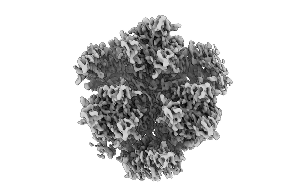 emd_11110.png emd_11110.png | 74 KB | ||
| Masks | emd_11110_msk_1.map | 30.5 MB | Mask map | |
| Others | emd_11110_half_map_1.map.gzemd_11110_half_map_2.map.gz | 23.2 MB 23.2 MB | ||
| Archive directory |  http://ftp.pdbj.org/pub/emdb/structures/EMD-11110ftp://ftp.pdbj.org/pub/emdb/structures/EMD-11110 http://ftp.pdbj.org/pub/emdb/structures/EMD-11110ftp://ftp.pdbj.org/pub/emdb/structures/EMD-11110 | HTTPS FTP |
-Related structure data


-Links
| EMDB pages | EMDB (EBI/PDBe) / EMDataResource |
|---|
-Map
| File | Download / File: emd_11110.map.gz / Format: CCP4 / Size: 30.5 MB / Type: IMAGE STORED AS FLOATING POINT NUMBER (4 BYTES) | ||||||||||||||||||||||||||||||||||||||||||||||||||||||||||||
|---|---|---|---|---|---|---|---|---|---|---|---|---|---|---|---|---|---|---|---|---|---|---|---|---|---|---|---|---|---|---|---|---|---|---|---|---|---|---|---|---|---|---|---|---|---|---|---|---|---|---|---|---|---|---|---|---|---|---|---|---|---|
| Annotation | Full unmasked volume | ||||||||||||||||||||||||||||||||||||||||||||||||||||||||||||
| Voxel size | X=Y=Z: 1.041 Å | ||||||||||||||||||||||||||||||||||||||||||||||||||||||||||||
| Density |
| ||||||||||||||||||||||||||||||||||||||||||||||||||||||||||||
| Symmetry | Space group: 1 | ||||||||||||||||||||||||||||||||||||||||||||||||||||||||||||
| Details | EMDB XML:
CCP4 map header:
| ||||||||||||||||||||||||||||||||||||||||||||||||||||||||||||
-Supplemental data
-Mask #1
| File | emd_11110_msk_1.map | ||||||||||||
|---|---|---|---|---|---|---|---|---|---|---|---|---|---|
| Projections & Slices |
| ||||||||||||
| Density Histograms |
 Z
Z Y
Y X
X


-Half map: Half Map 1
| File | emd_11110_half_map_1.map | ||||||||||||
|---|---|---|---|---|---|---|---|---|---|---|---|---|---|
| Annotation | Half Map 1 | ||||||||||||
| Projections & Slices |
| ||||||||||||
| Density Histograms |


-Half map: Half Map 2
| File | emd_11110_half_map_2.map | ||||||||||||
|---|---|---|---|---|---|---|---|---|---|---|---|---|---|
| Annotation | Half Map 2 | ||||||||||||
| Projections & Slices |
| ||||||||||||
| Density Histograms |


- Sample components
-Entire : Human adenovirus 41
| Entire | Name: Human adenovirus 41 |
|---|---|
| Components |
|
-Supramolecule #1: Human adenovirus 41
| Supramolecule | Name: Human adenovirus 41 / type: virus / ID: 1 / Parent: 0 / Macromolecule list: #1-#11 / NCBI-ID: 10524 / Sci species name: Human adenovirus 41 / Virus type: VIRION / Virus isolate: SEROTYPE / Virus enveloped: No / Virus empty: No |
|---|---|
| Host (natural) | Organism:  Homo sapiens (human) Homo sapiens (human) |
| Virus shell | Shell ID: 1 / Diameter: 900.0 Å |
-Experimental details
-Structure determination
| Method | cryo EM |
|---|---|
 Processing Processing | single particle reconstruction |
| Aggregation state | particle |
-Sample preparation
| Concentration | 4.2 mg/mL |
|---|---|
| Buffer | pH: 7.4 Details: PBS buffer made from comemrcial PBS tablets Medicargo, PBS tablets, 09-9400-100 |
| Vitrification | Cryogen name: ETHANE / Chamber humidity: 80 % / Chamber temperature: 295.15 K / Instrument: FEI VITROBOT MARK IV |
- Electron microscopy
| Microscope | FEI TITAN KRIOS |
|---|---|
| Electron beam | Acceleration voltage: 300 kV / Electron source: FIELD EMISSION GUN |
| Electron optics | C2 aperture diameter: 100.0 µm / Illumination mode: FLOOD BEAM / Imaging mode: BRIGHT FIELDBright-field microscopy / Nominal defocus max: 3.0 µm / Nominal defocus min: 0.5 µm / Nominal magnification: 130000 |
| Specialist optics | Energy filter - Name: GIF Bioquantum / Energy filter - Slit width: 20 eV |
| Sample stage | Specimen holder model: FEI TITAN KRIOS AUTOGRID HOLDER / Cooling holder cryogen: NITROGEN |
| Image recording | Film or detector model: GATAN K2 QUANTUM (4k x 4k) / Detector mode: COUNTING / Digitization - Frames/image: 2-20 / Number grids imaged: 2 / Number real images: 6929 / Average electron dose: 1.03 e/Å2 |
| Experimental equipment |  Model: Titan Krios / Image courtesy: FEI Company |
-Image processing
| Particle selection | Number selected: 1168320 |
|---|---|
| CTF correction | Software - Name: Gctf (ver. 1.18) / Software - details: icle CTF correction |
| Startup model | Type of model: EMDB MAP EMDB ID: |
| Initial angle assignment | Type: MAXIMUM LIKELIHOOD / Software - Name: RELION (ver. 3.0-beta) |
| Final 3D classification | Software - Name: RELION (ver. 3.0-beta) |
| Final angle assignment | Type: MAXIMUM LIKELIHOOD / Software - Name: RELION (ver. 3.0-beta) |
| Final reconstruction | Applied symmetry - Point group: C1 (asymmetric) / Resolution.type: BY AUTHOR / Resolution: 3.35 Å / Resolution method: FSC 0.143 CUT-OFF / Software - Name: RELION (ver. 3.0-beta) / Number images used: 467190 |
-Atomic model buiding 1
| Initial model | PDB ID: |
|---|---|
| Refinement | Space: REAL / Protocol: RIGID BODY FIT |
