 Movie
Movie Controller

[English] 日本語
 Yorodumi
- EMDB-11077: Structure of the influenza A matrix protein M1 from influenza A/H... -
+ Open data

- Basic information
| Entry | Database: EMDB / ID: EMD-11077 | ||||||||||||
|---|---|---|---|---|---|---|---|---|---|---|---|---|---|
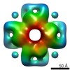
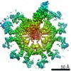
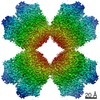
| Title | Structure of the influenza A matrix protein M1 from influenza A/Hong Kong/1/1968 virions | ||||||||||||
 Map data Map data | Structure of the influenza A matrix protein M1 from influenza A/Hong Kong/1968 virions | ||||||||||||
 Sample Sample |
| ||||||||||||
| Function / homology |  Function and homology information Function and homology information viral budding from plasma membrane / structural constituent of virion / host cell nucleus / virion membrane / RNA binding viral budding from plasma membrane / structural constituent of virion / host cell nucleus / virion membrane / RNA bindingSimilarity search - Function | ||||||||||||
| Biological species |   Influenza A virus Influenza A virus | ||||||||||||
| Method | subtomogram averaging / cryo EM / Resolution: 8.0 Å | ||||||||||||
 Authors Authors | Peukes J / Xiong X / Erlendsson S / Qu K / Wan W / Kraeusslich H-G / Briggs JAG | ||||||||||||
| Funding support | European Union,  Germany, Germany,  United Kingdom, 3 items United Kingdom, 3 items
| ||||||||||||
 Citation Citation | Journal: Nature / Year: 2020 Title: The native structure of the assembled matrix protein 1 of influenza A virus. Authors: Julia Peukes / Xiaoli Xiong / Simon Erlendsson / Kun Qu / William Wan / Leslie J Calder / Oliver Schraidt / Susann Kummer / Stefan M V Freund / Hans-Georg Kräusslich / John A G Briggs /    Abstract: Influenza A virus causes millions of severe cases of disease during annual epidemics. The most abundant protein in influenza virions is matrix protein 1 (M1), which mediates virus assembly by ...Influenza A virus causes millions of severe cases of disease during annual epidemics. The most abundant protein in influenza virions is matrix protein 1 (M1), which mediates virus assembly by forming an endoskeleton beneath the virus membrane. The structure of full-length M1, and how it oligomerizes to mediate the assembly of virions, is unknown. Here we determine the complete structure of assembled M1 within intact virus particles, as well as the structure of M1 oligomers reconstituted in vitro. We find that the C-terminal domain of M1 is disordered in solution but can fold and bind in trans to the N-terminal domain of another M1 monomer, thus polymerizing M1 into linear strands that coat the interior surface of the membrane of the assembling virion. In the M1 polymer, five histidine residues-contributed by three different monomers of M1-form a cluster that can serve as the pH-sensitive disassembly switch after entry into a target cell. These structures therefore reveal mechanisms of influenza virus assembly and disassembly. | ||||||||||||
| History |
|
- Structure visualization
| Movie |
Movie viewer |
|---|---|
| Structure viewer | EM map: SurfViewMolmilJmol/JSmol |
| Supplemental images |

- Downloads & links
-EMDB archive
| Map data | emd_11077.map.gz | 6.1 MB | EMDB map data format | |
|---|---|---|---|---|
| Header (meta data) | emd-11077-v30.xmlemd-11077.xml | 16.8 KB 16.8 KB | Display Display | EMDB header |
| FSC (resolution estimation) | emd_11077_fsc.xml | 4.4 KB | Display | FSC data file |
| Images | 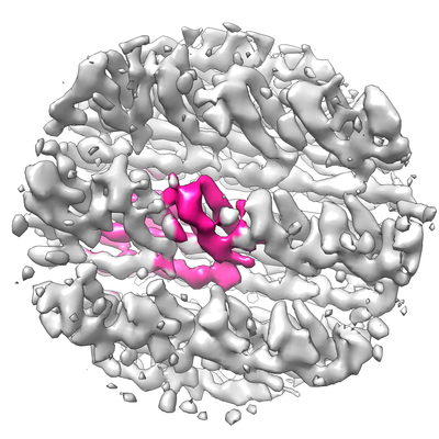 emd_11077.png emd_11077.png | 153.3 KB | ||
| Masks | emd_11077_msk_1.map | 6.6 MB | Mask map | |
| Others | emd_11077_half_map_1.map.gzemd_11077_half_map_2.map.gz | 5.9 MB 6.1 MB | ||
| Archive directory |  http://ftp.pdbj.org/pub/emdb/structures/EMD-11077ftp://ftp.pdbj.org/pub/emdb/structures/EMD-11077 http://ftp.pdbj.org/pub/emdb/structures/EMD-11077ftp://ftp.pdbj.org/pub/emdb/structures/EMD-11077 | HTTPS FTP |
-Related structure data


-Links
| EMDB pages | EMDB (EBI/PDBe) / EMDataResource |
|---|---|
| Related items in Molecule of the Month |


-Map
| File | Download / File: emd_11077.map.gz / Format: CCP4 / Size: 6.6 MB / Type: IMAGE STORED AS FLOATING POINT NUMBER (4 BYTES) | ||||||||||||||||||||||||||||||||||||||||||||||||||||||||||||||||||||
|---|---|---|---|---|---|---|---|---|---|---|---|---|---|---|---|---|---|---|---|---|---|---|---|---|---|---|---|---|---|---|---|---|---|---|---|---|---|---|---|---|---|---|---|---|---|---|---|---|---|---|---|---|---|---|---|---|---|---|---|---|---|---|---|---|---|---|---|---|---|
| Annotation | Structure of the influenza A matrix protein M1 from influenza A/Hong Kong/1968 virions | ||||||||||||||||||||||||||||||||||||||||||||||||||||||||||||||||||||
| Voxel size | X=Y=Z: 1.7 Å | ||||||||||||||||||||||||||||||||||||||||||||||||||||||||||||||||||||
| Density |
| ||||||||||||||||||||||||||||||||||||||||||||||||||||||||||||||||||||
| Symmetry | Space group: 1 | ||||||||||||||||||||||||||||||||||||||||||||||||||||||||||||||||||||
| Details | EMDB XML:
CCP4 map header:
| ||||||||||||||||||||||||||||||||||||||||||||||||||||||||||||||||||||
-Supplemental data
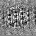
-Mask #1
| File | emd_11077_msk_1.map | ||||||||||||
|---|---|---|---|---|---|---|---|---|---|---|---|---|---|
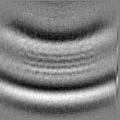
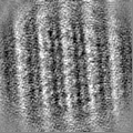
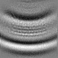
| Projections & Slices |
| ||||||||||||
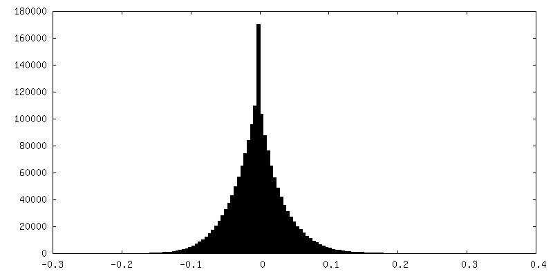
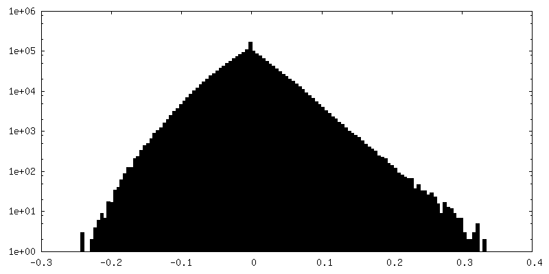
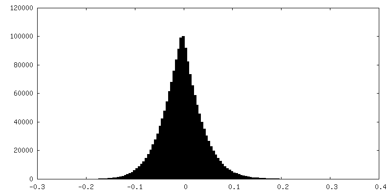
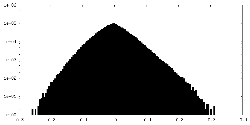
| Density Histograms |
 Z
Z Y
Y X
X


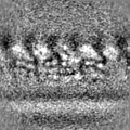
-Half map: #1
| File | emd_11077_half_map_1.map | ||||||||||||
|---|---|---|---|---|---|---|---|---|---|---|---|---|---|
| Projections & Slices |
| ||||||||||||
| Density Histograms |


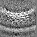
-Half map: #2
| File | emd_11077_half_map_2.map | ||||||||||||
|---|---|---|---|---|---|---|---|---|---|---|---|---|---|
| Projections & Slices |
| ||||||||||||
| Density Histograms |


- Sample components
-Entire : Influenza A virus
| Entire | Name: Influenza A virus |
|---|---|
| Components |
|
-Supramolecule #1: Influenza A virus
| Supramolecule | Name: Influenza A virus / type: virus / ID: 1 / Parent: 0 / Macromolecule list: #1 / NCBI-ID: 11320 / Sci species name: Influenza A virus / Sci species strain: A/Hong Kong/1/1968 (H3N2) / Virus type: VIRION / Virus isolate: OTHER / Virus enveloped: Yes / Virus empty: No |
|---|---|
| Host (natural) | Organism:  Homo sapiens (human) Homo sapiens (human) |
-Supramolecule #2: Matrix protein 1
| Supramolecule | Name: Matrix protein 1 / type: complex / ID: 2 / Parent: 1 / Macromolecule list: #1 Details: The M1 map was generated by subtomogram averaging of the M1 protein density in tomograms of influenza A virions. |
|---|---|
| Source (natural) | Organism: Influenza A virus / Strain: A/Hong Kong/1/1968 (H3N2) |
-Experimental details
-Structure determination
| Method | cryo EM |
|---|---|
 Processing Processing | subtomogram averaging |
| Aggregation state | particle |
-Sample preparation
| Buffer | pH: 7.5 |
|---|---|
| Grid | Model: Quantifoil R2/2 / Material: GOLD / Mesh: 200 / Support film - Material: CARBON |
| Vitrification | Cryogen name: ETHANE / Chamber humidity: 95 % / Instrument: LEICA EM GP |
- Electron microscopy
| Microscope | FEI TITAN KRIOS |
|---|---|
| Electron beam | Acceleration voltage: 300 kV / Electron source: FIELD EMISSION GUN |
| Electron optics | Illumination mode: FLOOD BEAM / Imaging mode: BRIGHT FIELDBright-field microscopy / Cs: 2.7 mm / Nominal defocus max: 4.0 µm / Nominal defocus min: 1.5 µm / Nominal magnification: 81000 |
| Specialist optics | Energy filter - Name: GIF Quantum LS / Energy filter - Slit width: 20 eV |
| Sample stage | Specimen holder model: FEI TITAN KRIOS AUTOGRID HOLDER / Cooling holder cryogen: NITROGEN |
| Image recording | Film or detector model: GATAN K2 QUANTUM (4k x 4k) / Detector mode: COUNTING / Average electron dose: 3.2 e/Å2 |
| Experimental equipment |  Model: Titan Krios / Image courtesy: FEI Company |
-Image processing
| Extraction | Number tomograms: 5 / Number images used: 100000 Method: Volumes picked semi-automatically along the particles membrane Software: (Name: TOM, MATLAB, UCSF Chimera)Details: Initial positions were generated by simulating the surface of a cylindricated volume around the manually determined central axis of each particle. Initial positions have been 2X oversampled. |
|---|---|
| CTF correction | Software: (Name: CTFFIND, NOVACTF) Details: CTF determination was performed using CTFFIND4. CTF correction was performed using 3D-CTF correction by CTF multiplication in NovaCTF. |
| Final angle assignment | Type: OTHER |
| Final reconstruction | Applied symmetry - Point group: C1 (asymmetric) / Resolution.type: BY AUTHOR / Resolution: 8.0 Å / Resolution method: FSC 0.143 CUT-OFF / Number subtomograms used: 12128 |
| FSC plot (resolution estimation) |  |
