 Movie
Movie Controller


+ Open data

- Basic information
| Entry | Database: EMDB / ID: EMD-10745 | |||||||||
|---|---|---|---|---|---|---|---|---|---|---|
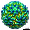
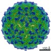
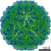
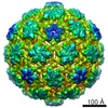
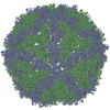
| Title | Leishmania RNA virus capsid with exogenous mRNA | |||||||||
 Map data Map data | ||||||||||
 Sample Sample |
| |||||||||
| Biological species |  Leishmania RNA virus 1 - LgM5313 Leishmania RNA virus 1 - LgM5313 | |||||||||
| Method |  single particle reconstruction / cryo EM / Resolution: 3.76 Å single particle reconstruction / cryo EM / Resolution: 3.76 Å | |||||||||
 Authors Authors | Prochazkova M | |||||||||
| Funding support |  Czech Republic, 1 items Czech Republic, 1 items
| |||||||||
 Citation Citation | Journal: J Virol / Year: 2021 Title: Capsid Structure of RNA Virus 1. Authors: Michaela Procházková / Tibor Füzik / Danyil Grybchuk / Francesco Luca Falginella / Lucie Podešvová / Vyacheslav Yurchenko / Robert Vácha / Pavel Plevka /  Abstract: parasites cause a variety of symptoms, including mucocutaneous leishmaniasis, which results in the destruction of the mucous membranes of the nose, mouth, and throat. The species of carrying RNA ... parasites cause a variety of symptoms, including mucocutaneous leishmaniasis, which results in the destruction of the mucous membranes of the nose, mouth, and throat. The species of carrying RNA virus 1 (LRV1), from the family , are more likely to cause severe disease and are less sensitive to treatment than those that do not contain the virus. Although the importance of LRV1 for the severity of leishmaniasis was discovered a long time ago, the structure of the virus remained unknown. Here, we present a cryo-electron microscopy reconstruction of the virus-like particle of LRV1 determined to a resolution of 3.65 Å. The capsid has icosahedral symmetry and is formed by 120 copies of a capsid protein assembled in asymmetric dimers. RNA genomes of viruses from the family are synthetized, but not capped at the 5' end, by virus RNA polymerases. To protect viral RNAs from degradation, capsid proteins of the L-A totivirus cleave the 5' caps of host mRNAs, creating decoys to overload the cellular RNA quality control system. Capsid proteins of LRV1 form positively charged clefts, which may be the cleavage sites for the 5' cap of mRNAs. The putative RNA binding site of LRV1 is distinct from that of the related L-A virus. The structure of the LRV1 capsid enables the rational design of compounds targeting the putative decapping site. Such inhibitors may be developed into a treatment for mucocutaneous leishmaniasis caused by LRV1-positive species of Twelve million people worldwide suffer from leishmaniasis, resulting in more than 30 thousand deaths annually. The disease has several variants that differ in their symptoms. The mucocutaneous form, which leads to disintegration of the nasal septum, lips, and palate, is caused predominantly by parasites carrying RNA virus 1 (LRV1). Here, we present the structure of the LRV1 capsid determined using cryo-electron microscopy. Capsid proteins of a related totivirus, L-A virus, protect viral RNAs from degradation by cleaving the 5' caps of host mRNAs. Capsid proteins of LRV1 may have the same function. We show that the LRV1 capsid contains positively charged clefts that may be sites for the cleavage of mRNAs of cells. The structure of the LRV1 capsid enables the rational design of compounds targeting the putative mRNA cleavage site. Such inhibitors may be used as treatments for mucocutaneous leishmaniasis. | |||||||||
| History |
|
- Structure visualization
| Movie |
 Movie viewer Movie viewer |
|---|---|
| Structure viewer | EM map: SurfViewMolmilJmol/JSmol |
| Supplemental images |

- Downloads & links
-EMDB archive
| Map data | emd_10745.map.gz | 76.6 MB | EMDB map data format | |
|---|---|---|---|---|
| Header (meta data) | emd-10745-v30.xmlemd-10745.xml | 14 KB 14 KB | Display Display | EMDB header |
| FSC (resolution estimation) | emd_10745_fsc.xml | 18.7 KB | Display | FSC data file |
| Images | 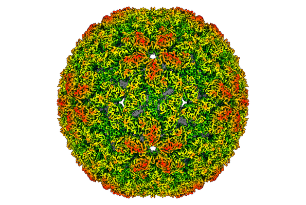 emd_10745.png emd_10745.png | 271.4 KB | ||
| Archive directory |  http://ftp.pdbj.org/pub/emdb/structures/EMD-10745ftp://ftp.pdbj.org/pub/emdb/structures/EMD-10745 http://ftp.pdbj.org/pub/emdb/structures/EMD-10745ftp://ftp.pdbj.org/pub/emdb/structures/EMD-10745 | HTTPS FTP |
-Related structure data


-Links
| EMDB pages | EMDB (EBI/PDBe) / EMDataResource |
|---|
-Map
| File | Download / File: emd_10745.map.gz / Format: CCP4 / Size: 620.9 MB / Type: IMAGE STORED AS FLOATING POINT NUMBER (4 BYTES) | ||||||||||||||||||||||||||||||||||||||||||||||||||||||||||||
|---|---|---|---|---|---|---|---|---|---|---|---|---|---|---|---|---|---|---|---|---|---|---|---|---|---|---|---|---|---|---|---|---|---|---|---|---|---|---|---|---|---|---|---|---|---|---|---|---|---|---|---|---|---|---|---|---|---|---|---|---|---|
| Voxel size | X=Y=Z: 1.063 Å | ||||||||||||||||||||||||||||||||||||||||||||||||||||||||||||
| Density |
| ||||||||||||||||||||||||||||||||||||||||||||||||||||||||||||
| Symmetry | Space group: 1 | ||||||||||||||||||||||||||||||||||||||||||||||||||||||||||||
| Details | EMDB XML:
CCP4 map header:
| ||||||||||||||||||||||||||||||||||||||||||||||||||||||||||||
-Supplemental data
- Sample components
-Entire : Leishmania RNA virus 1 - LgM5313
| Entire | Name: Leishmania RNA virus 1 - LgM5313 |
|---|---|
| Components |
|
-Supramolecule #1: Leishmania RNA virus 1 - LgM5313
| Supramolecule | Name: Leishmania RNA virus 1 - LgM5313 / type: virus / ID: 1 / Parent: 0 / Macromolecule list: #1 Details: Assembled particles mixed with isolated Leishmania mRNA NCBI-ID: 1285422 / Sci species name: Leishmania RNA virus 1 - LgM5313 / Virus type: VIRUS-LIKE PARTICLE / Virus isolate: OTHER / Virus enveloped: No / Virus empty: Yes |
|---|---|
| Host (natural) | Organism:  Leishmania guyanensis (eukaryote) / Strain: MHOM/BR/75/M4147 Leishmania guyanensis (eukaryote) / Strain: MHOM/BR/75/M4147 |
| Host system | Organism:  Escherichia coli BL21(DE3) (bacteria) Escherichia coli BL21(DE3) (bacteria) |
| Virus shell | Shell ID: 1 / Name: capsid / Diameter: 422.0 Å / T number (triangulation number): 2 |
-Experimental details
-Structure determination
| Method | cryo EM |
|---|---|
 Processing Processing | single particle reconstruction |
| Aggregation state | particle |
-Sample preparation
| Concentration | 15 mg/mL | |||||||||||||||
|---|---|---|---|---|---|---|---|---|---|---|---|---|---|---|---|---|
| Buffer | pH: 8 Component:
Details: Supplemented with isolated mRNA from Leishmania sp. | |||||||||||||||
| Grid | Model: Quantifoil R2/1 / Material: COPPER / Mesh: 300 / Support film - Material: CARBON / Support film - topology: HOLEY | |||||||||||||||
| Vitrification | Cryogen name: ETHANE / Chamber humidity: 70 % / Chamber temperature: 295 K / Instrument: FEI VITROBOT MARK II / Details: blot force 3 blot time 5 wait time 10. |
- Electron microscopy
| Microscope | FEI TITAN KRIOS |
|---|---|
| Electron beam | Acceleration voltage: 300 kV / Electron source: FIELD EMISSION GUN |
| Electron optics | C2 aperture diameter: 50.0 µm / Illumination mode: FLOOD BEAM / Imaging mode: BRIGHT FIELDBright-field microscopy / Nominal defocus max: 0.0005 µm / Nominal defocus min: -0.0005 µm / Nominal magnification: 75000 |
| Sample stage | Specimen holder model: FEI TITAN KRIOS AUTOGRID HOLDER / Cooling holder cryogen: NITROGEN |
| Image recording | Film or detector model: FEI FALCON III (4k x 4k) / Detector mode: COUNTING / Number grids imaged: 1 / Number real images: 8000 / Average exposure time: 0.5 sec. / Average electron dose: 21.0 e/Å2 |
| Experimental equipment |  Model: Titan Krios / Image courtesy: FEI Company |
-Image processing
| Particle selection | Number selected: 16715 |
|---|---|
| CTF correction | Software - Name: CTFFIND (ver. 4) |
| Startup model | Type of model: PDB ENTRY PDB model - PDB ID: Details: Initial model constructed. |
| Initial angle assignment | Type: ANGULAR RECONSTITUTION / Software - Name: RELION (ver. 2.1) |
| Final 3D classification | Number classes: 3 / Avg.num./class: 7000 / Software - Name: RELION |
| Final angle assignment | Type: PROJECTION MATCHING Projection matching processing - Angular sampling: 1.8 degrees Software - Name: RELION |
| Final reconstruction | Number classes used: 1 / Applied symmetry - Point group: I (icosahedral) / Resolution.type: BY AUTHOR / Resolution: 3.76 Å / Resolution method: FSC 0.143 CUT-OFF / Software - Name: RELION / Number images used: 9932 |
| FSC plot (resolution estimation) | 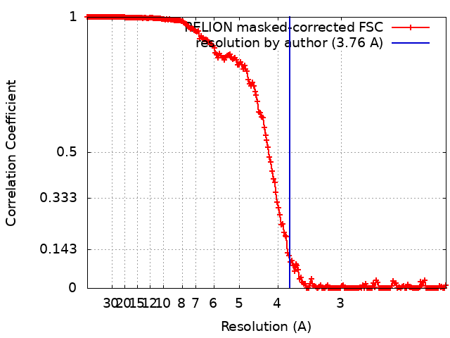 |
-Atomic model buiding 1
| Initial model | PDB ID: Chain - Chain ID: A |
|---|---|
| Details | PDB ID: 6Y83 fits the map |
| Refinement | Space: RECIPROCAL / Protocol: RIGID BODY FIT / Overall B value: 174 / Target criteria: R-factor |
