 Movie
Movie Controller

[English] 日本語
 Yorodumi
- EMDB-10593: Cryo-EM structure of Pf4 bacteriophage coat protein with single-s... -
+ Open data

- Basic information
| Entry | Database: EMDB / ID: EMD-10593 | |||||||||
|---|---|---|---|---|---|---|---|---|---|---|
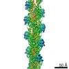
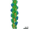
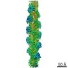
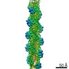
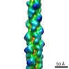
| Title | Cryo-EM structure of Pf4 bacteriophage coat protein with single-stranded DNA | |||||||||
 Map data Map data | ||||||||||
 Sample Sample |
| |||||||||
| Function / homology | Inovirus Coat protein B /  Capsid protein G8P / helical viral capsid / host cell membrane / membrane / Capsid protein G8P / Coat protein B of bacteriophage Pf1 Capsid protein G8P / helical viral capsid / host cell membrane / membrane / Capsid protein G8P / Coat protein B of bacteriophage Pf1 Function and homology information Function and homology information | |||||||||
| Biological species |  Pseudomonas virus Pf1 / Pseudomonas virus Pf1 /  Pseudomonas aeruginosa (strain ATCC 15692 / DSM 22644 / CIP 104116 / JCM 14847 / LMG 12228 / 1C / PRS 101 / PAO1) (bacteria) / Pseudomonas aeruginosa PAO1 (bacteria) Pseudomonas aeruginosa (strain ATCC 15692 / DSM 22644 / CIP 104116 / JCM 14847 / LMG 12228 / 1C / PRS 101 / PAO1) (bacteria) / Pseudomonas aeruginosa PAO1 (bacteria) | |||||||||
| Method | helical reconstruction / cryo EM / Resolution: 3.2 Å | |||||||||
 Authors Authors | Tarafder AK / von Kugelgen A / Bharat TAM | |||||||||
| Funding support |  United Kingdom, 1 items United Kingdom, 1 items
| |||||||||
 Citation Citation | Journal: Proc Natl Acad Sci U S A / Year: 2020 Title: Phage liquid crystalline droplets form occlusive sheaths that encapsulate and protect infectious rod-shaped bacteria. Authors: Abul K Tarafder / Andriko von Kügelgen / Adam J Mellul / Ulrike Schulze / Dirk G A L Aarts / Tanmay A M Bharat / Abstract: The opportunistic pathogen is a major cause of antibiotic-tolerant infections in humans. evades antibiotics in bacterial biofilms by up-regulating expression of a symbiotic filamentous inoviral ...The opportunistic pathogen is a major cause of antibiotic-tolerant infections in humans. evades antibiotics in bacterial biofilms by up-regulating expression of a symbiotic filamentous inoviral prophage, Pf4. We investigated the mechanism of phage-mediated antibiotic tolerance using biochemical reconstitution combined with structural biology and high-resolution cellular imaging. We resolved electron cryomicroscopy atomic structures of Pf4 with and without its linear single-stranded DNA genome, and studied Pf4 assembly into liquid crystalline droplets using optical microscopy and electron cryotomography. By biochemically replicating conditions necessary for antibiotic protection, we found that phage liquid crystalline droplets form phase-separated occlusive compartments around rod-shaped bacteria leading to increased bacterial survival. Encapsulation by these compartments was observed even when inanimate colloidal rods were used to mimic rod-shaped bacteria, suggesting that shape and size complementarity profoundly influences the process. Filamentous inoviruses are pervasive across prokaryotes, and in particular, several Gram-negative bacterial pathogens including , and harbor these prophages. We propose that biophysical occlusion mediated by secreted filamentous molecules such as Pf4 may be a general strategy of bacterial survival in harsh environments. | |||||||||
| History |
|
- Structure visualization
| Movie |
Movie viewer |
|---|---|
| Structure viewer | EM map: SurfViewMolmilJmol/JSmol |
| Supplemental images |

- Downloads & links
-EMDB archive
| Map data | emd_10593.map.gz | 227.1 MB | EMDB map data format | |
|---|---|---|---|---|
| Header (meta data) | emd-10593-v30.xmlemd-10593.xml | 18 KB 18 KB | Display Display | EMDB header |
| Images | 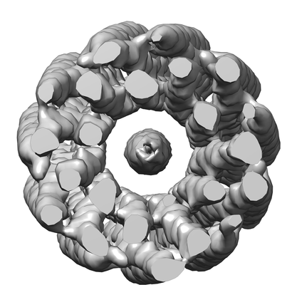 emd_10593.png emd_10593.png | 79.1 KB | ||
| Archive directory |  http://ftp.pdbj.org/pub/emdb/structures/EMD-10593ftp://ftp.pdbj.org/pub/emdb/structures/EMD-10593 http://ftp.pdbj.org/pub/emdb/structures/EMD-10593ftp://ftp.pdbj.org/pub/emdb/structures/EMD-10593 | HTTPS FTP |
-Related structure data
| Related structure data |  6tupMC  6tuqC M: atomic model generated by this map C: citing same article ( |
|---|---|
| Similar structure data |

-Links
| EMDB pages | EMDB (EBI/PDBe) / EMDataResource |
|---|---|
| Related items in Molecule of the Month |


-Map
| File | Download / File: emd_10593.map.gz / Format: CCP4 / Size: 244.1 MB / Type: IMAGE STORED AS FLOATING POINT NUMBER (4 BYTES) | ||||||||||||||||||||||||||||||||||||||||||||||||||||||||||||
|---|---|---|---|---|---|---|---|---|---|---|---|---|---|---|---|---|---|---|---|---|---|---|---|---|---|---|---|---|---|---|---|---|---|---|---|---|---|---|---|---|---|---|---|---|---|---|---|---|---|---|---|---|---|---|---|---|---|---|---|---|---|
| Voxel size | X=Y=Z: 1.38 Å | ||||||||||||||||||||||||||||||||||||||||||||||||||||||||||||
| Density |
| ||||||||||||||||||||||||||||||||||||||||||||||||||||||||||||
| Symmetry | Space group: 1 | ||||||||||||||||||||||||||||||||||||||||||||||||||||||||||||
| Details | EMDB XML:
CCP4 map header:
| ||||||||||||||||||||||||||||||||||||||||||||||||||||||||||||
-Supplemental data
- Sample components
-Entire : Pseudomonas virus Pf1
| Entire | Name: Pseudomonas virus Pf1 |
|---|---|
| Components |
|
-Supramolecule #1: Pseudomonas virus Pf1
| Supramolecule | Name: Pseudomonas virus Pf1 / type: virus / ID: 1 / Parent: 0 / Macromolecule list: all / Virus type: VIRION / Virus isolate: STRAIN / Virus enveloped: No / Virus empty: No |
|---|---|
| Host (natural) | Organism: Pseudomonas aeruginosa (strain ATCC 15692 / DSM 22644 / CIP 104116 / JCM 14847 / LMG 12228 / 1C / PRS 101 / PAO1) (bacteria) |
| Virus shell | Shell ID: 1 / Name: Coat protein B (CoaB) / Diameter: 62.0 Å |
-Supramolecule #2: Pseudomonas virus Pf1
| Supramolecule | Name: Pseudomonas virus Pf1 / type: complex / ID: 2 / Parent: 1 / Macromolecule list: #1 |
|---|---|
| Source (natural) | Organism: Pseudomonas virus Pf1 |
-Supramolecule #3: DNA
| Supramolecule | Name: DNA / type: complex / ID: 3 / Parent: 1 / Macromolecule list: #2 |
|---|---|
| Source (natural) | Organism: Pseudomonas aeruginosa (strain ATCC 15692 / DSM 22644 / CIP 104116 / JCM 14847 / LMG 12228 / 1C / PRS 101 / PAO1) (bacteria) |
-Macromolecule #1: Coat protein B of bacteriophage Pf1
| Macromolecule | Name: Coat protein B of bacteriophage Pf1 / type: protein_or_peptide / ID: 1 / Number of copies: 1 / Enantiomer: LEVO |
|---|---|
| Source (natural) | Organism: Pseudomonas virus Pf1 |
| Molecular weight | Theoretical: 4.612393 KDa |
| Sequence | String: GVIDTSAVES AITDGQGDMK AIGGYIVGAL VILAVAGLIY SMLRKA |
-Macromolecule #2: DNA (5'-D(P*AP*AP*AP*AP*AP*A)-3')
| Macromolecule | Name: DNA (5'-D(P*AP*AP*AP*AP*AP*A)-3') / type: dna / ID: 2 / Details: Model of pf4 single-stranded DNA genome / Number of copies: 1 / Classification: DNA |
|---|---|
| Source (natural) | Organism: Pseudomonas aeruginosa PAO1 (bacteria) |
| Molecular weight | Theoretical: 1.834283 KDa |
| Sequence | String: (DA)(DA)(DA)(DA)(DA)(DA) |
-Experimental details
-Structure determination
| Method | cryo EM |
|---|---|
 Processing Processing | helical reconstruction |
| Aggregation state | filament |
-Sample preparation
| Concentration | 5 mg/mL | |||||||||||||||
|---|---|---|---|---|---|---|---|---|---|---|---|---|---|---|---|---|
| Buffer | pH: 7.4 Component:
Details: 1x Phosphate buffered saline | |||||||||||||||
| Grid | Model: Quantifoil R2/2 / Material: COPPER/RHODIUM / Mesh: 200 / Support film - Material: CARBON / Support film - topology: HOLEY / Pretreatment - Type: GLOW DISCHARGE / Pretreatment - Atmosphere: AIR / Pretreatment - Pressure: 0.03 kPa Details: 20 second glow discharge at 15 mA in a LeicaEM ACE200 | |||||||||||||||
| Vitrification | Cryogen name: ETHANE / Chamber humidity: 100 % / Chamber temperature: 283 K / Instrument: FEI VITROBOT MARK IV Details: Samples for cryo-EM were prepared by pipetting 2.5 ul of the sample onto freshly glow-discharged Quantifoil grids (Cu/Rh R2/2, 200 mesh). Grids were blotted for 2.5 seconds with a blot force ...Details: Samples for cryo-EM were prepared by pipetting 2.5 ul of the sample onto freshly glow-discharged Quantifoil grids (Cu/Rh R2/2, 200 mesh). Grids were blotted for 2.5 seconds with a blot force of -15, 0.5 second drain and 0 second wait times.. | |||||||||||||||
| Details | Filamentous phage purified from source |
- Electron microscopy
| Microscope | FEI TITAN KRIOS |
|---|---|
| Electron beam | Acceleration voltage: 300 kV / Electron source: FIELD EMISSION GUN |
| Electron optics | C2 aperture diameter: 50.0 µm / Calibrated defocus max: -3.0 µm / Calibrated defocus min: -1.0 µm / Calibrated magnification: 105000 / Illumination mode: FLOOD BEAM / Imaging mode: BRIGHT FIELDBright-field microscopy / Cs: 2.7 mm / Nominal defocus max: -3.0 µm / Nominal defocus min: -1.0 µm / Nominal magnification: 105000 |
| Specialist optics | Energy filter - Name: GIF Bioquantum / Energy filter - Slit width: 20 eV |
| Sample stage | Specimen holder model: FEI TITAN KRIOS AUTOGRID HOLDER / Cooling holder cryogen: NITROGEN |
| Temperature | Min: 80.0 K / Max: 80.0 K |
| Image recording | Film or detector model: GATAN K2 SUMMIT (4k x 4k) / Detector mode: COUNTING / Digitization - Frames/image: 1-40 / Number grids imaged: 2 / Number real images: 4110 / Average exposure time: 10.0 sec. / Average electron dose: 43.0 e/Å2 |
| Experimental equipment |  Model: Titan Krios / Image courtesy: FEI Company |
-Image processing
| Segment selection | Number selected: 351381 / Software - Name: RELION (ver. 2.0) |
|---|---|
| CTF correction | Software - Name: RELION (ver. 2.0) Details: Wiener filter implemented in the RELION refinement algorithm |
| Startup model | Type of model: NONE / Details: Fourier-Bessel indexing |
| Final angle assignment | Type: NOT APPLICABLE / Software - Name: RELION (ver. 2.0) |
| Final reconstruction | Number classes used: 1 Applied symmetry - Helical parameters - Δz: 3.14 Å Applied symmetry - Helical parameters - Δ&Phi: 65.9 ° Applied symmetry - Helical parameters - Axial symmetry: C1 (asymmetric) Algorithm: FOURIER SPACE / Resolution.type: BY AUTHOR / Resolution: 3.2 Å / Resolution method: FSC 0.143 CUT-OFF / Software - Name: RELION (ver. 3.0) / Number images used: 185002 |
-Atomic model buiding 1
| Details | The helical filament symmetry is only applied to the Pf4 coat protein and not the DNA. The reason for this is that the DNA bases are not resolved clearly due to averaging along the ssDNA genome of the phage, meaning that refinement of the protein into the map is of a higher quality than the ssDNA, where a poly-adenine model has been built. |
|---|---|
| Refinement | Space: REAL / Protocol: BACKBONE TRACE / Overall B value: 93.74 / Target criteria: Correlation coefficient |
| Output model | PDB-6tup: |
