 Movie
Movie Controller

[English] 日本語
 Yorodumi
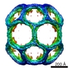
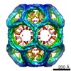
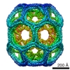
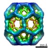
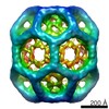
- EMDB-0116: Cryo-EM structure of the 36 triskelia D6 barrel clathrin coat complex -
+ Open data

- Basic information
| Entry | Database: EMDB / ID: EMD-0116 | |||||||||
|---|---|---|---|---|---|---|---|---|---|---|
| Title | Cryo-EM structure of the 36 triskelia D6 barrel clathrin coat complex | |||||||||
 Map data Map data | ||||||||||
 Sample Sample |
| |||||||||
| Biological species |   Sus scrofa (pig) / pig (pig) Sus scrofa (pig) / pig (pig) | |||||||||
| Method | single particle reconstruction / cryo EM / Resolution: 12.18 Å | |||||||||
 Authors Authors | Morris KL / Smith CJ | |||||||||
 Citation Citation | Journal: Nat Struct Mol Biol / Year: 2019 Title: Cryo-EM of multiple cage architectures reveals a universal mode of clathrin self-assembly. Authors: Kyle L Morris / Joseph R Jones / Mary Halebian / Shenping Wu / Michael Baker / Jean-Paul Armache / Amaurys Avila Ibarra / Richard B Sessions / Alexander D Cameron / Yifan Cheng / Corinne J Smith /   Abstract: Clathrin forms diverse lattice and cage structures that change size and shape rapidly in response to the needs of eukaryotic cells during clathrin-mediated endocytosis and intracellular trafficking. ...Clathrin forms diverse lattice and cage structures that change size and shape rapidly in response to the needs of eukaryotic cells during clathrin-mediated endocytosis and intracellular trafficking. We present the cryo-EM structure and molecular model of assembled porcine clathrin, providing insights into interactions that stabilize key elements of the clathrin lattice, namely, between adjacent heavy chains, at the light chain-heavy chain interface and within the trimerization domain. Furthermore, we report cryo-EM maps for five different clathrin cage architectures. Fitting structural models to three of these maps shows that their assembly requires only a limited range of triskelion leg conformations, yet inherent flexibility is required to maintain contacts. Analysis of the protein-protein interfaces shows remarkable conservation of contact sites despite architectural variation. These data reveal a universal mode of clathrin assembly that allows variable cage architecture and adaptation of coated vesicle size and shape during clathrin-mediated vesicular trafficking or endocytosis. | |||||||||
| History |
|
- Structure visualization
| Movie |
 Movie viewer Movie viewer |
|---|---|
| Structure viewer | EM map: SurfViewMolmilJmol/JSmol |
| Supplemental images |

- Downloads & links
-EMDB archive
| Map data | emd_0116.map.gz | 72.8 MB | EMDB map data format | |
|---|---|---|---|---|
| Header (meta data) | emd-0116-v30.xmlemd-0116.xml | 22.1 KB 22.1 KB | Display Display | EMDB header |
| FSC (resolution estimation) | emd_0116_fsc.xml | 17.9 KB | Display | FSC data file |
| Images | 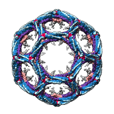 emd_0116.png emd_0116.png | 186.1 KB | ||
| Masks | emd_0116_msk_1.map | 476.8 MB | Mask map | |
| Others | emd_0116_additional.map.gzemd_0116_additional_1.map.gzemd_0116_half_map_1.map.gzemd_0116_half_map_2.map.gz | 376.3 MB 376.3 MB 378.8 MB 379 MB | ||
| Archive directory |  http://ftp.pdbj.org/pub/emdb/structures/EMD-0116ftp://ftp.pdbj.org/pub/emdb/structures/EMD-0116 http://ftp.pdbj.org/pub/emdb/structures/EMD-0116ftp://ftp.pdbj.org/pub/emdb/structures/EMD-0116 | HTTPS FTP |
-Related structure data
| Related structure data | 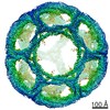 0114C 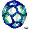 0115C 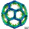 0118C  0120C 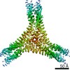 0121C 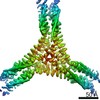 0122C 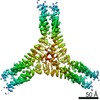 0123C 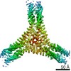 0124C 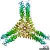 0125C 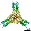 0126C 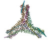 6sctC C: citing same article ( |
|---|---|
| Similar structure data | |
| EM raw data | EMPIAR-10294 (Title: Single particle cryo-EM dataset of clathrin cages with phase flipping suitable for refinement Data size: 11.9 Data #1: Clathrin cage particle images with phase flipping [picked particles - single frame - processed]) EMPIAR-10295 (Title: Single particle cryo-EM dataset of clathrin cages suitable for subparticle extractionData size: 19.9 Data #1: Clathrin cage particle images - 36 D6 barrel [picked particles - multiframe - unprocessed] Data #2: Clathrin cage particle images - 32 sweet potato [picked particles - multiframe - unprocessed] Data #3: Clathrin cage particle images - 28 mini coat [picked particles - multiframe - unprocessed] Data #4: Clathrin cage particle images - 36 tennis ball [picked particles - multiframe - unprocessed] Data #5: Clathrin cage particle images - 37 big apple [picked particles - multiframe - unprocessed]) |
-Links
| EMDB pages | EMDB (EBI/PDBe) / EMDataResource |
|---|
-Map
| File | Download / File: emd_0116.map.gz / Format: CCP4 / Size: 476.8 MB / Type: IMAGE STORED AS FLOATING POINT NUMBER (4 BYTES) | ||||||||||||||||||||||||||||||||||||||||||||||||||||||||||||
|---|---|---|---|---|---|---|---|---|---|---|---|---|---|---|---|---|---|---|---|---|---|---|---|---|---|---|---|---|---|---|---|---|---|---|---|---|---|---|---|---|---|---|---|---|---|---|---|---|---|---|---|---|---|---|---|---|---|---|---|---|---|
| Voxel size | X=Y=Z: 1.705 Å | ||||||||||||||||||||||||||||||||||||||||||||||||||||||||||||
| Density |
| ||||||||||||||||||||||||||||||||||||||||||||||||||||||||||||
| Symmetry | Space group: 1 | ||||||||||||||||||||||||||||||||||||||||||||||||||||||||||||
| Details | EMDB XML:
CCP4 map header:
| ||||||||||||||||||||||||||||||||||||||||||||||||||||||||||||
-Supplemental data
-Mask #1
| File | emd_0116_msk_1.map | ||||||||||||
|---|---|---|---|---|---|---|---|---|---|---|---|---|---|
| Projections & Slices |
| ||||||||||||
| Density Histograms |
 Z
Z Y
Y X
X


-Additional map: Whole cage autorefine and map used for mask creation.
| File | emd_0116_additional.map | ||||||||||||
|---|---|---|---|---|---|---|---|---|---|---|---|---|---|
| Annotation | Whole cage autorefine and map used for mask creation. | ||||||||||||
| Projections & Slices |
| ||||||||||||
| Density Histograms |


-Additional map: Whole cage autorefine and map used for mask creation.
| File | emd_0116_additional_1.map | ||||||||||||
|---|---|---|---|---|---|---|---|---|---|---|---|---|---|
| Annotation | Whole cage autorefine and map used for mask creation. | ||||||||||||
| Projections & Slices |
| ||||||||||||
| Density Histograms |


-Half map: Phase flipped whole cage half map.
| File | emd_0116_half_map_1.map | ||||||||||||
|---|---|---|---|---|---|---|---|---|---|---|---|---|---|
| Annotation | Phase flipped whole cage half map. | ||||||||||||
| Projections & Slices |
| ||||||||||||
| Density Histograms |


-Half map: Phase flipped whole cage half map.
| File | emd_0116_half_map_2.map | ||||||||||||
|---|---|---|---|---|---|---|---|---|---|---|---|---|---|
| Annotation | Phase flipped whole cage half map. | ||||||||||||
| Projections & Slices |
| ||||||||||||
| Density Histograms |


- Sample components
-Entire : Assembly of clathrin heavy and light chain into coat complexes
| Entire | Name: Assembly of clathrin heavy and light chain into coat complexes |
|---|---|
| Components |
|
-Supramolecule #1: Assembly of clathrin heavy and light chain into coat complexes
| Supramolecule | Name: Assembly of clathrin heavy and light chain into coat complexes type: complex / ID: 1 / Parent: 0 / Macromolecule list: all |
|---|---|
| Source (natural) | Organism: Sus scrofa (pig) / Organ: BRAIN |
| Molecular weight | Theoretical: 22.8 MDa |
-Macromolecule #1: clathrin heavy chain
| Macromolecule | Name: clathrin heavy chain / type: protein_or_peptide / ID: 1 / Enantiomer: LEVO |
|---|---|
| Source (natural) | Organism: pig (pig) / Organ: BRAIN |
| Sequence | String: MAQILPIRFQ EHLQLQNLGI NPANIGFSTL TMESDKFICI REKVGEQAQV VIIDMNDPSN PIRRPISADS AIMNPASKVI ALKAGKTLQI FNIEMKSKMK AHTMTDDVTF WKWISLNTVA LVTDNAVYHW SMEGESQPVK MFDRHSSLAG CQIINYRTDA KQKWLLLTGI ...String: MAQILPIRFQ EHLQLQNLGI NPANIGFSTL TMESDKFICI REKVGEQAQV VIIDMNDPSN PIRRPISADS AIMNPASKVI ALKAGKTLQI FNIEMKSKMK AHTMTDDVTF WKWISLNTVA LVTDNAVYHW SMEGESQPVK MFDRHSSLAG CQIINYRTDA KQKWLLLTGI SAQQNRVVGA MQLYSVDRKV SQPIEGHAAS FAQFKMEGNA EESTLFCFAV RGQAGGKLHI IEVGTPPTGN QPFPKKAVDV FFPPEAQNDF PVAMQISEKH DVVFLITKYG YIHLYDLETG TCIYMNRISG ETIFVTAPHE ATAGIIGVNR KGQVLSVCVE EENIIPYITN VLQNPDLALR MAVRNNLAGA EELFARKFNA LFAQGNYSEA AKVAANAPKG ILRTPDTIRR FQSVPAQPGQ TSPLLQYFGI LLDQGQLNKY ESLELCRPVL QQGRKQLLEK WLKEDKLECS EELGDLVKSV DPTLALSVYL RANVPNKVIQ CFAETGQVQK IVLYAKKVGY TPDWIFLLRN VMRISPDQGQ QFAQMLVQDE EPLADITQIV DVFMEYNLIQ QCTAFLLDAL KNNRPSEGPL QTRLLEMNLM HAPQVADAIL GNQMFTHYDR AHIAQLCEKA GLLQRALEHF TDLYDIKRAV VHTHLLNPEW LVNYFGSLSV EDSLECLRAM LSANIRQNLQ ICVQVASKYH EQLSTQSLIE LFESFKSFEG LFYFLGSIVN FSQDPDVHFK YIQAACKTGQ IKEVERICRE SNCYDPERVK NFLKEAKLTD QLPLIIVCDR FDFVHDLVLY LYRNNLQKYI EIYVQKVNPS RLPVVIGGLL DVDCSEDVIK NLILVVRGQF STDELVAEVE KRNRLKLLLP WLEARIHEGC EEPATHNALA KIYIDSNNNP ERFLRENPYY DSRVVGKYCE KRDPHLACVA YERGQCDLEL INVCNENSLF KSLSRYLVRR KDPELWGSVL LESNPYRRPL IDQVVQTALS ETQDPEEVSV TVKAFMTADL PNELIELLEK IVLDNSVFSE HRNLQNLLIL TAIKADRTRV MEYINRLDNY DAPDIANIAI SNELFEEAFA IFRKFDVNTS AVQVLIEHIG NLDRAYEFAE RCNEPAVWSQ LAKAQLQKGM VKEAIDSYIK ADDPSSYMEV VQAANTSGNW EELVKYLQMA RKKARESYVE TELIFALAKT NRLAELEEFI NGPNNAHIQQ VGDRCYDEKM YDAAKLLYNN VSNFGRLAST LVHLGEYQAA VDGARKANST RTWKEVCFAC VDGKEFRLAQ MCGLHIVVHA DELEELINYY QDRGYFEELI TMLEAALGLE RAHMGMFTEL AILYSKFKPQ KMREHLELFW SRVNIPKVLR AAEQAHLWAE LVFLYDKYEE YDNAIITMMN HPTDAWKEGQ FKDIITKVAN VELYYRAIQF YLEFKPLLLN DLLMVLSPRL DHTRAVNYFS KVKQLPLVKP YLRSVQNHNN KSVNESLNNL FITEEDYQAL RTSIDAYDNF DNISLAQRLE KHELIEFRRI AAYLFKGNNR WKQSVELCKK DSLYKDAMQY ASESKDTELA EELLQWFLQE EKRECFGACL FTCYDLLRPD VVLETAWRHN IMDFAMPYFI QVMKEYLTKV DKLDASESLR KEEEQATETQ PIVYGQPQLM LTAGPSVAVP PQAPFGYGYT APPYGQPQPG FGYSM |
-Macromolecule #2: clathrin light chain b
| Macromolecule | Name: clathrin light chain b / type: protein_or_peptide / ID: 2 / Enantiomer: LEVO |
|---|---|
| Source (natural) | Organism: pig (pig) / Organ: BRAIN |
| Sequence | String: MADDFGFFSS SESGAPEVAE EDPAAAFLAQ QESEIAGIEN DEGFGAPAGS QAALAQPGPA SGAGPEDMGT TVNGDVFQDA NGPADGYAAI AQADRLTQEP ESIRKWREEQ RKRLQELDAA SKVTEQEWRE KAKKDLEEWN QRQSEQVEKN KINNRIADKA FYQQPDADII ...String: MADDFGFFSS SESGAPEVAE EDPAAAFLAQ QESEIAGIEN DEGFGAPAGS QAALAQPGPA SGAGPEDMGT TVNGDVFQDA NGPADGYAAI AQADRLTQEP ESIRKWREEQ RKRLQELDAA SKVTEQEWRE KAKKDLEEWN QRQSEQVEKN KINNRIADKA FYQQPDADII GYVASEEAFV KESKEETPGT EWEKVAQLCD FNPKSSKQCK DVSRLRSVLM SLKQTPLSR |
-Macromolecule #3: clathrin light chain a
| Macromolecule | Name: clathrin light chain a / type: protein_or_peptide / ID: 3 / Enantiomer: LEVO |
|---|---|
| Source (natural) | Organism: pig (pig) / Organ: BRAIN |
| Sequence | String: MADLDPFGAP AGPSLGNGVA GEEDPAAAFL AQQESEIAGI ENDEAFAILD GGAPGPQPHG EPPGGPDAV DGVMNGEYYQ ESNGPTDSYA AISQVDRLQS EPESIRKWRE EQTERLEALD A NSRKQEAE WKEKAIKELE EWYARQDEQL QKTKANNRVA DEAFYKQPFA ...String: MADLDPFGAP AGPSLGNGVA GEEDPAAAFL AQQESEIAGI ENDEAFAILD GGAPGPQPHG EPPGGPDAV DGVMNGEYYQ ESNGPTDSYA AISQVDRLQS EPESIRKWRE EQTERLEALD A NSRKQEAE WKEKAIKELE EWYARQDEQL QKTKANNRVA DEAFYKQPFA DVIGYVAAEE AF VNDIEES SPGTEWERVA RLCDFNPKSS KQAKDVSRMR SVLISLKQAP LVH |
-Experimental details
-Structure determination
| Method | cryo EM |
|---|---|
 Processing Processing | single particle reconstruction |
| Aggregation state | particle |
-Sample preparation
| Concentration | 6.32 mg/mL | |||||||||||||||
|---|---|---|---|---|---|---|---|---|---|---|---|---|---|---|---|---|
| Buffer | pH: 6.4 Component:
| |||||||||||||||
| Vitrification | Cryogen name: ETHANE-PROPANE / Instrument: HOMEMADE PLUNGER Details: 3 uL applied to a grid at room temperature and humidity. 3 second hand blot and plunge.. | |||||||||||||||
| Details | Clathrin coat complexes, end point assembly exhibiting architectural heterogeneity |
- Electron microscopy
| Microscope | FEI TITAN KRIOS |
|---|---|
| Electron beam | Acceleration voltage: 300 kV / Electron source: FIELD EMISSION GUN |
| Electron optics | C2 aperture diameter: 70.0 µm / Calibrated magnification: 82111 / Illumination mode: FLOOD BEAM / Imaging mode: BRIGHT FIELDBright-field microscopy / Cs: 2.7 mm / Nominal defocus max: 3.2 µm / Nominal defocus min: 1.4 µm |
| Sample stage | Specimen holder model: FEI TITAN KRIOS AUTOGRID HOLDER / Cooling holder cryogen: NITROGEN |
| Image recording | Film or detector model: FEI FALCON II (4k x 4k) / Detector mode: INTEGRATING / Digitization - Dimensions - Width: 4096 pixel / Digitization - Dimensions - Height: 4096 pixel / Average exposure time: 3.0 sec. / Average electron dose: 53.0 e/Å2 |
| Experimental equipment |  Model: Titan Krios / Image courtesy: FEI Company |
-Image processing
| Particle selection | Number selected: 12785 |
|---|---|
| CTF correction | Software - Name: Gctf (ver. 1.06) |
| Startup model | Type of model: OTHER / Details: Map from supervised 3D classification |
| Initial angle assignment | Type: MAXIMUM LIKELIHOOD / Software - Name: RELION (ver. 2.1) Details: Initial model and angles come from localised reconstruction of the hub (Ilca 2015) |
| Final angle assignment | Type: MAXIMUM LIKELIHOOD / Software - Name: RELION (ver. 2.1) |
| Final reconstruction | Applied symmetry - Point group: D6 (2x6 fold dihedral) / Algorithm: BACK PROJECTION / Resolution.type: BY AUTHOR / Resolution: 12.18 Å / Resolution method: FSC 0.143 CUT-OFF / Software - Name: RELION (ver. 2.1) / Number images used: 1160 |
| FSC plot (resolution estimation) |  |
-Atomic model buiding 1
| Refinement | Space: REAL / Protocol: FLEXIBLE FIT / Target criteria: Cross-correlation coefficient |
|---|
