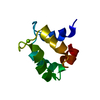
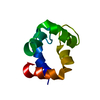
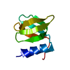
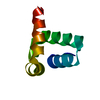
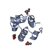
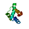
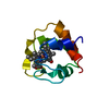
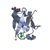
Journal: Proteins / Year: 2013 Title: Solution structures of polcalcin Phl p 7 in three ligation states: Apo-, hemi-Mg2+-bound, and fully Ca2+-bound. Authors: Michael T Henzl / Arthur G Sirianni / Wei G Wycoff / Anmin Tan / John J Tanner / Abstract: Polcalcins are small EF-hand proteins believed to assist in regulating pollen-tube growth. Phl p 7, from timothy grass (Phleum pratense), crystallizes as a domain-swapped dimer at low pH. This study ...Polcalcins are small EF-hand proteins believed to assist in regulating pollen-tube growth. Phl p 7, from timothy grass (Phleum pratense), crystallizes as a domain-swapped dimer at low pH. This study describes the solution structures of the recombinant protein in buffered saline at pH 6.0, containing either 5.0 mM EDTA, 5.0 mM Mg(2+), or 100 μM Ca(2+). Phl p 7 is monomeric in all three ligation states. In the apo-form, both EF-hand motifs reside in the closed conformation, with roughly antiparallel N- and C-terminal helical segments. In 5.0 mM Mg(2+), the divalent ion is bound by EF-hand 2, perturbing interhelical angles and imposing more regular helical structure. The structure of Ca(2+)-bound Phl p 7 resembles that previously reported for Bet v 4-likewise exposing apolar surface to the solvent. Occluded in the apo- and Mg(2+)-bound forms, this surface presumably provides the docking site for Phl p 7 targets. Unlike Bet v 4, EF-hand 2 in Phl p 7 includes five potential anionic ligands, due to replacement of the consensus serine residue at -x (residue 55 in Phl p 7) with aspartate. In the Phl p 7 crystal structure, D55 functions as a helix cap for helix D. In solution, however, D55 apparently serves as a ligand to the bound Ca(2+). When Mg(2+) resides in site 2, the D55 carboxylate withdraws to a distance consistent with a role as an outer-sphere ligand. (15)N relaxation data, collected at 600 MHz, indicate that backbone mobility is limited in all three ligation states.
Contact author
John Tanner (Mizzou, University of Missouri-Columbia, Columbia, MO, USA)
Instrument name: Advanced Light Source (ALS) 12.3.1 (SIBYLS) City: Berkeley, CA / 国: USA / Type of source: X-ray synchrotronSynchrotron / Wavelength: 0.1127 Å
Detector
Name: MAR 165 CCD
Scan
Title: Calcium-bound Phl p 7 / Measurement date: Dec 8, 2011 / Cell temperature: 10 °C / Unit: 1/A /
Min
Max
Q
0.021
0.3256
Distance distribution function P(R)
Sofotware P(R): GNOM 5.0 / Number of points: 502 /
Min
Max
Q
0.020978
0.325586
P(R) point
1
502
R
0
36.19
Result
Type of curve: single_conc /
Experimental
Porod
MW
8.545 kDa
8.5 kDa
Volume
-
13.6 nm3
P(R)
Guinier
Guinier error
Forward scattering, I0
52.19
52.54
0.075
Radius of gyration, Rg
1.27 nm
1.29 nm
0.003
Min
Max
D
-
3.62
Guinier point
1
132
+
About Yorodumi
-
News
-
Feb 9, 2022. New format data for meta-information of EMDB entries
New format data for meta-information of EMDB entries
Version 3 of the EMDB header file is now the official format.
The previous official version 1.9 will be removed from the archive.
In the structure databanks used in Yorodumi, some data are registered as the other names, "COVID-19 virus" and "2019-nCoV". Here are the details of the virus and the list of structure data.
Jan 31, 2019. EMDB accession codes are about to change! (news from PDBe EMDB page)
EMDB accession codes are about to change! (news from PDBe EMDB page)
The allocation of 4 digits for EMDB accession codes will soon come to an end. Whilst these codes will remain in use, new EMDB accession codes will include an additional digit and will expand incrementally as the available range of codes is exhausted. The current 4-digit format prefixed with “EMD-” (i.e. EMD-XXXX) will advance to a 5-digit format (i.e. EMD-XXXXX), and so on. It is currently estimated that the 4-digit codes will be depleted around Spring 2019, at which point the 5-digit format will come into force.
The EM Navigator/Yorodumi systems omit the EMD- prefix.
Related info.:Q: What is EMD? / ID/Accession-code notation in Yorodumi/EM Navigator
Yorodumi is a browser for structure data from EMDB, PDB, SASBDB, etc.
This page is also the successor to EM Navigator detail page, and also detail information page/front-end page for Omokage search.
The word "yorodu" (or yorozu) is an old Japanese word meaning "ten thousand". "mi" (miru) is to see.
Related info.:EMDB / PDB / SASBDB / Comparison of 3 databanks / Yorodumi Search / Aug 31, 2016. New EM Navigator & Yorodumi / Yorodumi Papers / Jmol/JSmol / Function and homology information / Changes in new EM Navigator and Yorodumi
 Movie
Movie Controller

 Open data
 Basic information
 Sample
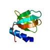
 EF-hand, calcium binding motif /
  Function and homology information
 Citation
 Contact author
 Structure visualization
 Downloads & links
 SASDDJ2


 Search similar-shape structures of this assembly by Omokage search (details)


