 Movie
Movie Controller


+ Open data

- Basic information
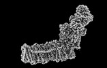
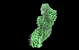
| Entry | Database: PDB / ID: 8e9h | |||||||||||||||
|---|---|---|---|---|---|---|---|---|---|---|---|---|---|---|---|---|
| Title | Mycobacterial respiratory complex I, fully-inserted quinone | |||||||||||||||
 Components Components |
| |||||||||||||||
 Keywords Keywords |  MEMBRANE PROTEIN / Complex / Oxidative Phosphorylation / NADH-quinone oxidoreductase / Iron-sulfur Protein MEMBRANE PROTEIN / Complex / Oxidative Phosphorylation / NADH-quinone oxidoreductase / Iron-sulfur Protein | |||||||||||||||
| Function / homology |  Function and homology informationNADH dehydrogenase (quinone) / Translocases; Catalysing the translocation of protons; Linked to oxidoreductase reactions / NADH:ubiquinone reductase (non-electrogenic) activity / respiratory chain complex I / molybdopterin cofactor binding / oxidoreductase activity, acting on NAD(P)H, quinone or similar compound as acceptor / phosphorelay signal transduction system / NADH dehydrogenase (ubiquinone) activity / quinone binding / ATP synthesis coupled electron transport ...NADH dehydrogenase (quinone) / Translocases; Catalysing the translocation of protons; Linked to oxidoreductase reactions / NADH:ubiquinone reductase (non-electrogenic) activity / respiratory chain complex I / molybdopterin cofactor binding / oxidoreductase activity, acting on NAD(P)H, quinone or similar compound as acceptor / phosphorelay signal transduction system / NADH dehydrogenase (ubiquinone) activity / quinone binding / ATP synthesis coupled electron transport / 2 iron, 2 sulfur cluster binding / NAD binding / FMN binding / 4 iron, 4 sulfur cluster binding / oxidoreductase activity / iron ion binding / membrane / metal ion binding / plasma membrane Function and homology informationNADH dehydrogenase (quinone) / Translocases; Catalysing the translocation of protons; Linked to oxidoreductase reactions / NADH:ubiquinone reductase (non-electrogenic) activity / respiratory chain complex I / molybdopterin cofactor binding / oxidoreductase activity, acting on NAD(P)H, quinone or similar compound as acceptor / phosphorelay signal transduction system / NADH dehydrogenase (ubiquinone) activity / quinone binding / ATP synthesis coupled electron transport ...NADH dehydrogenase (quinone) / Translocases; Catalysing the translocation of protons; Linked to oxidoreductase reactions / NADH:ubiquinone reductase (non-electrogenic) activity / respiratory chain complex I / molybdopterin cofactor binding / oxidoreductase activity, acting on NAD(P)H, quinone or similar compound as acceptor / phosphorelay signal transduction system / NADH dehydrogenase (ubiquinone) activity / quinone binding / ATP synthesis coupled electron transport / 2 iron, 2 sulfur cluster binding / NAD binding / FMN binding / 4 iron, 4 sulfur cluster binding / oxidoreductase activity / iron ion binding / membrane / metal ion binding / plasma membraneSimilarity search - Function | |||||||||||||||
| Biological species |  Mycolicibacterium smegmatis MC2 155 (bacteria) Mycolicibacterium smegmatis MC2 155 (bacteria) | |||||||||||||||
| Method | ELECTRON MICROSCOPY / single particle reconstruction / cryo EM / Resolution: 2.7 Å | |||||||||||||||
 Authors Authors | Liang, Y. / Rubinstein, J.L. | |||||||||||||||
| Funding support |  Canada, 4items Canada, 4items
| |||||||||||||||
 Citation Citation | Journal: Proc Natl Acad Sci U S A / Year: 2023 Title: Structure of mycobacterial respiratory complex I. Authors: Yingke Liang / Alicia Plourde / Stephanie A Bueler / Jun Liu / Peter Brzezinski / Siavash Vahidi / John L Rubinstein /  Abstract: Oxidative phosphorylation, the combined activity of the electron transport chain (ETC) and adenosine triphosphate synthase, has emerged as a valuable target for the treatment of infection by and ...Oxidative phosphorylation, the combined activity of the electron transport chain (ETC) and adenosine triphosphate synthase, has emerged as a valuable target for the treatment of infection by and other mycobacteria. The mycobacterial ETC is highly branched with multiple dehydrogenases transferring electrons to a membrane-bound pool of menaquinone and multiple oxidases transferring electrons from the pool. The proton-pumping type I nicotinamide adenine dinucleotide (NADH) dehydrogenase (Complex I) is found in low abundance in the plasma membranes of mycobacteria in typical in vitro culture conditions and is often considered dispensable. We found that growth of in carbon-limited conditions greatly increased the abundance of Complex I and allowed isolation of a rotenone-sensitive preparation of the enzyme. Determination of the structure of the complex by cryoEM revealed the "orphan" two-component response regulator protein MSMEG_2064 as a subunit of the assembly. MSMEG_2064 in the complex occupies a site similar to the proposed redox-sensing subunit NDUFA9 in eukaryotic Complex I. An apparent purine nucleoside triphosphate within the NuoG subunit resembles the GTP-derived molybdenum cofactor in homologous formate dehydrogenase enzymes. The membrane region of the complex binds acyl phosphatidylinositol dimannoside, a characteristic three-tailed lipid from the mycobacterial membrane. The structure also shows menaquinone, which is preferentially used over ubiquinone by gram-positive bacteria, in two different positions along the quinone channel, comparable to ubiquinone in other structures and suggesting a conserved quinone binding mechanism. #1: Journal: Biorxiv / Year: 2022Title: Structure of mycobacterial respiratory Complex I Authors: Liang, Y. / Plourde, A. / Bueler, S.A. / Liu, J. / Brzezinski, P. / Vahidi, S. / Rubinstein, J.L. | |||||||||||||||
| History |
|
- Structure visualization
| Structure viewer | Molecule: MolmilJmol/JSmol |
|---|
- Downloads & links
-Download
| PDBx/mmCIF format | 8e9h.cif.gz | 831.7 KB | Display | PDBx/mmCIF format |
|---|---|---|---|---|
| PDB format | pdb8e9h.ent.gz | 666.3 KB | Display | PDB format |
| PDBx/mmJSON format | 8e9h.json.gz | Tree view | PDBx/mmJSON format | |
| Others |  Other downloads Other downloads |
-Validation report
| Arichive directory | https://data.pdbj.org/pub/pdb/validation_reports/e9/8e9hftp://data.pdbj.org/pub/pdb/validation_reports/e9/8e9h | HTTPS FTP |
|---|
-Related structure data
| Related structure data | 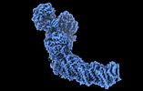 27964MC 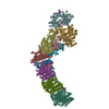 8e9gC  8e9iC M: map data used to model this data C: citing same article ( |
|---|---|
| Similar structure data |
-Links
 PDBj
PDBj


- Assembly
| Deposited unit | 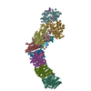
|
|---|---|
| 1 |
|
-Components
-Protein , 1 types, 1 molecules O
| #1: Protein | Two-component regulatory system Mass: 14119.161 Da / Num. of mol.: 1 / Source method: isolated from a natural source Source: (natural) Mycolicibacterium smegmatis MC2 155 (bacteria)Strain: ATCC 700084 / mc(2)155 / References: UniProt: A0QU37 |
|---|
-NADH-quinone oxidoreductase subunit ... , 12 types, 12 molecules BACDEGFIHJKN
| #2: Protein | NADH dehydrogenase (quinone) / NADH dehydrogenase I subunit B / NDH-1 subunit B Mass: 20123.703 Da / Num. of mol.: 1 / Source method: isolated from a natural source Source: (natural) Mycolicibacterium smegmatis MC2 155 (bacteria)Strain: ATCC 700084 / mc(2)155 References: UniProt: A0QU35, Translocases; Catalysing the translocation of protons; Linked to oxidoreductase reactions |
|---|---|
| #3: Protein | NADH dehydrogenase (quinone) Mass: 13628.287 Da / Num. of mol.: 1 / Source method: isolated from a natural source Source: (natural) Mycolicibacterium smegmatis MC2 155 (bacteria)References: UniProt: A0QU36 |
| #4: Protein | NADH dehydrogenase (quinone) / NADH dehydrogenase I subunit C / NDH-1 subunit C Mass: 26610.689 Da / Num. of mol.: 1 / Source method: isolated from a natural source Source: (natural) Mycolicibacterium smegmatis MC2 155 (bacteria)Strain: ATCC 700084 / mc(2)155 References: UniProt: A0QU34, Translocases; Catalysing the translocation of protons; Linked to oxidoreductase reactions |
| #5: Protein | NADH dehydrogenase (quinone) Mass: 48556.750 Da / Num. of mol.: 1 / Source method: isolated from a natural source Source: (natural) Mycolicibacterium smegmatis MC2 155 (bacteria)References: UniProt: A0QU33 |
| #6: Protein | NADH dehydrogenase (quinone) Mass: 25746.746 Da / Num. of mol.: 1 / Source method: isolated from a natural source Source: (natural) Mycolicibacterium smegmatis MC2 155 (bacteria)Strain: ATCC 700084 / mc(2)155 / References: UniProt: A0QU32, NADH dehydrogenase (quinone) |
| #7: Protein | NADH dehydrogenase (quinone) Mass: 83894.773 Da / Num. of mol.: 1 / Source method: isolated from a natural source Source: (natural) Mycolicibacterium smegmatis MC2 155 (bacteria)Strain: ATCC 700084 / mc(2)155 References: UniProt: A0QU30, Translocases; Catalysing the translocation of protons; Linked to oxidoreductase reactions |
| #8: Protein | NADH dehydrogenase (quinone) Mass: 47831.078 Da / Num. of mol.: 1 / Source method: isolated from a natural source Source: (natural) Mycolicibacterium smegmatis MC2 155 (bacteria)Strain: ATCC 700084 / mc(2)155 References: UniProt: A0QU31, Translocases; Catalysing the translocation of protons; Linked to oxidoreductase reactions |
| #9: Protein | NADH dehydrogenase (quinone) Mass: 19860.510 Da / Num. of mol.: 1 / Source method: isolated from a natural source Source: (natural) Mycolicibacterium smegmatis MC2 155 (bacteria)References: UniProt: A0QU28 |
| #10: Protein | NADH dehydrogenase (quinone) / NADH dehydrogenase I subunit H / NDH-1 subunit H Mass: 44477.293 Da / Num. of mol.: 1 / Source method: isolated from a natural source Source: (natural) Mycolicibacterium smegmatis MC2 155 (bacteria)Strain: ATCC 700084 / mc(2)155 References: UniProt: A0QU29, Translocases; Catalysing the translocation of protons; Linked to oxidoreductase reactions |
| #11: Protein | NADH dehydrogenase (quinone) Mass: 26398.791 Da / Num. of mol.: 1 / Source method: isolated from a natural source Source: (natural) Mycolicibacterium smegmatis MC2 155 (bacteria)Strain: ATCC 700084 / mc(2)155 References: UniProt: A0QU27, Translocases; Catalysing the translocation of protons; Linked to oxidoreductase reactions |
| #12: Protein | NADH dehydrogenase (quinone) Mass: 10876.923 Da / Num. of mol.: 1 / Source method: isolated from a natural source Source: (natural) Mycolicibacterium smegmatis MC2 155 (bacteria)References: UniProt: A0QU26 |
| #14: Protein | NADH dehydrogenase (quinone) Mass: 54013.266 Da / Num. of mol.: 1 / Source method: isolated from a natural source Source: (natural) Mycolicibacterium smegmatis MC2 155 (bacteria)References: UniProt: A0QU23 |
-NADH-quinone oxidoreductase, ... , 2 types, 2 molecules LM
| #13: Protein | NADH dehydrogenase (quinone) Mass: 65446.609 Da / Num. of mol.: 1 / Source method: isolated from a natural source Source: (natural) Mycolicibacterium smegmatis MC2 155 (bacteria)Strain: ATCC 700084 / mc(2)155 / References: UniProt: A0QU25, NADH dehydrogenase (quinone) |
|---|---|
| #15: Protein | NADH dehydrogenase (quinone) Mass: 56338.316 Da / Num. of mol.: 1 / Source method: isolated from a natural source Source: (natural) Mycolicibacterium smegmatis MC2 155 (bacteria)Strain: ATCC 700084 / mc(2)155 / References: UniProt: A0QU24, NADH dehydrogenase (quinone) |
-Non-polymers , 7 types, 14 molecules 
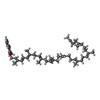


| #16: Chemical | ChemComp-SF4 / Iron–sulfur cluster Mass: 351.640 Da / Num. of mol.: 7 / Source method: obtained synthetically / Formula: Fe4S4 / Feature type: SUBJECT OF INVESTIGATION Mass: 351.640 Da / Num. of mol.: 7 / Source method: obtained synthetically / Formula: Fe4S4 / Feature type: SUBJECT OF INVESTIGATION#17: Chemical | ChemComp-MQ9 / | Vitamin K2 Mass: 785.233 Da / Num. of mol.: 1 / Source method: obtained synthetically / Formula: C56H80O2 / Feature type: SUBJECT OF INVESTIGATION Mass: 785.233 Da / Num. of mol.: 1 / Source method: obtained synthetically / Formula: C56H80O2 / Feature type: SUBJECT OF INVESTIGATION#18: Chemical | Iron–sulfur cluster Mass: 175.820 Da / Num. of mol.: 2 / Source method: obtained synthetically / Formula: Fe2S2 / Feature type: SUBJECT OF INVESTIGATION Mass: 175.820 Da / Num. of mol.: 2 / Source method: obtained synthetically / Formula: Fe2S2 / Feature type: SUBJECT OF INVESTIGATION#19: Chemical | ChemComp-GTP / | Guanosine triphosphate Mass: 523.180 Da / Num. of mol.: 1 / Source method: obtained synthetically / Formula: C10H16N5O14P3 / Feature type: SUBJECT OF INVESTIGATION / Comment: GTP, energy-carrying molecule*YM Mass: 523.180 Da / Num. of mol.: 1 / Source method: obtained synthetically / Formula: C10H16N5O14P3 / Feature type: SUBJECT OF INVESTIGATION / Comment: GTP, energy-carrying molecule*YM#20: Chemical | ChemComp-FMN / | Flavin mononucleotide Mass: 456.344 Da / Num. of mol.: 1 / Source method: obtained synthetically / Formula: C17H21N4O9P / Feature type: SUBJECT OF INVESTIGATION Mass: 456.344 Da / Num. of mol.: 1 / Source method: obtained synthetically / Formula: C17H21N4O9P / Feature type: SUBJECT OF INVESTIGATION#21: Chemical | ChemComp-ZN / |  Mass: 65.409 Da / Num. of mol.: 1 / Source method: obtained synthetically / Formula: Zn / Feature type: SUBJECT OF INVESTIGATION Mass: 65.409 Da / Num. of mol.: 1 / Source method: obtained synthetically / Formula: Zn / Feature type: SUBJECT OF INVESTIGATION#22: Chemical | ChemComp-XP2 / ( |  Mass: 1121.243 Da / Num. of mol.: 1 / Source method: obtained synthetically / Formula: C51H93O24P / Feature type: SUBJECT OF INVESTIGATION Mass: 1121.243 Da / Num. of mol.: 1 / Source method: obtained synthetically / Formula: C51H93O24P / Feature type: SUBJECT OF INVESTIGATION |
|---|
-Details
| Has ligand of interest | Y |
|---|
-Experimental details
-Experiment
| Experiment | Method: ELECTRON MICROSCOPY |
|---|---|
| EM experiment | Aggregation state: PARTICLE / 3D reconstruction method: single particle reconstruction |
- Sample preparation
| Component | Name: Mycobacterial respiratory complex I, fully-inserted quinone Type: COMPLEX / Entity ID: #1-#15 / Source: NATURAL |
|---|---|
| Molecular weight | Value: 0.56 MDa / Experimental value: NO |
| Source (natural) | Organism: Mycolicibacterium smegmatis MC2 155 (bacteria) / Cellular location: Inner membrane |
| Buffer solution | pH: 6 |
| Specimen | Conc.: 2 mg/ml / Embedding applied: NO / Shadowing applied: NO / Staining applied: NO / Vitrification applied: YES |
| Specimen support | Grid material: COPPER/RHODIUM / Grid mesh size: 400 divisions/in. / Grid type: Homemade |
| Vitrification | Instrument: LEICA EM GP / Cryogen name: ETHANE / Humidity: 100 % / Chamber temperature: 277 K |
- Electron microscopy imaging
| Experimental equipment |  Model: Titan Krios / Image courtesy: FEI Company |
|---|---|
| Microscopy | Model: FEI TITAN KRIOS |
| Electron gun | Electron source: FIELD EMISSION GUN / Accelerating voltage: 300 kV / Illumination mode: FLOOD BEAM |
| Electron lens | Mode: BRIGHT FIELDBright-field microscopy / Nominal magnification: 75000 X / Nominal defocus max: 2200 nm / Nominal defocus min: 800 nm |
| Specimen holder | Cryogen: NITROGEN / Specimen holder model: FEI TITAN KRIOS AUTOGRID HOLDER |
| Image recording | Electron dose: 47 e/Å2 / Film or detector model: FEI FALCON IV (4k x 4k) |
- Processing
| Software | Name: PHENIX / Version: 1.19.2_4158: / Classification: refinement | ||||||||||||||||||||||||
|---|---|---|---|---|---|---|---|---|---|---|---|---|---|---|---|---|---|---|---|---|---|---|---|---|---|
| EM software |
| ||||||||||||||||||||||||
| CTF correction | Type: PHASE FLIPPING AND AMPLITUDE CORRECTION | ||||||||||||||||||||||||
| Symmetry | Point symmetry: C1 (asymmetric) | ||||||||||||||||||||||||
| 3D reconstruction | Resolution: 2.7 Å / Resolution method: FSC 0.143 CUT-OFF / Num. of particles: 45342 / Symmetry type: POINT | ||||||||||||||||||||||||
| Refine LS restraints |
|
