 Movie
Movie Controller


+ Open data

- Basic information
| Entry | Database: PDB / ID: 8bym | ||||||
|---|---|---|---|---|---|---|---|
| Title | Outer membrane attachment porin OmpM1 from Veillonella parvula | ||||||
 Components Components | S-layer homology domain-containing protein | ||||||
 Keywords Keywords |  MEMBRANE PROTEIN / Porin / outer membrane / attachment / peptidoglycan-binding MEMBRANE PROTEIN / Porin / outer membrane / attachment / peptidoglycan-binding | ||||||
| Function / homology | S-layer / S-layer homology domain / S-layer homology domain / S-layer homology (SLH) domain profile. / S-layer homology domain-containing protein Function and homology information Function and homology information | ||||||
| Biological species |  Veillonella parvula (bacteria) Veillonella parvula (bacteria) | ||||||
| Method | ELECTRON MICROSCOPY / single particle reconstruction / cryo EM / Resolution: 3.15 Å | ||||||
 Authors Authors | Silale, A. / van den Berg, B. | ||||||
| Funding support |  United Kingdom, 1items United Kingdom, 1items
| ||||||
 Citation Citation | Journal: Nat Commun / Year: 2023 Title: Dual function of OmpM as outer membrane tether and nutrient uptake channel in diderm Firmicutes. Authors: Augustinas Silale / Yiling Zhu / Jerzy Witwinowski / Robert E Smith / Kahlan E Newman / Satya P Bhamidimarri / Arnaud Baslé / Syma Khalid / Christophe Beloin / Simonetta Gribaldo / Bert van den Berg /  Abstract: The outer membrane (OM) in diderm, or Gram-negative, bacteria must be tethered to peptidoglycan for mechanical stability and to maintain cell morphology. Most diderm phyla from the Terrabacteria ...The outer membrane (OM) in diderm, or Gram-negative, bacteria must be tethered to peptidoglycan for mechanical stability and to maintain cell morphology. Most diderm phyla from the Terrabacteria group have recently been shown to lack well-characterised OM attachment systems, but instead have OmpM, which could represent an ancestral tethering system in bacteria. Here, we have determined the structure of the most abundant OmpM protein from Veillonella parvula (diderm Firmicutes) by single particle cryogenic electron microscopy. We also characterised the channel properties of the transmembrane β-barrel of OmpM and investigated the structure and PG-binding properties of its periplasmic stalk region. Our results show that OM tethering and nutrient acquisition are genetically linked in V. parvula, and probably other diderm Terrabacteria. This dual function of OmpM may have played a role in the loss of the OM in ancestral bacteria and the emergence of monoderm bacterial lineages. | ||||||
| History |
|
- Structure visualization
| Structure viewer | Molecule: MolmilJmol/JSmol |
|---|
- Downloads & links
-Download
| PDBx/mmCIF format | 8bym.cif.gz | 208.6 KB | Display | PDBx/mmCIF format |
|---|---|---|---|---|
| PDB format | pdb8bym.ent.gz | 151.1 KB | Display | PDB format |
| PDBx/mmJSON format | 8bym.json.gz | Tree view | PDBx/mmJSON format | |
| Others |  Other downloads Other downloads |
-Validation report
| Arichive directory | https://data.pdbj.org/pub/pdb/validation_reports/by/8bymftp://data.pdbj.org/pub/pdb/validation_reports/by/8bym | HTTPS FTP |
|---|
-Related structure data
| Related structure data |  16328MC  8bysC 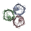 8bytC  8bz2C M: map data used to model this data C: citing same article ( |
|---|---|
| Similar structure data |


-Links
 PDBj
PDBj- Assembly
| Deposited unit | 
|
|---|---|
| 1 |
|
-Components
| #1: Protein | Mass: 47555.062 Da / Num. of mol.: 3 Source method: isolated from a genetically manipulated source Source: (gene. exp.) Veillonella parvula (bacteria) / Strain: SKV38Gene: D3219_09385, DWV36_08570, FNLLGLLA_01389, GL281_07935, HMPREF1865_01844 Production host: Escherichia coli BL21(DE3) (bacteria) / References: UniProt: A0A100YN03 |
|---|
-Experimental details
-Experiment
| Experiment | Method: ELECTRON MICROSCOPY |
|---|---|
| EM experiment | Aggregation state: PARTICLE / 3D reconstruction method: single particle reconstruction |
- Sample preparation
| Component | Name: Veillonella parvula OmpM1 outer membrane attachment porin trimer Type: COMPLEX / Entity ID: all / Source: RECOMBINANT |
|---|---|
| Molecular weight | Value: 0.130 MDa / Experimental value: NO |
| Source (natural) | Organism: Veillonella parvula (bacteria) / Strain: SKV38 |
| Source (recombinant) | Organism: Escherichia coli BL21(DE3) (bacteria) |
| Buffer solution | pH: 7.5 Details: 10 mM HEPES-NaOH pH 7.5, 100 mM NaCl, 0.12% decyl maltoside (DM) |
| Specimen | Conc.: 11.6 mg/ml / Embedding applied: NO / Shadowing applied: NO / Staining applied: NO / Vitrification applied: YES |
| Specimen support | Grid material: COPPER / Grid mesh size: 200 divisions/in. / Grid type: Quantifoil R1.2/1.3 |
| Vitrification | Instrument: FEI VITROBOT MARK IV / Cryogen name: ETHANE / Humidity: 100 % / Chamber temperature: 277 K |
- Electron microscopy imaging
| Microscopy | Model: TFS GLACIOS |
|---|---|
| Electron gun | Electron source: FIELD EMISSION GUN / Accelerating voltage: 200 kV / Illumination mode: FLOOD BEAM |
| Electron lens | Mode: BRIGHT FIELDBright-field microscopy / Nominal magnification: 240000 X / Nominal defocus max: 2000 nm / Nominal defocus min: 800 nm |
| Image recording | Electron dose: 50.1 e/Å2 / Film or detector model: FEI FALCON IV (4k x 4k) / Num. of grids imaged: 1 / Num. of real images: 4284 |
- Processing
| Software |
| ||||||||||||||||||||||||||||||||||||
|---|---|---|---|---|---|---|---|---|---|---|---|---|---|---|---|---|---|---|---|---|---|---|---|---|---|---|---|---|---|---|---|---|---|---|---|---|---|
| EM software |
| ||||||||||||||||||||||||||||||||||||
| CTF correction | Type: PHASE FLIPPING AND AMPLITUDE CORRECTION | ||||||||||||||||||||||||||||||||||||
| Symmetry | Point symmetry: C1 (asymmetric) | ||||||||||||||||||||||||||||||||||||
| 3D reconstruction | Resolution: 3.15 Å / Resolution method: FSC 0.143 CUT-OFF / Num. of particles: 96280 / Symmetry type: POINT | ||||||||||||||||||||||||||||||||||||
| Atomic model building | Protocol: AB INITIO MODEL / Space: REAL | ||||||||||||||||||||||||||||||||||||
| Refinement | Cross valid method: NONE |
