 Movie
Movie Controller

[English] 日本語
 Yorodumi
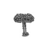
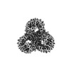
- EMDB-16332: Outer membrane attachment porin OmpM1 from Veillonella parvula, native -
+ Open data

- Basic information
| Entry |  | |||||||||
|---|---|---|---|---|---|---|---|---|---|---|
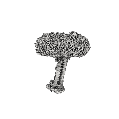
| Title | Outer membrane attachment porin OmpM1 from Veillonella parvula, native | |||||||||
 Map data Map data | B-factor sharpened (-97.5 angstrom sq.) map from non-uniform refinement | |||||||||
 Sample Sample |
| |||||||||
 Keywords Keywords | Outer membrane attachment /  porin / peptidoglycan-binding / nutrient transport / MEMBRANE PROTEIN porin / peptidoglycan-binding / nutrient transport / MEMBRANE PROTEIN | |||||||||
| Function / homology | S-layer / S-layer homology domain / S-layer homology domain / S-layer homology (SLH) domain profile. / S-layer homology domain-containing protein Function and homology information Function and homology information | |||||||||
| Biological species |  Veillonella parvula (bacteria) Veillonella parvula (bacteria) | |||||||||
| Method | single particle reconstruction / cryo EM / Resolution: 3.28 Å | |||||||||
 Authors Authors | Silale A / van den Berg B | |||||||||
| Funding support |  United Kingdom, 1 items United Kingdom, 1 items
| |||||||||
 Citation Citation | Journal: Nat Commun / Year: 2023 Title: Dual function of OmpM as outer membrane tether and nutrient uptake channel in diderm Firmicutes. Authors: Augustinas Silale / Yiling Zhu / Jerzy Witwinowski / Robert E Smith / Kahlan E Newman / Satya P Bhamidimarri / Arnaud Baslé / Syma Khalid / Christophe Beloin / Simonetta Gribaldo / Bert van den Berg /  Abstract: The outer membrane (OM) in diderm, or Gram-negative, bacteria must be tethered to peptidoglycan for mechanical stability and to maintain cell morphology. Most diderm phyla from the Terrabacteria ...The outer membrane (OM) in diderm, or Gram-negative, bacteria must be tethered to peptidoglycan for mechanical stability and to maintain cell morphology. Most diderm phyla from the Terrabacteria group have recently been shown to lack well-characterised OM attachment systems, but instead have OmpM, which could represent an ancestral tethering system in bacteria. Here, we have determined the structure of the most abundant OmpM protein from Veillonella parvula (diderm Firmicutes) by single particle cryogenic electron microscopy. We also characterised the channel properties of the transmembrane β-barrel of OmpM and investigated the structure and PG-binding properties of its periplasmic stalk region. Our results show that OM tethering and nutrient acquisition are genetically linked in V. parvula, and probably other diderm Terrabacteria. This dual function of OmpM may have played a role in the loss of the OM in ancestral bacteria and the emergence of monoderm bacterial lineages. | |||||||||
| History |
|
- Structure visualization
| Supplemental images |
|---|
- Downloads & links
-EMDB archive
| Map data | emd_16332.map.gz | 97.4 MB | EMDB map data format | |
|---|---|---|---|---|
| Header (meta data) | emd-16332-v30.xmlemd-16332.xml | 16.7 KB 16.7 KB | Display Display | EMDB header |
| FSC (resolution estimation) | emd_16332_fsc.xml | 9.9 KB | Display | FSC data file |
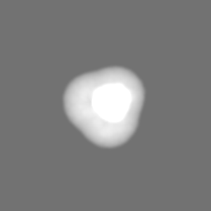
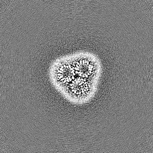
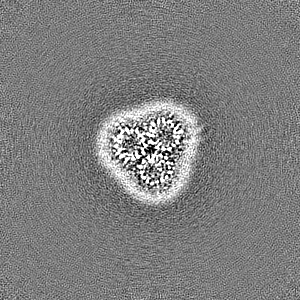
| Images |  emd_16332.png emd_16332.png | 52.9 KB | ||
| Masks | emd_16332_msk_1.map | 103 MB | Mask map | |
| Filedesc metadata | emd-16332.cif.gz | 5.9 KB | ||
| Others | emd_16332_half_map_1.map.gzemd_16332_half_map_2.map.gz | 95.5 MB 95.5 MB | ||
| Archive directory |  http://ftp.pdbj.org/pub/emdb/structures/EMD-16332ftp://ftp.pdbj.org/pub/emdb/structures/EMD-16332 http://ftp.pdbj.org/pub/emdb/structures/EMD-16332ftp://ftp.pdbj.org/pub/emdb/structures/EMD-16332 | HTTPS FTP |
-Related structure data
| Related structure data |  8bysMC  8bymC  8bytC  8bz2C C: citing same article ( M: atomic model generated by this map |
|---|---|
| Similar structure data |
-Links
| EMDB pages | EMDB (EBI/PDBe) / EMDataResource |
|---|
-Map
| File | Download / File: emd_16332.map.gz / Format: CCP4 / Size: 103 MB / Type: IMAGE STORED AS FLOATING POINT NUMBER (4 BYTES) | ||||||||||||||||||||
|---|---|---|---|---|---|---|---|---|---|---|---|---|---|---|---|---|---|---|---|---|---|
| Annotation | B-factor sharpened (-97.5 angstrom sq.) map from non-uniform refinement | ||||||||||||||||||||
| Voxel size | X=Y=Z: 1.148 Å | ||||||||||||||||||||
| Density |
| ||||||||||||||||||||
| Symmetry | Space group: 1 | ||||||||||||||||||||
| Details | EMDB XML:
|
-Supplemental data
-Mask #1
| File | emd_16332_msk_1.map | ||||||||||||
|---|---|---|---|---|---|---|---|---|---|---|---|---|---|
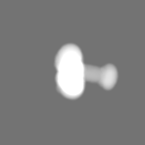
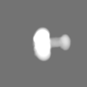
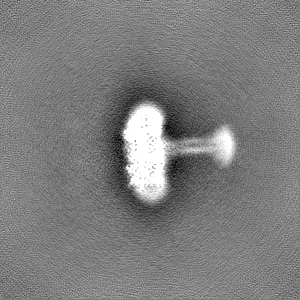
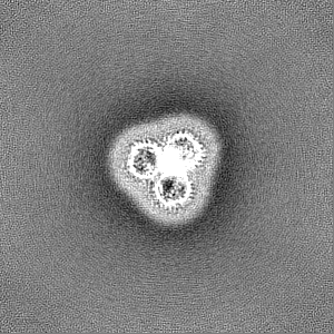
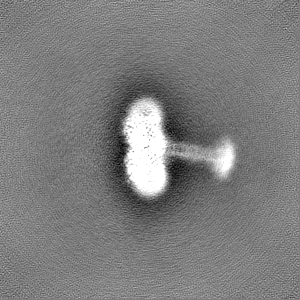
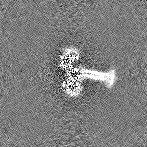
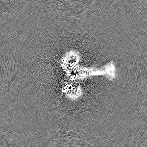
| Projections & Slices |
| ||||||||||||
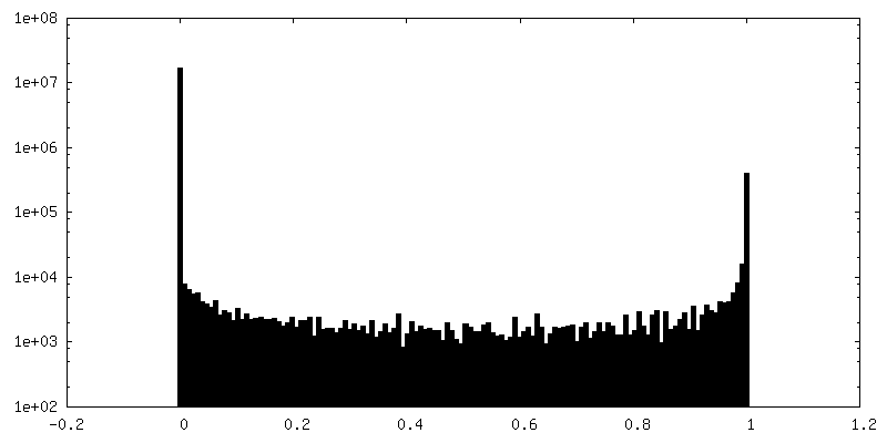
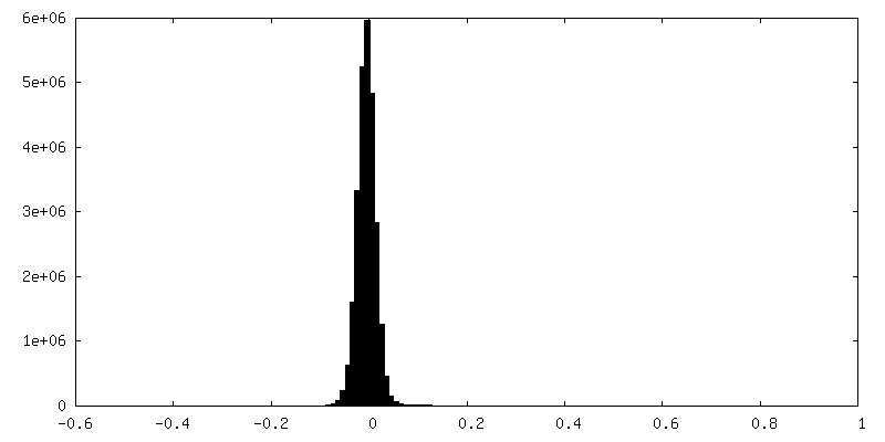
| Density Histograms |
 Z
Z Y
Y X
X


-Half map: Half map 1
| File | emd_16332_half_map_1.map | ||||||||||||
|---|---|---|---|---|---|---|---|---|---|---|---|---|---|
| Annotation | Half map 1 | ||||||||||||
| Projections & Slices |
| ||||||||||||
| Density Histograms |


-Half map: Half map 2
| File | emd_16332_half_map_2.map | ||||||||||||
|---|---|---|---|---|---|---|---|---|---|---|---|---|---|
| Annotation | Half map 2 | ||||||||||||
| Projections & Slices |
| ||||||||||||
| Density Histograms |


- Sample components
-Entire : Outer membrane attachment porin OmpM1 trimer
| Entire | Name: Outer membrane attachment porin OmpM1 trimer |
|---|---|
| Components |
|
-Supramolecule #1: Outer membrane attachment porin OmpM1 trimer
| Supramolecule | Name: Outer membrane attachment porin OmpM1 trimer / type: complex / ID: 1 / Parent: 0 / Macromolecule list: all |
|---|---|
| Source (natural) | Organism: Veillonella parvula (bacteria) / Strain: SKV38 |
| Molecular weight | Theoretical: 130 KDa |
-Macromolecule #1: S-layer homology domain-containing protein
| Macromolecule | Name: S-layer homology domain-containing protein / type: protein_or_peptide / ID: 1 / Number of copies: 3 / Enantiomer: LEVO |
|---|---|
| Source (natural) | Organism: Veillonella parvula (bacteria) / Strain: SKV38 |
| Molecular weight | Theoretical: 46.547863 KDa |
| Recombinant expression | Organism: Veillonella parvula (bacteria) |
| Sequence | String: MKKQFATMLA ATAVLGVTTA FAANPFSDVT PDSWAYQAVS QLAQAGIVNG YPDGTFKGQN NITRYEMAQM VAKAMANQDR ANAEQQAMI NRLADEFSNE LNNLGVRVSR LEDRVGNVKV TGDARIRYQG SEDKGVYKAN SKSLTDGRAR VQFNANVNDK T QAVVRVKG ...String: MKKQFATMLA ATAVLGVTTA FAANPFSDVT PDSWAYQAVS QLAQAGIVNG YPDGTFKGQN NITRYEMAQM VAKAMANQDR ANAEQQAMI NRLADEFSNE LNNLGVRVSR LEDRVGNVKV TGDARIRYQG SEDKGVYKAN SKSLTDGRAR VQFNANVNDK T QAVVRVKG NYEFGDSTKG SQATIDRAYV DHKFGSNVSA KAGRFQQTIG GGLMYDDTFD GAQLNVGNDK VQVQGAYGYM ID GAADGNS KSDNPSVSYV GLKGKVGKES SVGGFYSRLS SGNLNHNGVT VNSDKQDVYG FNADFRKNKL WAGGEWLKAS NVD NSQAWT AGLGYGNYDI AKKGTWDVKG QYFNQKANAP IVSSTWDQAY DLTNTSNGYK GYMASVDYAV QDNVGLSAGY GFNS KDQSG NDLSDFYRAE LNYKFGGHHH HHH UniProtKB: S-layer homology domain-containing protein |
-Experimental details
-Structure determination
| Method | cryo EM |
|---|---|
 Processing Processing | single particle reconstruction |
| Aggregation state | particle |
-Sample preparation
| Concentration | 8 mg/mL |
|---|---|
| Buffer | pH: 7.5 Details: 10 mM HEPES-NaOH, 100 mM NaCl, 0.12% decyl maltoside |
| Grid | Model: Quantifoil R1.2/1.3 / Material: COPPER / Mesh: 200 / Support film - Material: CARBON / Support film - topology: HOLEY / Pretreatment - Type: GLOW DISCHARGE / Pretreatment - Atmosphere: AIR |
| Vitrification | Cryogen name: ETHANE / Chamber humidity: 100 % / Chamber temperature: 277 K |
- Electron microscopy
| Microscope | TFS GLACIOS |
|---|---|
| Electron beam | Acceleration voltage: 200 kV / Electron source: FIELD EMISSION GUN |
| Electron optics | Illumination mode: FLOOD BEAM / Imaging mode: BRIGHT FIELDBright-field microscopy / Nominal defocus max: 2.0 µm / Nominal defocus min: 1.0 µm / Nominal magnification: 240000 |
| Image recording | Film or detector model: FEI FALCON IV (4k x 4k) / Number grids imaged: 1 / Number real images: 6505 / Average electron dose: 50.0 e/Å2 |
-Image processing
| Startup model | Type of model: INSILICO MODEL |
|---|---|
| Initial angle assignment | Type: MAXIMUM LIKELIHOOD / Software - Name: cryoSPARC (ver. 3.3.2) |
| Final angle assignment | Type: MAXIMUM LIKELIHOOD / Software - Name: cryoSPARC (ver. 3.3.2) |
| Final reconstruction | Applied symmetry - Point group: C1 (asymmetric) / Resolution.type: BY AUTHOR / Resolution: 3.28 Å / Resolution method: FSC 0.143 CUT-OFF / Software - Name: cryoSPARC (ver. 3.3.2) / Number images used: 144245 |
| FSC plot (resolution estimation) | 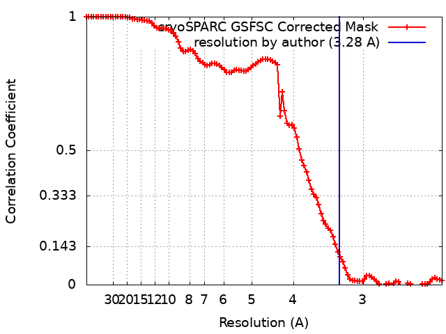 |
-Atomic model buiding 1
| Refinement | Space: REAL / Protocol: AB INITIO MODEL |
|---|---|
| Output model | PDB-8bys: |
